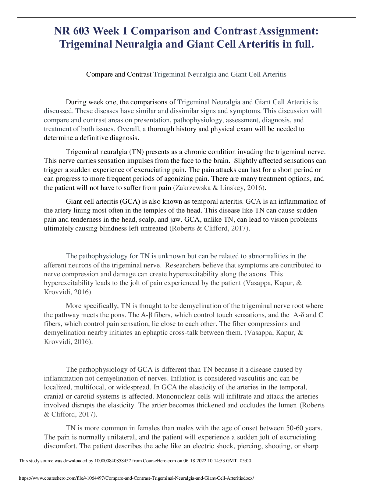
Compare and Contrast Trigeminal Neuralgia and Giant Cell Arteritis
During week one, the comparisons of Trigeminal Neuralgia and Giant Cell Arteritis is
discussed. These diseases have similar and dissimilar signs and sy
...
Compare and Contrast Trigeminal Neuralgia and Giant Cell Arteritis
During week one, the comparisons of Trigeminal Neuralgia and Giant Cell Arteritis is
discussed. These diseases have similar and dissimilar signs and symptoms. This discussion will
compare and contrast areas on presentation, pathophysiology, assessment, diagnosis, and
treatment of both issues. Overall, a thorough history and physical exam will be needed to
determine a definitive diagnosis.
Trigeminal neuralgia (TN) presents as a chronic condition invading the trigeminal nerve.
This nerve carries sensation impulses from the face to the brain. Slightly affected sensations can
trigger a sudden experience of excruciating pain. The pain attacks can last for a short period or
can progress to more frequent periods of agonizing pain. There are many treatment options, and
the patient will not have to suffer from pain (Zakrzewska & Linskey, 2016).
Giant cell arteritis (GCA) is also known as temporal arteritis. GCA is an inflammation of
the artery lining most often in the temples of the head. This disease like TN can cause sudden
pain and tenderness in the head, scalp, and jaw. GCA, unlike TN, can lead to vision problems
ultimately causing blindness left untreated (Roberts & Clifford, 2017).
The pathophysiology for TN is unknown but can be related to abnormalities in the
afferent neurons of the trigeminal nerve. Researchers believe that symptoms are contributed to
nerve compression and damage can create hyperexcitability along the axons. This
hyperexcitability leads to the jolt of pain experienced by the patient (Vasappa, Kapur, &
Krovvidi, 2016).
More specifically, TN is thought to be demyelination of the trigeminal nerve root where
the pathway meets the pons. The A-β fibers, which control touch sensations, and the A-δ and C
fibers, which control pain sensation, lie close to each other. The fiber compressions and
demyelination nearby initiates an ephaptic cross-talk between them. (Vasappa, Kapur, &
Krovvidi, 2016).
The pathophysiology of GCA is different than TN because it a disease caused by
inflammation not demyelination of nerves. Inflation is considered vasculitis and can be
localized, multifocal, or widespread. In GCA the elasticity of the arteries in the temporal,
cranial or carotid systems is affected. Mononuclear cells will infiltrate and attack the arteries
involved disrupts the elasticity. The artier becomes thickened and occludes the lumen (Roberts
& Clifford, 2017).
TN is more common in females than males with the age of onset between 50-60 years.
The pain is normally unilateral, and the patient will experience a sudden jolt of excruciating
discomfort. The patient describes the ache like an electric shock, piercing, shooting, or sharp
like a knife stab characteristics. The discomfort will normally last for a couple of seconds or
This study source was downloaded by 100000831988016 from CourseHero.com on 04-18-2022 07:30:01 GMT -05:00
https://www.coursehero.com/file/41064497/Compare-and-Contrast-Trigeminal-Neuralgia-and-Giant-Cell-Arteritisdocx/
can become routine with episodes lasting days, week, or months. The locations are normally in
the teeth, jaw, cheeks, and will be less common in the forehead and eye, unlike GCA. In TN
there are not associated symptoms as a complication (Vasappa, Kapur, & Krovvidi, 2016).
Like TN, GCA is also more common in females than males. The age is older being with
onsets of problems greater than 50 years of age with the mean age being 71 years. The pain is
experienced unilaterally like TN but is experienced bilaterally. The pain has a similar sudden
onset, but the patient will experience symptoms like a headache. The person will experience
pain in the temporal and occipital areas and will not be felt in the teeth or jaws. The frequency
of pain can vary unlike the sudden unknown onset of the TN. Som complications of GCA can
lead to permanent blindness, double vision, scalp tenderness, and overall joint aching (Roberts
& Clifford, 2017).
To diagnosis TN the provider typically uses history and clinical features to determine the
cause. A neurological exam will be used to detect trigger areas. Investigations around the
trigeminal nerve or fifth cranial nerve are examined. The patient's description of pain is a great
diagnosis tool to support the presence of TN. An MRI is used to help rule out secondary cases of
TN. This secondary causes could be like MS or a tumor causing decompression of
nerves(Vasappa, Kapur, & Krovvidi, 2016).
GCA is more difficult to diagnosis than TN because it can resemble other conditions.
Like with TN the provider will ask about symptoms, medical history and conduct a thorough
exam. The exam will focus more on temporal arteries. One or both of the temporal arteries will
present with tenderness, a reduced pulse, and will feel hard to touch(Koster, Matteson, &
Warrington, 2018.
GCA will require blood work, unlike TN, and these test will be erythrocyte sedimentation
rate, and C-reactive protein (CRP). The sed rate shows how fast red blood cells fall to the bottom
of the collection tube which can indicate some inflammation in the body. The CRP measure
elements produced by the liver that indicative of swelling (Koster, Matteson, & Warrington,
2018.
The imaging used to diagnosis GCA is Doppler ultrasound, magnetic resonance
angiography (MRA), and positron emission tomography (PET). The doppler can be used to see
how well the blood is flowing in the blood vessels. A slow movement would be indicative of
closed lumen or edema. The MRA is used with an MRI to show contrast pictures inside the blood
vessels. The PET scan is also similar to an MRA but uses a tracer solution that creates a detailed
picture of the patient’s larger vessels and illuminates areas of swelling. GCA requires more blood
work and imaging than TN. To ultimately diagnosis GCA a biopsy will produce a specimen
including abnormally large cells creating inflammation Matteson, & Warrington, 2018).
The treatment for TN revolves around anticonvulsants and neuropathic pain medications.
Examples are carbamazepine, gabapentin, pregabalin, and amitriptyline. Carbamazepine is
known to prevent synaptic triggering in the trigeminal nucleus. The other medications focus on
treating TN like a neuropathic pain condition. If medications fail then surgical procedures are
needed (Vasappa, Kapur, & Krovvidi, 2016).
This study source was downloaded by 100000831988016 from CourseHero.com on 04-18-2022 07:30:01 GMT -05:00
https://www.coursehero.com/file/41064497/Compare-and-Contrast-Trigeminal-Neuralgia-and-Giant-Cell-Arteritisdocx/
The treatment for GCA is different than TN because it focuses around reducing
inflammation. High doses of corticosteroids like prednisone are used to reduce the pain by
lessening the irritation. The patient should feel symptoms subside in a few days after treatment is
started. When the patient is tapering off the steroid, headaches can be present, and they may
develop signs of polymyalgia rheumatica. These flare-ups may require an added boost of
steroids. GCA does not require a surgical route to eliminate symptoms like TN Matteson, &
Warrington, 2018).
The comparison and contrast of GCA and TN are interesting to review. Both disease
present with sudden pain, one originates in the jaw and the other as a headache. They are very
different in pathology because TN relates to nerve compression as GCA is a result of
inflammation in the blood vessels. Providers will assess both issues by a collection of medical
history, symptoms present, and a detailed assessment focusing on neurological deficits. The
diagnosis is simpler for TN and GCA requires imagining and blood work to identify. In the end,
both diseases require different treatment and
[Show More]



 F21.png)


.png)
.png)



.png)

.png)

