Anth 1120 Review Questions and Answers latest 2023,100% CORRECT
Biology > LECTURE SLIDES/NOTES > PORTAGE LEARNING. BIO 152. AP2 Lab4.docx. 100% comprehension. APPROVED PASS RATE (All)
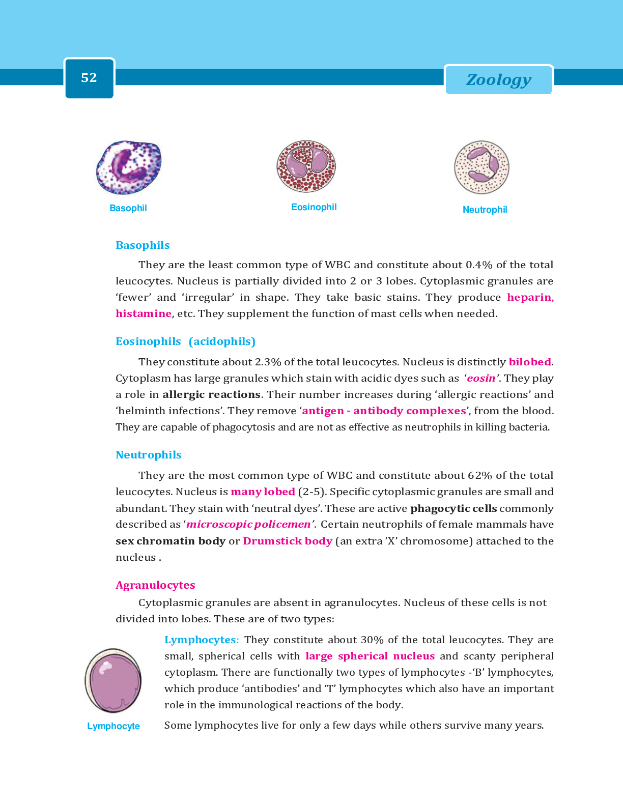
Circulatory System-Blood Flow Tracings Circulatory system- how the blood from the heart is circulated throughout the body. General blood flow through the body after it leaves the heart follows a map ... ping system. From the heart, it flows through arteries, then capillaries, and comes back to the heart by way of veins. BLOOD VESSELS IN THE CIRCULATORY SYSTEM: Arteries- referred to as efferent blood vessel in the circulatory system o They always carry blood away from the heart. o Typically carry oxygenated blood. (except in the pulmonary artery) o 1. Conducting arteries: very large arteries and typically exit from the heart They have the need/ability to expand when the heart beats so they withstand the splurge. So, with every heartbeat, blood surges out of the heart into these large vessels. To do this, they must have a layer of elastic tissue that gives it the ability to stretch and recoil when the heart is relaxing. EX: aorta and pulmonary trunk o Atherosclerosis- when arteries age and stiffen because plaque builds up in the arteries, they loose that elasticity and the ability to stretch when blood surges into them. It results in an increase in pressure in those vessels which can lead to an aneurism- a weak point in a artery and with each heartbeat, that little thin walled area will pulsate and it can further and further weaken and eventually rupture which can lead to death or stroke. o 2. Distributing arteries: A.k.a. medium or muscular arteries They are direct branches from the conducting arteries Have a very thick muscular wall (up to 40 layers of smooth muscle) The muscular wall in a distributing artery makes up 75% of the arterial wall itself. EX: brachial artery, femoral artery (named for area where they are distributing their blood) o 3. Resistance arteries: A.k.a. small arteries Too numerous to name o 4. Metarterioles: Very short vessels that will link the arteriole system to the capillaries. AFTER BLOOD FLOWS THROUGH THE ARTERIAL SYSTEM, IT THEN FLOWS INTO A SERIES OF CAPILLARY BEDS. o 5. Capillaries: Known as the exchange vessels because they have very thin walls that exchange oxygen, glucose, and other nutrients in the body. They connect the smallest arteries to the smallest veins. Veins- afferent blood vessels because they bring blood back to the heart o They typically carry deoxygenated blood (except the pulmonary vein) o They are called Capacitance Vessels because they typically control a large amount of volume. o Can stretch more easily than arteries and have thin, flaccid walls and can therefore accommodate a larger volume of blood. o Comparing arteries to veins: In a resting person, about 11% of the blood is found in the arteries while 54% is found in the veins. Veins are subject to much lower blood pressure because they are further away from the heart. Because of that, they can have thinner walls compared to the arteries that need more muscular support. In the arteriole system, we go from large to small vessels and the venous system is opposite. o 6. Post capillary vein: Very small veins called venules. o 7. Muscular Venules: Very small and too numerous to name o 8. Medium veins: They drain blood from specific areas of the body, organs and muscles. EX: radial and ulnar veins (drain blood out of the forearm) Unique: they contain valves- flaps of tissue that extend into the lumen and point upwards towards the heart. Because veins have a very low blood pressure, they don’t have the strength to pump the blood back to the heart against the pull of gravity. Skeletal muscles that surround the veins also help to pump the blood o EX: when calve muscles contract, they squeeze around the vein and push blood up through the valve. o Kind of like a “milking action” known as the skeletal muscle pump. When the valves weaken and can’t perform their job, varicose veins will result. They are visible through the skin due to the blood pooling and backflowing into the venous system. o 9. Venous Sinuses: Large vessels with very thin walls and very large lumens EX: coronary sinus of the heart where blood that flows through the heart, will dump into the coronary sinus before entering back into the vena cava. EX: Dural sinuses in the brain. They collect the blood flow from the brain before they return to the heart. o 10. Large veins EX: Superior/Inferior Vena Cava- will bring blood back to the heart and finally start the system all over again. REVIEW OF BLOOD FLOW THROUGH THE HEART Inferior Vena Cava: drains from anywhere below the diaphragm Superior Vena Cava: drains from anywhere above the diaphragm They both bring blood into the right atrium of the heart Tricuspid valve Right ventricle Pulmonary semilunar valve----pulmonary trunk-----left and right pulmonary arteries----- flow to the lungs Blood picks up oxygen in the lungs and then to the Pulmonary veins Left atrium Bicuspid valve Left ventricle Aortic semilunar valve Ascending aorta: Off of the ascending aorta, the branches for the [Show More]
Last updated: 3 years ago
Preview 1 out of 36 pages

Buy this document to get the full access instantly
Instant Download Access after purchase
Buy NowInstant download
We Accept:

Can't find what you want? Try our AI powered Search
Connected school, study & course
About the document
Uploaded On
Apr 28, 2022
Number of pages
36
Written in
All
This document has been written for:
Uploaded
Apr 28, 2022
Downloads
0
Views
250
Scholarfriends.com Online Platform by Browsegrades Inc. 651N South Broad St, Middletown DE. United States.
We're available through e-mail, Twitter, Facebook, and live chat.
FAQ
Questions? Leave a message!
Copyright © Scholarfriends · High quality services·

.png)