BIOD152 Anatomy and Physiology II
Quiz #1:Module 1
Question 1
Not yet graded / 0 pts
Anatomy and Physiology of the Nervous System: Introduction
1. What is the difference between anatomy and physiology?
2. T
...
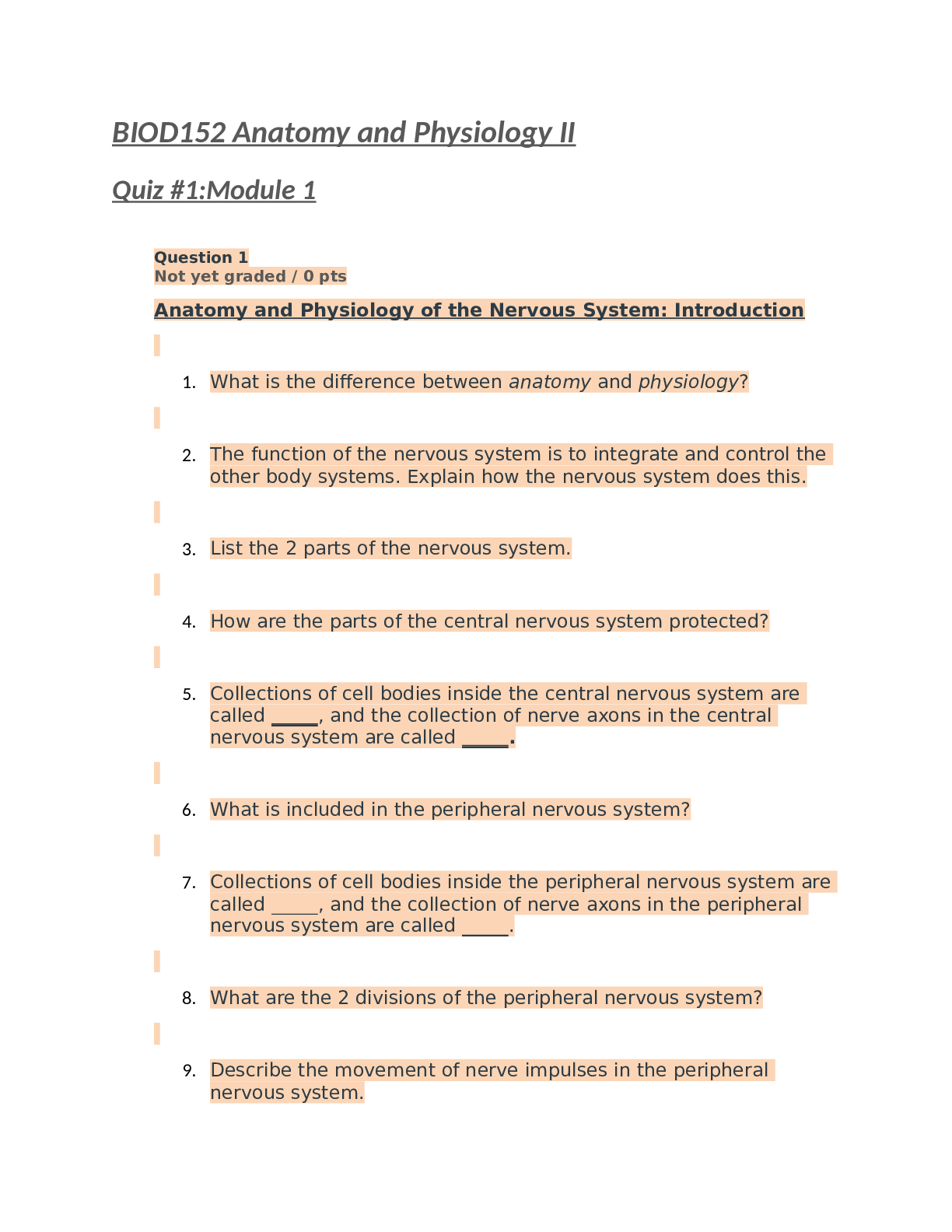
BIOD152 Anatomy and Physiology II
Quiz #1:Module 1
Question 1
Not yet graded / 0 pts
Anatomy and Physiology of the Nervous System: Introduction
1. What is the difference between anatomy and physiology?
2. The function of the nervous system is to integrate and control the other body systems. Explain how the nervous system does this.
3. List the 2 parts of the nervous system.
4. How are the parts of the central nervous system protected?
5. Collections of cell bodies inside the central nervous system are called , and the collection of nerve axons in the central nervous system are called .
6. What is included in the peripheral nervous system?
7. Collections of cell bodies inside the peripheral nervous system are called , and the collection of nerve axons in the peripheral nervous system are called .
8. What are the 2 divisions of the peripheral nervous system?
9. Describe the movement of nerve impulses in the peripheral nervous system.
10. What are the 2 divisions of the efferent division of the peripheral nervous system?
11. What is controlled by the somatic and autonomic nervous systems?
12. What are the 2 divisions of the autonomic nervous system?
13. What is the function of the sympathetic nervous system?
14. What is the function of the parasympathetic nervous system? Your Answer:
1.
Anatomy is the physical aspects of the body, while physiology is the function and inner workings of the body's systems.
2.
The nervous system utilizes electrical impulses and signals to receive, process, and control or respond to stimuli.
3.
Central and peripheral nervous system 4.
The spinal cord is protected by the vertebrae and the brain is protected by the skull.
5.
Nuclei, Tracts 6.
This includes every nerve not in the brain of spinal cord, so all cranial and spinal nerves.
7.
Ganglia, nerves
8.
Afferent (sensory) and efferent (motor) 9.
Peripheral nervous system receives impulses from sensory nerves via afferent division and forwards onto the central nervous system. The impulses from the central nervous system then signals the muscles through efferent signaling.
10.
SOmatic nervous system: Generally under conscious control, so contorl of muscles skin and joints.
Autonomic Nervous system: The involuntary portion, controls smooth muscles and glands/endocrine ssytem.
11.
Somatic controls skeletal muscles, skin and joints. voluntary movement control
Autonomic controls smooth muscle (digestion, unconscious muscles) and organs/glands
12.
Symapathetic nervous system: the "Fight or flight", prepares for stress, emergencies, or strenuous activity.
Parasympathetic nervous System: conserves energy "rest and Digest", and supports digestion.
13.
Sympathetic nervous system is the Fight or Flight, and activates for stress, activity, or emergencies.
14.
Parasympathetic Nervous System: "rest and Digest" activates in normal resting circumstances, to conserve energy and promote normal body functions when energy is readily available for digestion.
1. Anatomy focuses on the structure of the
body. Physiology looks at how the body parts function together. The physiology of the body depends on the anatomy of the body.
2. The nervous system receives and processes information and sends out signals to the muscles and glands to elicit an
appropriate response. In this way, the nervous system integrates and controls the other systems of the body.
3. CNS and PNS (central nervous system and peripheral nervous system)
4. The skull protects the brain and the vertebrae protect the spinal cord.
5. nuclei, tracts.
6. The PNS includes the cranial nerves and the spinal nerves.
7. ganglia, nerves.
8. Afferent, efferent
9. The peripheral nervous system receives impulses from the sensory organs via the afferent division and then relays signals or impulses from the central nervous system to muscles and glands via the motor or efferent division.
10. Somatic system and the autonomic system.
11. The somatic system controls the movements of skeletal muscles, skin, and joints. The autonomic system nerves control the glands and smooth muscles of the internal organs.
12. sympathetic and parasympathetic nervous systems.
13. The sympathetic nervous system activates and prepares the body for vigorous muscular activity, stress, and emergencies.
14. The parasympathetic nervous system generally operates during normal situations, permits digestion, and conserves energy.
Question 2
Not yet graded / 0 pts
Neurons
15. Label the components of a neuron.
16. List 3 unusual characteristics of neurons.
17. List the 3 parts that all neurons contain.
18. Describe the structure and function of the neuron cell body.
19. Describe the structure and function of the dendrite.
20. Describe the axon, including the number in each neuron, function, structure and organelles.
21. Describe the function and site of synthesis and storage of neurotransmitters.
22. What is a synapse?
23. A post-synaptic neuron is a neuron that is found the synapse.
24. neurons have three or more extensions from the cell body and have one axon and many dendrites.
25. neurons have a central cell body with two extensions.
26. neurons have one extension of f the cell body which branches into two: one central process running to the CNS and another peripheral process running to the sensory receptor.
27. neurons are unipolar and function to carry information from the peripheral to the central nervous system.
28. These types of neurons are also called association neurons.
29. neurons send messages from the central nervous system to the peripheral.
Your Answer: 15.
Cell body or Soma Axon
Dendrites
nucleus Axon Hillock
Myelin Sheath Nodes of Ranvier Axon terminal 16.
do not undergo Mitosis, Can only survive a few minutes without oxygen, can survive an entire lifetime, dead cells in CNS cannot be replaced.
17.
Cell body or Soma Axon
Dendrites 18.
Neuron Cell body houses all organelles and is the location where all nerve cell materials are stored and created. Does NOT include centrioles, which allow for mitosis. main function is to produce neurotransmitters, which are stored in the axon terminal vesicles.
19.
Dendrites are branch-like structures branching out from the cell body that serve to receive signals from other nerve cells to propogate the elecrtical impulses.
20.
Axon send signals from the nerve cell to other cells through axon terminals. Axons contain cell components like the cell body, however do not contain endoplasmic reticulum. Requires proteins from the cell body to be sent down for neurotransmitters. There is always a single Axon transmitting signals away from the cell body.
Contain Schwann cells whiuch make up the Myelin Sheath surrounding axons. Gaps between the Schwann cells are called nodes of ranvier, where signals jump between nodes through Saltatory conduction.
21.
Neurotransmitters are used to transmit signals between nerve cells, and are created by the cell body as proteins and transported to the axon terminal vesicles for storage.
22.
The gap between two neurons where neurotransmitters are used to propogate the signal.
23.
The neuron found after the synapse. Presynaptic the the neron before the synapse transmitting the signal.
24.
Multipolar Neuron 25.
Bi-polar Neuron 26.
Unipolar Neuron 27.
Sensory Neurons 28.
interneurons 29.
Motor Neurons
15. See figure in module
16. Neurons do not undergo mitosis (cell division), require enormous amounts of fuel being able to survive just minutes without oxygen and can last an entire human lifetime.
17. The dendrites, the cell body, and the axon.
18. Synthesizes all nerve cell products, consists of a large nucleus with surrounding cytoplasm containing the normal organelles
19. Numerous short extensions that emanate from the cell body which receive information from other neurons conducting those nerve impulses toward the cell body.
20. Conducts nerve impulses away from the cell body to its axon terminals where it is emitted across a synapse to the dendrite of another neuron. Axons are composed of cells like the cell body but lack rough endoplasmic reticulum.
21. One of the main functions of the cell body is to manufacture neurotransmitters, which are chemicals stored in secretory vesicles at the end of axon terminals.When neurotransmitters are released by the axon terminal vesicles, they carry the transmission of the nerve impulse from one neuron to another.
22. A synapse is a gap between two neurons.
23. After
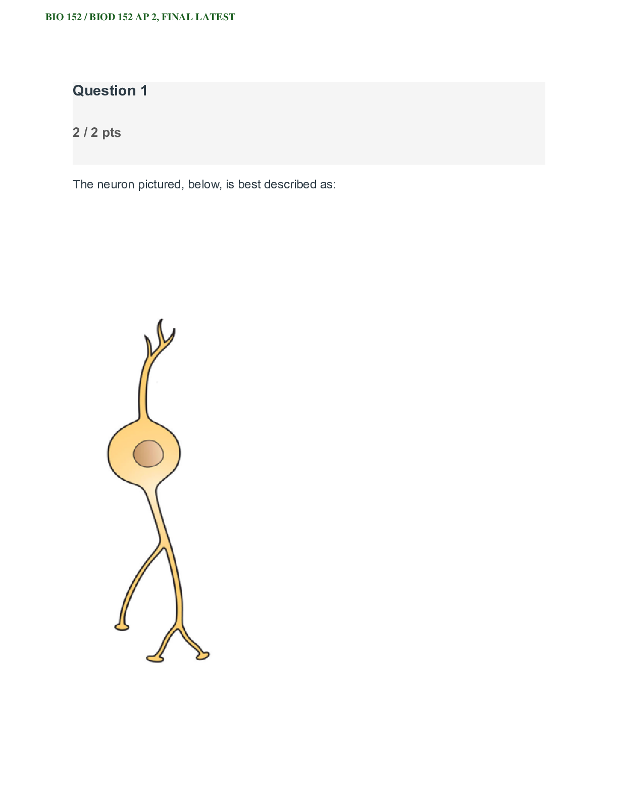
24. Multipolar
25. Bipolar
26. Unipolar (pseudounipolar)
27. Sensory
28. Interneurons
29. Motor
Question 3
Not yet graded / 0 pts
Neuroglial Cells
30. What is the function of neuroglial cells?
31. What are the peripheral nervous system neuroglial cell types?
32. True or False: Axons cannot regenerate in the peripheral nervous system.
33. True or False: Myelin sheath is continuous and has no gaps.
34. List the four types of support neuroglial cells in the central system and a function of each.
Your Answer:
30.
Neuroglial cells are "support'" cells that help maintain the ebst possible environment for nerve signal conduction and survivability.
31.
Schwann Cells surround the axon and increases the accuracy and speed of electrical impulses (fatty tissue)
Satellite Cells help regulate environment for the nerve cell by surrounding and protecting them.
32. F 33. F 34.
Ependymal Cells, which circulate cerebrospinal fluid and allow fluiid exchange between spinal cord, brain and CSF.
Oligodendrocytes insulate central nervous system axons.
Astrocytes manage chemical levels of blood and capilaries surrounding the nerves. (Blood Brain Barrier)
Microglial Cells offer protection by removing dead cells or cell materials and fighting infection.
30. Neuroglial cells are support cells, helping to support neurons to enable them to thrive in their needed environment.
31. Schwann cells; satellite cells
32. False
33. False
34. Ependymal cells circulate cerebrospinal fluid and allow fluid exchange between brain, spinal cord and cerebrospinal fluid (CSF). Oligodendrocytes act as the insulation for central nervous system axons. Astrocytes control chemical environment of neurons by wrapping around the blood capillaries, forming the blood brain barrier. Microglial cells protect the CNS by scavenging dead cells and infectious microorganisms.
Question 4
Not yet graded / 0 pts
Action Potentials
35. What is the technical term used to describe a nerve impulse and what causes the impulse?
36. An axon's membrane is polarized with a resting potential of -70 mV. Explain what this means and what maintains this resting potential.
37. What are the four steps of an action potential in order?
38. Describe what happens to the charges on the axon cell membrane during depolarization and what causes this to happen.
38. Describe what happens to the charges on the axon cell membrane during repolarization and what causes this to happen.
39. Describe what happens during afterpolarization.
40. What causes the difference in intensity of a sensation?
41. True or False: An impulse from a neuron moves in both directions.
42. What is meant by neuron signals being electrochemical in nature?
43. What is the chemical portion of neuron signal transmission?
44. How is an impulse passed from one nerve cell to another?
45. What prevents continuous stimulation of a nerve synapse and how is this accomplished?
46. What neurotransmitter helps regulate emotional responses and muscle tone?
47. What neurotransmitter is found at the neuromuscular junctions?
48. Once ACh is released in the NMJ, what happens to cause muscle contraction to occur?
Your Answer:
35.
Action potential is the process of conducting electrical impulse which occurs due to an unequal distribution of ions on either side of the membrane.
36.
This means the axon at rest is polarized, where the inside of the axon cell body is engative or has a lower charge than the outside. This is known as resting potential.
37.
Starts at Resting potential where all sodium and potassium gates are closed.
Deploarizes when sodium gates open and charge increases to +40
Repolarizes as the sodium gates close and potassium gates open allowing positive charge to leave axon.
Afterpolarization opr hyperpolarization potassium gates close slowly and allows voltage to drop below -70 before returning to -70 at resting potential.
38.
During depolarization, changes in the charge will allow sodium gates to open and rush positive charged sodium iuons into the cell.
39.
The shift in charge in the interior of the cell leads to external sodium gates closing and the internal potassium gates opening to allow potassium to leave the cell and lower intracellular charge.
40.
The potassium gates are slower to close, so more positive charge will elave the cell resulting in the charge dropping lower than -70 briefly, before the potassium gates close and the cell averages back out to -70
41.
The number of nerve cells being innervated and frequency.
42. F 43.
The eletrical signal is transmitted down the cell body while chemical neurotransmitters must pass the signal between axon and dendrite, or through the synaptic gap.
44.
Neruotransmitters 45.
neruotransmitters accross the synaptic gap. 46.
the cells inhibit the neurotransmitters through enzymes, packaging in vesicles, or through resorbing the neurotransmitters.
47.
DOpamine 48.
Acetylcholine 49.
Acetylcholine binds to receptors in muscles to open sodium channels, which triggers a sarcoplasmic reticulum action potential and releases Calcium, which results in muscle contraction.
35. A nerve impulse is called an action potential and is caused by the movement of unequally distributed ions on either side of an axon’s plasma membrane.
36. The axon plasma membrane is polarized, meaning that one side has a different charge than the other side. This difference called a resting potential means that the charge on the inside of the axon's cell membrane is 70 millivolts less than the outside of the membrane. A sodium- potassium pump using active transport carries ions across the plasma membrane and because three Na+ ions are pumped out as two K+ ions are pumped in a relative positive charge develops and is maintained on the outside of the membrane.
37. Resting Potential, Depolarization, Repolarization, Afterpolarization (hyperpolarization)
38. Sodium gates open and sodium rushes into the axon and the inside becomes more positive than the outside causing the membrane potential to become more positive.
39. The sodium gates close and potassium gates open allowing potassium to rush out of the axon. This returns a negative charge to the inside of the axon re-establishing the negative potential.
40. The potassium gates that open during repolarization are slow to close and there is an afterpolarization undershoot of the potential.
41. Due to the number of neurons stimulated and the frequency with which they are stimulated.
42. False
43. The signal moves from electrical (through the neuron) to chemical (in the synapse) to electrical again once the signal reaches the next neuron.
44. Neurotransmitters.
45. There is a minute fluid-filled space, called a synapse, between the axon terminal of the sending neuron and the dendrite of the receiving neuron. When a nerve impulse reaches the end of an axon, neurotransmitters are released into the synapse. These bind with a receptor on the next neuron, opening Na+ gates in the receiving dendrite which causes depolarization and the impulse is carried.
46. The short existence of neurotransmitters in the synapse prevents continuous stimulation. Some synapses contain enzymes that rapidly inactivate neurotransmitters and other synapses rapidly absorb the neurotransmitter.
47. Dopamine
48. Acetylcholine
49. Acetylcholine binds to receptors on the muscle fiber that cause sodium channels to open. Sodium rushes out of the muscle cell, triggering an action potential which reaches the sarcoplasmic reticulum. Calcium ions are released from the sarcoplasmic reticulum of the muscle cell causing the muscle to contract.
Question 5
Not yet graded / 0 pts
Reflexes
50. Define the term reflex. Give an internal and an external example.
51. Sensory information travels into the spinal cord via the of a nerve
52. What is the gray and white matter of the spinal cord?
53. What does the DRG contain?
54. Sensory neurons synapse on cells in the of the spinal cord.
55. Motor neuron cell bodies are in the of the spinal cord.
56. Motor neurons (axons) leave the spinal cord via the .
57. When does a ventral root transition to a spinal nerve?
58. Label the components of the cross section of a spinal cord.
59. Why is a spinal reflex faster than a conscious decision to move by the brain?
60. List the 5 components of a reflex arc.
61. The stretch reflex utilizes what type of specialized receptor to detect over-stretch?
62. What is the purpose of the stretch reflex? Your Answer:
50.
These are instantaneous involuntary responses from the nervous system. an internal example would be the regulation of certain organ or endocrine functions, like release of hormones due to specific bodily signals. External is like shivering due to external lower temperatures.
51.
Dorsal Root. 52.
Gray matter contains cell bodies, white matter is the axons of neurons. 53.
the dorsal root ganglia contains cell bodies of sensory neurons. 54.
interneurons. 55.
Anterior Horn 56.
Ventral Root 57.
Once it connects back to the dorsal root. 58.
59.
They only have to travel to the spinal cord and back and not all the way to the brain, so less nerves are needed to transmit the signal and the reaction time is lower.
60.
Receptor reacts to stimuli
Afferent neurons pass signal towards Central Nervous System. Integration center in CNS passes signal through synapse Efferent neuron passes signal along efferent pathway to effector.
effector responds by contracting muscle or producing chemical or hormone.
61.
Muscle Spindles 62.
protects the muscles from over exerting themselves to the point they tear or become damaged.
50. Reflexes are nearly instantaneous, automatic, involuntary motor responses to stimuli occurring inside or outside of the body. A subconscious reflex is the regulation of blood sugar by the hormones. An external example is touching a very hot object and immediately withdrawing your hand.
51. Dorsal root
52. Gray matter contains the cell bodies of neurons. The white matter of the spinal cord contains the axons of neurons.
53. Contains the cell bodies of sensory neurons. 54. Posterior horn
55. Anterior horn
56. Ventral root
57. A spinal nerve contains both sensory and motor neurons from the ventral and dorsal roots.
58. See figures in module.
59. Spinal reflexes are faster not only because they involve fewer neurons, but also because the electrical signal does not have to travel to the brain and back. Spinal reflexes only travel to the spinal cord and back which is a much shorter distance.
60. The receptor, the afferent neuron, the integration center, the efferent neuron and the effector.
61. Muscle spindles
62. Stretch reflexes are a special type of muscle reflex which protect the muscle against increases in length which may tear or damage muscle fibers.
[Show More]


.png)