*NURSING > QUESTIONS & ANSWERS > NURSING Practice Questions with accurate answers. 99% pass rate. Graded A+ (All)
NURSING Practice Questions with accurate answers. 99% pass rate. Graded A+
Document Content and Description Below
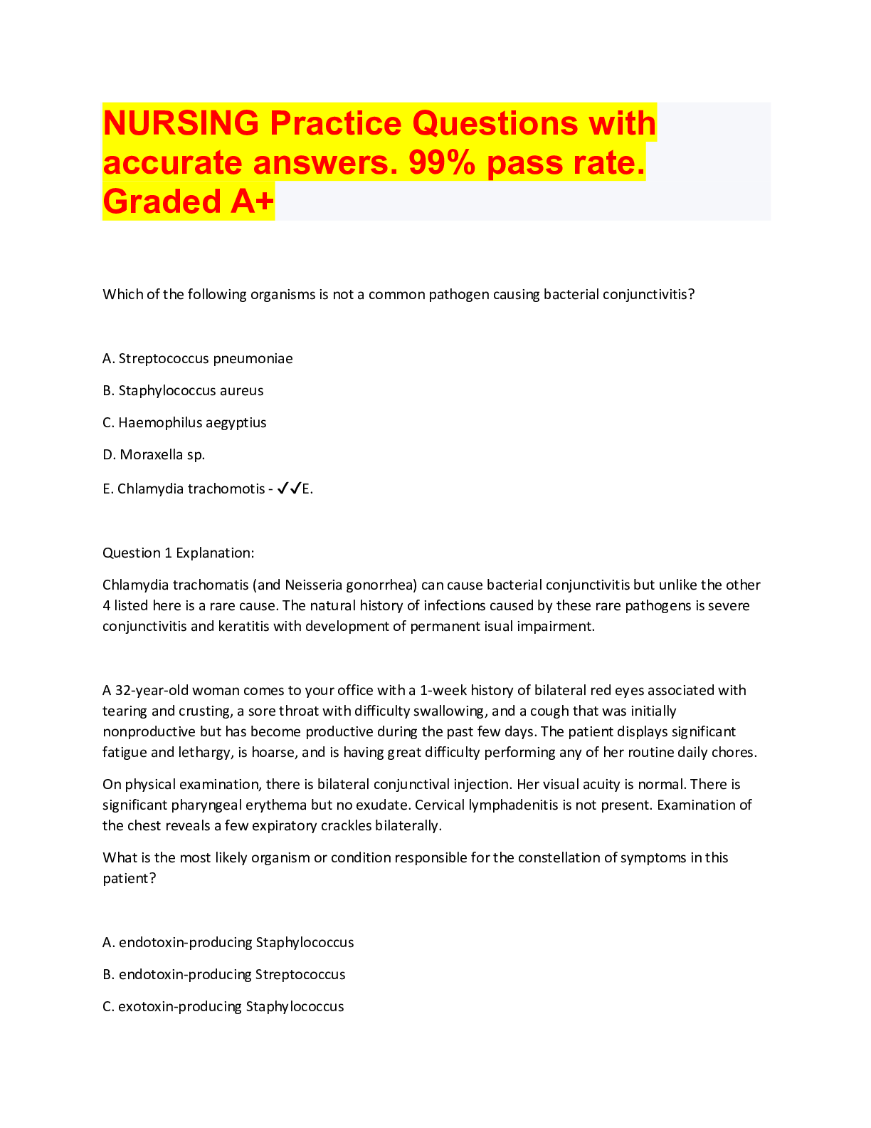
NURSING Practice Questions with accurate answers. 99% pass rate. Graded A+ Which of the following organisms is not a common pathogen causing bacterial conjunctivitis? A. Streptococcus pneumoni... ae B. Staphylococcus aureus C. Haemophilus aegyptius D. Moraxella sp. E. Chlamydia trachomotis - ✔✔E. Question 1 Explanation: Chlamydia trachomatis (and Neisseria gonorrhea) can cause bacterial conjunctivitis but unlike the other 4 listed here is a rare cause. The natural history of infections caused by these rare pathogens is severe conjunctivitis and keratitis with development of permanent isual impairment. A 32-year-old woman comes to your office with a 1-week history of bilateral red eyes associated with tearing and crusting, a sore throat with difficulty swallowing, and a cough that was initially nonproductive but has become productive during the past few days. The patient displays significant fatigue and lethargy, is hoarse, and is having great difficulty performing any of her routine daily chores. On physical examination, there is bilateral conjunctival injection. Her visual acuity is normal. There is significant pharyngeal erythema but no exudate. Cervical lymphadenitis is not present. Examination of the chest reveals a few expiratory crackles bilaterally. What is the most likely organism or condition responsible for the constellation of symptoms in this patient? A. endotoxin-producing Staphylococcus B. endotoxin-producing Streptococcus C. exotoxin-producing Staphylococcus D. activation of the autoimmune system E. none of the above - ✔✔E. Question 2 Explanation: This picture is completely consistent with adenovirus infection and a primary viral conjunctivitis. Viral agents, especially adenovirus, produce signs and symptoms of upper respiratory tract infection, with the red eye being prominent among those symptoms. With adenovirus, there is often associated conjunctival hyperemia, eyelid edema, and a serous or seropurulent discharge. Viral conjunctivitis is self-limited, lasting 1 to 3 weeks. If the conjunctivitis is definitely caused by a virus, no antibiotic treatment is necessary. There is no indication to perform a throat culture or any other test at this time. The only theoretical concerns are the "rales" that are present in both lung bases; you could argue that if the patient is sick enough, a chest radiograph may be indicated. A 7 year old boy presents to your clinic with thick purulent discharge from her right eye which was significantly worse this morning. What should you do next? A. take a sample of the discharge and send it to the lab for culture. B. send the patient home to perform warm compresses for 20 minutes 3 times daily tell them to come back if symptoms become worse C. moxifloxacin 0.5% drops tid for 7 to 10 days D. topical 1% prednisolone acetate qid - ✔✔C. Question 3 Explanation: If neither gonococcal nor chlamydial infection is suspected, most clinicians treat presumptively with moxifloxacin 0.5% drops tid for 7 to 10 days or another fluoroquinolone or trimethoprim/polymyxin B qid. A poor clinical response after 2 or 3 days indicates that the cause is resistant bacteria, a virus, or an allergy. Culture and sensitivity studies should then be done (if not done previously); results direct subsequent treatment. A 12 year old present to your office with red eyes, itching and tearing bilaterally. He has a past medical history significant for asthma. As you examine the inner eyelid what finding do you expect to see? A. cobblestone mucosa B. Kayser-Fleischer rings C. mucopurulent discharge D. dendritic ulcerations - ✔✔A. Question 4 Explanation: A classic finding of allergic conjunctivitis is cobblestone mucosa on the inner/upper eyelid. A 17-year-old girl comes to your office with a 1-day history of red eye. She describes not being able to open her right eye in the morning because of crusting and discharge. The right eye feels swollen and uncomfortable, although there is no pain. On examination, she has a significant redness and injection of the right bulbar and palpebral conjunctivae. There is a mucopurulent discharge present. No other abnormalities are present on physical examination. Her visual acuity is normal. A. bacterial conjunctivitis B. viral conjunctivitis C. allergic conjunctivitis D. autoimmune conjunctivitis - ✔✔A. Question 5 Explanation: This patient has a primary bacterial conjunctivitis. Unlike viral conjunctivitis, bacterial conjunctivitis will produce a mucopurulent discharge from the beginning. Symptoms are more often unilateral, and associated eye discomfort is common. In bacterial conjunctivitis, normal visual acuity is always maintained. There is usually uniform engorgement of all the conjunctival blood vessels. There is no staining of the cornea with fluorescein. Bacterial conjunctivitis should be treated with antibiotic drops such as sodium sulfacetamide, gentamicin, or fluoroquinolones. A 12 year-old presents with complaint of both eyes "watering." He also complains of sinus congestion and sneezing for two weeks. On exam vital signs are T-38°C, P-80/minute, and RR-20/minute. The eyes reveal mild conjunctival injection bilaterally, clear watery discharge, and no matting. Pupils are equal, round, and reactive to light and accommodation. The extraocular movements are intact. The funduscopic exam shows normal disc and vessels. The TMs are normal and the canals are clear. The nasal mucosa is boggy, with clear rhinorrhea. Which of the following is the most helpful pharmacologic agent? A.Artificial tears B. Tobramycin drops C. Erythromycin ointment D. Naphazoline (Naphcon-A) drops (Antihistamine) - ✔✔D. Question 6 Explanation: Naphazoline is a topical antihistamine that relieves symptoms of allergic conjunctivitis. A patient is evaluated in the office with a red eye. The patient awoke with redness and a watery discharge from the eye. The eyelids were not matted together. Examination reveals a palpable preauricular node. Which of the following is the most likely diagnosis? A. bacterial conjunctivitis B. viral conjunctivitis C. allergic conjunctivitis D. gonococcal conjunctivitis - ✔✔B. Question 7 Explanation: Viral conjunctivitis is associated with copious watery discharge and preauricular adenopathy. A 23 year-old sexually active female presents with a 4 day history of painless bilateral eye exudates which she describes as copious. Visual acuity is 20/20, generalized conjunctival inflammation with sparing of the cornea is noted on physical examination. Gram stain of the exudate reveals gram negative diplococci. Appropriate management of this case is A. ceftriaxone (Rocephin) B. polymyxin ophthalmic drops (Aerosporin) C. ciprofloxacin (Cipro) D. doxycycline (Doryx) - ✔✔A. Question 8 Explanation: With sparing of the cornea, as in this case, a single 1 gram IM dose of ceftriaxone is sufficient treatment for ophthalmic gonorrhea. If the cornea is involved, 5 days of IM ceftriaxone would be required. A patient presents complaining of left eye discharge and eyes that were matted shut this morning. The patient denies changes in visual acuity, but states that he is afraid to put his contacts in. On physical examination you note erythematous conjunctivae and mucopurulent discharge of the left eye. The cornea is clear. Which of the following topical agents is the treatment of choice in this patient? A. Aminoglycoside (Tobrex) (Fluoroquinolone) B. Olopatadine (Patanol) C. Cycloplegic (Steroid) D. Prednisolone acetate - ✔✔A. Question 9 Explanation: Topical aminoglycoside or fluoroquinolones are indicated in contact lens wearers with conjunctivitis to cover for Pseudomonas infection. A 9 year-old patient presents with conjunctivitis after swimming at the local pool. On examination, there is visible lid edema with redness of the palpebral conjunctiva, copious watery discharge, and scanty exudate. The sanitation system of the public pool is through the use of a salt water system; therefore, the possibility of a chemical induced conjunctivitis is almost non-existent. Which of the following should be instituted to prevent the sequalae of the condition A. Ketorolac tromethamine (Acular) B. Dexamethasone ophthalmic C. Naphazoline HCL (Naphcon A) D. Sulfacetamide ophthalmic - ✔✔D. Question 10 Explanation: One of the most common causes of viral conjunctivitis is adenovirus type 3. Contaminated swimming pools can be source of infection. Topical sulfonamides prevent secondary bacterial infection. Other than age, which of the following are known risk factors for developing cataracts? A. Alcohol B. Tobacco use C. Metabolic syndrome D. Sunlight exposure E. all of the above - ✔✔E. Question 1 Explanation: Cataracts are the most common cause of vision loss in people over age 40 and is the principal cause of blindness in the world. Cataracts occur with aging. Other risk factors may include the following: Trauma (sometimes causing cataracts years later), Smoking, Alcohol use, Exposure to x-rays, Heat from infrared exposure, Systemic disease (eg, diabetes), Uveitis, Systemic drugs (eg, corticosteroids), Undernutrition, Chronic ultraviolet light exposure. Which of the following findings is most consistent with cataracts? A. conjunctival injection B. poorly visualized optic disc C. central visual field loss D. arcus senilis - ✔✔B. Question 2 Explanation: Cataracts are caused by opacification of the crystalline lens, and this decreases the amount of light that enters the eye. It is difficult to see through the lens from either direction, and thus, the optic disc is poorly visualized on examination. A 66 year-old male presents complaining of 6 month history of progressive blurred vision without associated pain. On examination there is no erythema or injection of the sclera. On funduscopic examination there is an absent red reflex and a cloudy lens. Which of the following is the most likely diagnosis? A. Retinal detachment B. Chronic glaucoma C. Age-related macular degeneration D. Cataract - ✔✔D. Question 3 Explanation: Cataracts present with blurred vision that progress over months to years. On examination the red reflex becomes increasingly difficult to visualize until it is finally absent and the pupil is white. A patient presents with eye pain and blurred vision. Snellen testing reveals vision of 20/200 in the affected eye and 20/20 in the unaffected eye. Fluorescein staining reveals the presence of a dendritic ulcer. Which of the following is the most likely diagnosis? A. Viral keratitis B. Fungal corneal ulcer C. Acanthamoeba keratitis D. Bacterial corneal ulcer - ✔✔A. Question 1 Explanation: Herpes Simplex virus is a common cause of dendritic ulceration noted on fluorescein staining RF = CONTACT LENSES A 29-year-old man presents to the emergency department with bilateral eye pain. The patient states it has slowly been worsening over the past 48 hours. He admits to going out this past weekend and drinking large amounts of alcohol and having unprotected sex but cannot recall a predisposing event. The patient's vitals are within normal limits. Physical exam is notable for bilateral painful and red eyes with opacification and ulceration of each cornea. The patient's contact lenses are removed and a slit lamp exam is performed and shows bilateral corneal ulceration. Which of the following is the best treatment for this patient? A. Acyclovir B. Erythromycin ointment C. Gatifloxacin eye drops D. Intravitreal vancomycin and ceftazidime E. Topical dexamethasone and refrain from wearing contacts - ✔✔C. Question 1 Explanation: This patient is presenting with painful, red eyes with opacification and ulceration of each cornea in the setting of contact lens use suggesting a diagnosis of contact lens-associated infectious keratitis. Treatment for contact lens-associated infectious keratitis includes topical broad-spectrum antibiotics (such as gatifloxacin eye drops). Bacterial possibilities = Staph A. Pseudomonas, Coag-Negative Staph, Diphtheroids, Strep Pneumo A 37 year old male presents with itching and redness at the base of his eyelids. On examination you find scaley patches and red skin at the naso-labial folds. What is the most likely diagnosis? A. Ectropion B. Orbital cellulitus C. Pterygium D. blepharitis with seborrheic dermatitis - ✔✔D. Question 1 Explanation: A patient with blepharitis will present with eyelid changes that include crusting, scaling, red-rimming of eyelid and eyelash flaking. It is associated with with seborrhea and rosacea. The underlying pathology causing blepharitis is A. A dysfunction in the meibomian glands B. Impaired aqueous outflow through the trabecular meshwork C. Infectious obstruction of the nasolacrimal gland D. Inflammation or infection of the outer membrane of the eyeball and the inner eyelid. - ✔✔A. Question 2 Explanation: Blepharitis is caused by the oil glands at the base of the eyelashes (meibomian glands) becoming clogged, a bacterial infection, allergies, or other conditions. The treatment of choice for blepharitis includes: A. Antibiotic ointment (eg, bacitracin/polymyxin B, erythromycin, or gentamicin 0.3% qid for 7 to 10 days) B. Warm compresses over the closed eyelid C. Tear supplements during the day D. Gentle cleansing of the eyelid margin 2 times a day with a cotton swab dipped in a dilute solution of baby shampoo (2 to 3 drops in ½ cup of warm water) E. all of the above - ✔✔E. Chronic disease is treated with tear supplements, warm compresses, and occasionally oral antibiotics (eg, a tetracycline) for meibomian gland dysfunction or with eyelid hygiene and tear supplements for seborrheic blepharitis. Gentle cleansing of the eyelid margin 2 times a day with a cotton swab dipped in a dilute solution of baby shampoo (2 to 3 drops in ½ cup of warm water) Crusyt eyelids in AM, ABx in the event of Staph Infection Which of the following is a staphylococcal infection characterized by a localized red swollen and acutely tender abscess of the upper or lower eyelid? A. Hordeolum B. Uveitis C. Chalazion D. Dacryocystitis - ✔✔A. Question 1 Explanation: Hordeolum (stye) is a staphylococcal infection characterized by a localized red swollen and acutely tender abscess of the upper or lower eyelid. Hordeolum are infections of the glands of the eyelid while chalazion is a sterile, chronic inflammation that results from a blocked meibomian gland TX: Hot compresses, if no improvement I&D. If cellulitis present start oral abx The best way to differentiate between a Chalazion and a Hordeolum is A. The presence of pus B. hordeolum only appear on the lower eyelid C. pain D. visual changes E. both A and C - ✔✔E Question 1 Explanation: Chalazion are relatively painless lesions (in comparison to a hordeolum which is a "hot", painful lesion). They are characterized by their insidious onset with minimal irritation. A chalazion is noninfectious obstruction of a meibomian gland causing extravasation of irritating lipid material in the eyelid soft tissues with focal secondary granulomatous inflammation. A hordeolum (stye) is an acute, localized swelling of the eyelid that may be external or internal and usually is a pyogenic (typically staphylococcal) infection or abscess. Which of the following is not effective in the treatment of chalazion? A. Systemic antibiotics B. Warm compresses C. Incision and curettage D. corticosteroid therapy injection - ✔✔A. Question 2 Explanation: Hot compresses for 5 to 10 min 2 or 3 times a day can be used to hasten resolution of chalazia. Incision and curettage or intrachalazion corticosteroid therapy (0.05 to 0.2 mL triamcinolone 25 mg/mL) may be indicated if chalazia are large, unsightly, and persist for more than several weeks despite conservative therapy. Unlike Hordeolum, chalazion are not infectious and will not benefit from the use of oral antibiotic therapy, unless there is concern for surrounding infection. A patient presents with a nontender, painless, nodule involving a meibomian gland. Which of the following is the most likely diagnosis? A. Chalazion B. Dacryocystitis C. Entropion D. Hordeolum - ✔✔A. Question 3 Explanation: Chalazion is characterized by a hard, nontender swelling on the upper or lower lid with redness and swelling of the adjacent conjunctiva and is due to granulomatous inflammation of a meibomian gland. PAINLESS LID NODULE. (C = Chalazion = Chronic and "cold") Non-infectious (not hot) Which of the following clinical findings differentiates periorbital from orbital cellulitis? A. erythema B. fever C. lid edema D. worsening pain with eye movements E. development of a rash on the face - ✔✔D. Periorbital cellulits is characterized by warmth, redness, swelling, and tenderness over the affected eye, along with conjunctival injection, eyelid swelling, chemosis, and fever. Orbital cellulitis includes all the symptoms of periorbital (preseptal) cellulitis with the addition of ocular pain and limitation of eye movement. Other physical examination findings may include lid edema, proptosis, marked tenderness to the globe, decreased visual acuity, and pupillary paralysis. The most common organism isolated in periorbital cellulitis in vaccinated children in the absence of trauma is A. H. influenzae type B B. Streptococcus pneumoniae C. Moraxella catarrhalis D. Staphylococcus aureus E. Pseudomonas aeruginosa - ✔✔B. Question 2 Explanation: Periorbital and orbital cellulitis may be caused by trauma (e.g., a wound, an insect bite), an associated infection (e.g., sinusitis), or seeding from bacteremia. Before widespread immunization, Haemophilus influenzae type B was the most common cause secondary to bacteremia (about 80% of cases) and remains so in nonimmunized populations. Streptococcus pneumoniae accounted for most of the remaining 20% of cases. S. pneumoniae is the most likely agent in Haemophilus influenzae type B-vaccinated patients when sinusitis is present. The most common pathogens associated with external foci (trauma) are Staphylococcus aureus and Streptococcus pyogenes, but these are seldom isolated from the blood. In general, a bacterial pathogen is isolated from the blood in < 33% of patients with periorbital cellulitis. CT W/ contrast to evaluate abscess formation and extent of disease - Admit, Start Vanco to cover MRSA Which of the following does the macula provide? A. Night vision B. Color vision C. Peripheral vision D. Central vision acuity - ✔✔D. Question 1 Explanation: The macula is responsible for central visual acuity A patient with type 2 diabetes mellitus presents for a yearly eye exam. Ophthalmoscopic exam reveals neovascularization. Which of the following is the most likely complication related to this finding? A. Glaucoma B. Cataracts C. Vitreous hemorrhage D. Optic Neuritis - ✔✔C. Question 2 Explanation: Proliferative retinopathy, as evidenced by neovascularization, is associated with an increased risk of vitreous hemorrhage. Which of the following is the leading cause of permanent visual loss in a patient over the age of 75? A. Blepharitis B. Cataracts C. Central retinal artery occlusion D. Macular degeneration - ✔✔D. Question 3 Explanation: Age-related macular degeneration is the leading cause of permanent visual loss in the older population. The exact cause is unknown, but the prevalence increases with each decade over age 50 years. Picture showing normal retina / dry / wet - ✔✔Drusen = DRY Wet = Neovascularization A 59 year-old male complains of "flashing lights behind my eye" followed by sudden loss of vision, stating that it was "like a curtain across my eye." He denies trauma. He takes Glucophage for his diabetes mellitus and atenolol for his hypertension. He has no other complaints. On funduscopic exam, the retina appears to be out of focus. Which of the following is the most likely diagnosis? A. Central retinal vein occlusion B. Retinal artery occlusion C. Retinal detachment D. Hyphema - ✔✔C. Question 1 Explanation: Patients with retinal detachment frequently complain of flashes of light or floaters that occur during traction on the retina as it detaches. This is followed by loss of vision. In small detachments, the retina may appear out of focus, but with larger detachments, a retinal fold may be identified. A 75 year-old patient with history of macular degeneration and hypertension presents with complaint of sudden onset of visual loss in the left eye. The patient denies pain. On examination you note a dome-shaped retina and subretinal fluid that shifts with position changes. Which of the following is the most likely diagnosis in this patient? A. Central retinal vein occlusion B. Acute Angle-closure glaucoma c. Acute nongranulomatous anterior uveitis D. Serous retinal detachment - ✔✔D. An 85-year-old nursing home patient was seen in a local physician's office during the day for a corneal abrasion. The patient had antibiotic drops instilled, and the eye was patched. At 10: 00 p.m., the nursing staff calls reporting the patient is very confused. The most appropriate action is to A. Remove the eye patch B. Prescribe haloperidol C. Have the patient taken to the emergency room D. Reassure the nursing staff and see the patient the next day - ✔✔A. Question 1 Explanation: Sensory deprivation, such as patching an elderly patient's eyes, may lead to an acute case of delirium. Even small alterations in the elderly patient's environment can lead to confusion. In cases of corneal abrasions, an elderly patient should receive topical ophthalmic antibiotics. Although eye patching traditionally has been recommended in the treatment of corneal abrasions, multiple well-designed studies show that patching does not help and may hinder. A 57 year-old male was working on his farm, when some manure was slung hitting his left eye. He presents several days after with a red, tearing, painful eye. Fluorescein stain reveals uptake over the cornea looking like a shallow crater. Which of the following interventions would be harmful? A. Ophthalmic antibiotics B. Pressure patch C. Examination for visual acuity D. Copious irrigation - ✔✔B. Question 2 Explanation: Patching of the eye after abrasion associated with organic material contamination is contraindicated due to increased risk of fungal infection. A 16 year-old male involved in a fight sustained a laceration to his right upper eyelid. He is unable to open his eye, and a possible laceration of the globe is suspected. Which of the following is the next step? A. Use a slit lamp to determine the extent of the injury. B. Use fluorescein strips to determine the extent of injury. C. Apply a metal eye shield and refer to an ophthalmologist. D. Apply antibiotic ointment to the lid and recheck in 24 hours. - ✔✔C. Question 3 Explanation: Protect the eye from any pressure with a rigid metal eye shield and refer for immediate ophthalmologic consultation. Avoid unnecessary actions that would delay treatment or cause further injury. A patient presents with the complaint of irritation of the left eye one day after gardening. He states "I think there is something in my eye." Which of the following findings is consistent with your suspected diagnosis? A. increased intraocular pressure B. rust ring C. hazy cornea D. fluorescein uptake - ✔✔D. Question 4 Explanation: Fluorescein dye uptake is diagnostic for corneal abrasion Which of the following is a potential complication of a traumatic hyphema? A. retinal detachment B. glaucoma C. cataract formation D. chronic conjunctivitis - ✔✔B. Question 1 Explanation: If the trabecular network becomes obstructed from the hyphema then glaucoma may occur. In a patient with amaurosis fugax (transient loss of vision uni/bil) what is the most appropriate initial diagnostic study? A. Ophthalmoscopy B. Schiotz tonometry C. MR angiography D. Carotid ultrasound - ✔✔D. Question 1 Explanation: The most common cause of amaurosis fugax is an atherosclerotic plaque in the carotid artery which can be identified with ultrasound. 45-year-old woman presents to the ED with acute painless loss of vision, photophobia associated with a smaller unilateral pupil on the involved side. Which of the following is the most likely diagnosis? A. central retinal vein occlusion (CRVO) B. iritis/uveitis C. retrobulbar hemorrhage or hematoma D. hyphema E. central retinal artery occlusion - ✔✔E. Question 2 Explanation: Central retinal artery occlusion is characterized by acute visual loss usually attributed to ischemic or thrombus to the major retinal arterial blood supply. Typically, the patient presents with sudden, painless onset of markedly decreased unilateral loss of vision. Physical examination findings include significant decrease in visual acuity, relative afferent pupillary defect (ie, Marcus Gunn pupil), and a pale retina with a red spot that is visible on funduscopic examination. Central retinal vein occlusion (CRVO) is characterized by painless, unilateral vision loss of varying severity, slower onset of decreased vision than with arterial occlusion, retinal hemorrhages, cotton wool spots, and macular edema. Physical examination findings include ciliary flush (ie, circumcorneal perilimbal injection of the episcleritis and scleral vessels) conjunctival injection and cells may be present in the anterior chamber. The pupil on the affected side is often small and irregular. Direct and consensual light reflex will cause pain on the affected side to increase. Retrobulbar hemorrhage is associated with decreased ocular range of motion, decreased vision, ptosis of the lid, and increased pressure in the globe raising intraocular pressure. The high pressure decreases retinal artery perfusion, which results in retinal ischemia. The patient presents with decreased visual acuity, proptosis, and a dilated nonreactive pupil. A hyphema is caused by bleeding from the vasculature of the iris usually precipitated by trauma. Blood is often visualized in the anterior chamber and can be seen via slit lamp evaluation. Symptoms usually consist of pain, photophobia, and decreased vision. Intraocular pressures may increase as well. The major clinical consideration is the potential of reoccurring bleeding. Monocular vision loss with amaurosis fugax THINK THESE - Retinal artery occlusion - Optic neuritis - GCA - May be related to ruptured plaque from ipsilateral carotid artery - Atrial fibrillation A 64 year-old woman complains of headache and left eye pain for about a day. She says it started yesterday as a dull ache and now is throbbing. She also complains of nausea and vomiting, which she attributes to the popcorn she ate at the movie theater yesterday afternoon. On exam, the left pupil is mid-dilated and nonreactive. The cornea is hazy. A ciliary flush is noted. Which of the following is the most likely diagnosis? A. Migraine headache B. Temporal arteritis C. Acute glaucoma D. Retinal artery occlusion - ✔✔C. Question 1 Explanation: Acute glaucoma often presents with abdominal complaints that may delay diagnosis. Findings of ciliary flush, mid-dilated and nonreactive pupil, and hazy cornea in a patient with severe eye pain are consistent with acute angle closure glaucoma. Use of systemic corticosteroids can cause which of the following adverse effects in the eye? A. Cortical blindness B. Optic atrophy C. Glaucoma D. Papilledema - ✔✔C. Question 2 Explanation: Glaucoma can be caused by the long-term use of steroids. Which of the following may precipitate acute angle-closure glaucoma? A. metoclopramide B. timolol C. glyburide D. acetazolamide - ✔✔A. Question 3 Explanation: Metoclopramide and other drugs with high anticholinergic effects may precipitate acute angle-closure glaucoma from pupillary dilation A 56 year-old female presents complaining of intense left eye pain associated with unilateral headache, nausea, and colored rings around lights. On examination you note decreased visual acuity, a pupil that is fixed and mid-dilated, and ciliary flushing. Which of the following is the most likely diagnosis? A. Acute glaucoma B. Migraine C. Episcleritis D. Acute uveitis - ✔✔A. Question 4 Explanation: Acute glaucoma is an ocular emergency that presents as an acutely painful eye and elevated intraocular pressure. Patients typically complain of acute eye pain associated with unilateral headache, nausea/vomiting, cloudy vision, and colored rings around lights. On exam the pupil is fixed and mid dilated with prominent ciliary flush. You are evaluating a 67-year-old Asian male in the emergency room for acute onset right eye pain. He states he was at the evening premier of a newly released movie when the pain started. He had acute, profound visual loss in the affected eye. The pain was intense enough for him to leave the theatre before the movie's conclusion, and present to your location. On examination, the eye appears injected (red) and the cornea appears hazy. His pupils are 6 mm on the affected side and 3 mm on the unaffected side. They respond to light on the unaffected side but not on the affected side. On palpation, the globe feels tense. What history question is most relevant to support the diagnosis? A. Contact lens use B. Past sexual contacts C. Recent URI symptoms D Visualizing halos around street lights - ✔✔D. Question 5 Explanation: Primary acute angle-closure glaucoma occurs only with closure of a pre existing narrow anterior chamber angle found in older age groups, hyperopes, inuites, and Asians. Angle closure may be precipitated by pupillary dilation and thus can occur from sitting in a darkened room, at times of stress or, rarely, from pharmacologic mydriasis. The symptoms given are classic for acute angle-closure glaucoma (older age group, Asian, rapid onset of severe pain and profound visual loss with halos around light, red eye, steamy cornea, dilated pupil, hard eye to palpation). A 39-year-old female lifeguard presents with a slow-growing lesion on her eye that has increased in size over the past few years. The patient has no visual changes or discomfort. What is the appropriate management at this time? A. Surgical resection B. Observation C. Scleral buckle D. Topical antifungal drops - ✔✔B. Observation Patient has a pterygium which no vision changes or discomfort. What is the most common cause of permanent legal blindnes and visual loss in elderly patients 75 years and older? A. Macular degeneration B. Diabetic retinopathy C. Chronic open angle glaucoma D. Acute angle closure glaucoma - ✔✔A. Macular degeneration A patient presents with monocular pain, photophobia, tearing and decreased visual acuity. A branching lesion detected with fluorescein staining is pictured below. What is the most likely diagnosis? A. Bacterial conjunctivitis B. Herpes Simplex virus (HSV) keratitis C. Anterior uveitis D. Optic neuritis - ✔✔B. Herpes Simplex Virus (HSV) Keratitis A 26-year-old male presents to the emergency room after a physical altercation. The patient reports being struck multiple times in the face. He denies a loss of consciousness but complains of severe left-sided periorbital pain and monocular diplopia when he attempts to look upward. The patient also complains of blurred vision in the left eye and numbness of the left cheek bone. On physical exam, the left eye ball is posteriorly displaced. There is subcutaneous emphysema of the left zygoma as well as slight mobility. Sensation of the anteromedial aspect of the zygoma is significntly decreased. Extraocular movement of the left eye is impaired. X-ray of the facial bones deonstrates a "tear-drop sign". Based on the X-ray and physical exam findings, damage to what nerve is causing this patient's zygoma paresthesia? A. Supraorbital B. Supratrochlear C. Infraorbital D. Zygomaticotemporal - ✔✔C. Infraorbital On fundoscopic exam of a patient with sudden acute painless monocular visual loss, you observe extensive retinal hemorrhages that have a "blood and thunder appearance". Which of the following is a known risk factor of central retinal vein occlusion? A. Age > 50 B. Genetic predisposition C. Hypertension D. Cigarette smoking - ✔✔C. HTN A 29-year-old female presents to urgent care with the complaint of a painful, warm, red and swollen lump on her right upper eyelid x1 day. SHe denies associated trauma, visual changes, discharge or fevers. On exam you observe a 3mm area of induration, warmth and erythema localized to the lateral upper lid. There is no surrounding periorbital cellulitis. EOM's are intact and painless with movement, visual acuity is grossly normal. based on history and exam findings, what is the next appropriate initial course of action? A. Warm compresses to affected area x4 daily B. Perform I&D w/ cx C. PO abx D. Immediate emergent referral to ophthalmology - ✔✔A. A 2-year old child presents with right eye swelling. CT shows inflammation of the eyelid, but not the orbit. What is the recommended treatment? A. Ocular decompression alone B. Ednoscopic sinus surgery and IV abx C. PO abx w/ close FUP D. IV abx and heparin infusion E. IV abx, heparin infusion and endoscopic sinus surgery - ✔✔C. PO abx w/ close FUP A 44-year-old female presents with persistent right eye irritation for 3 months. The patient complains of excessive tearing and a slight FBS. She denies visual changes, trauma and contact-lens use. Complete ocular exam reveals no abnormality other than her bottom eyelid is turned inward. What is this condition called? A. Ectropion B. Entropion C. Exotropia D. Esotropia - ✔✔Entropion A 60-year-old female presents with painless, atraumatic vision loss in her left-eye since last evening. The patient states that she initially experienced flashes of light in her eye and floaters and that these symptoms progressed to vision loss. At first, she lost vision in the periphery, like "a curtain coming down" over her eye. The patient's visual loss then progressed centrally. Fundoscopic exam reveals a postiive Schaffer's sign. What is the most likely diagnosis? - ✔✔A. Retinal detachment A pterygium can be distinguishe dfrom a pinguecula based on history alone. What is the most distinguishing factor between these conditions? A. Pinguecula enlarges slowly, but pterygium size is constant. B. Pinguecula does not enlarge over time, but pterygium does C. Both conditions grow rapidly. D. Pinguecula grows rapidly, but pterygium size is constant - ✔✔B. Pinguecula does not enlarge over time, but pterygium does A 55 year old female presents with acute painless visual loss in the right eye that occurred just prior to arrival. The patient reports that she was watching television and her right eye "went black". She denies any headache, recent trauma, dizziness, palpitations or photophobia. Her past medical history is significant for CAD, HTN, HLD. On exam, the patient has no perception to light or hand motion in the right eye. The left eye is 20/20. A dilated fundoscopic exam is performed using a slit lamp and the following is seen (Flip card to see picture DONT CHEAT). What are the two distinguishing findings from the image that are pathogonomic for the appropriate diagnosis? A. Cherry retina and pale macula B. Cherry red macula and pale retina C. Retinal hemorrhages with "blood and thunder" and pale retina D. Cupping of the optic nerve and cherry red retina - ✔✔B. Cherry red macula and pale retina Correct answer: (B) Cherry red macula and pale retina. Explanation: The above fundoscopic exam findings and history are consistent with Central Retinal Artery Occlusion (CRAO). CRAO is thrombus or embolus of the central retinal artery most commonly caused by artherosclerotic disease in patients 50-80 years old. CRAO presents with acute, sudden onset of painless monocular vision loss. Diagnosis is based off history and fundoscopy which reveals a pale retina with a cherry -red macula (red spot) which occurs due to obstruction of retinal blood flow. Veins may also be segmented having a "box car appearance". Emboli may be visualized in 20% of cases and is absent of hemorrhage. Treatment; this condition is an ophthalmological emergency and immediate consult should be obtained. No treatments have been shown effective, however decreasing the intraocular pressure to prevent anterior chamber involvement should occur using acetazolamide. Other treatments include laying the patient flat on their back and massaging the orbit in an attempt to dislodge the clot. Vessel dilation may also be performed as well. A 9-year-old male is brought into the office for "pink eye". He developed the symptoms 3 days ago while vacationing with his grandparents. The child reports symptoms began a day after going to the local water park. The child has had no fevers, upper respiratory infection symptoms or visual changes. He complains of watery discharge and irritation. Physical exam reveals bilaterally injected conjunctiva and copious watery discharge. EOM's are intact and painless with movement, PERRLA and visual acuity is within normal limits. Bilateral preauricular adenopathy is present as well. The remainder of the HEENT exam is unremarkable. What is the most likely diagnosis? A. Chalazion B. Viral conjunctivitis C. Bacterial conjuncitvitis D. Dacrocystitis - ✔✔B. [Show More]
Last updated: 2 years ago
Preview 1 out of 81 pages
Buy this document to get the full access instantly
Instant Download Access after purchase
Buy NowInstant download
We Accept:

Reviews( 0 )
$10.00
Can't find what you want? Try our AI powered Search
Document information
Connected school, study & course
About the document
Uploaded On
Aug 16, 2022
Number of pages
81
Written in
Seller

Reviews Received
Additional information
This document has been written for:
Uploaded
Aug 16, 2022
Downloads
0
Views
92









.png)

.png)
