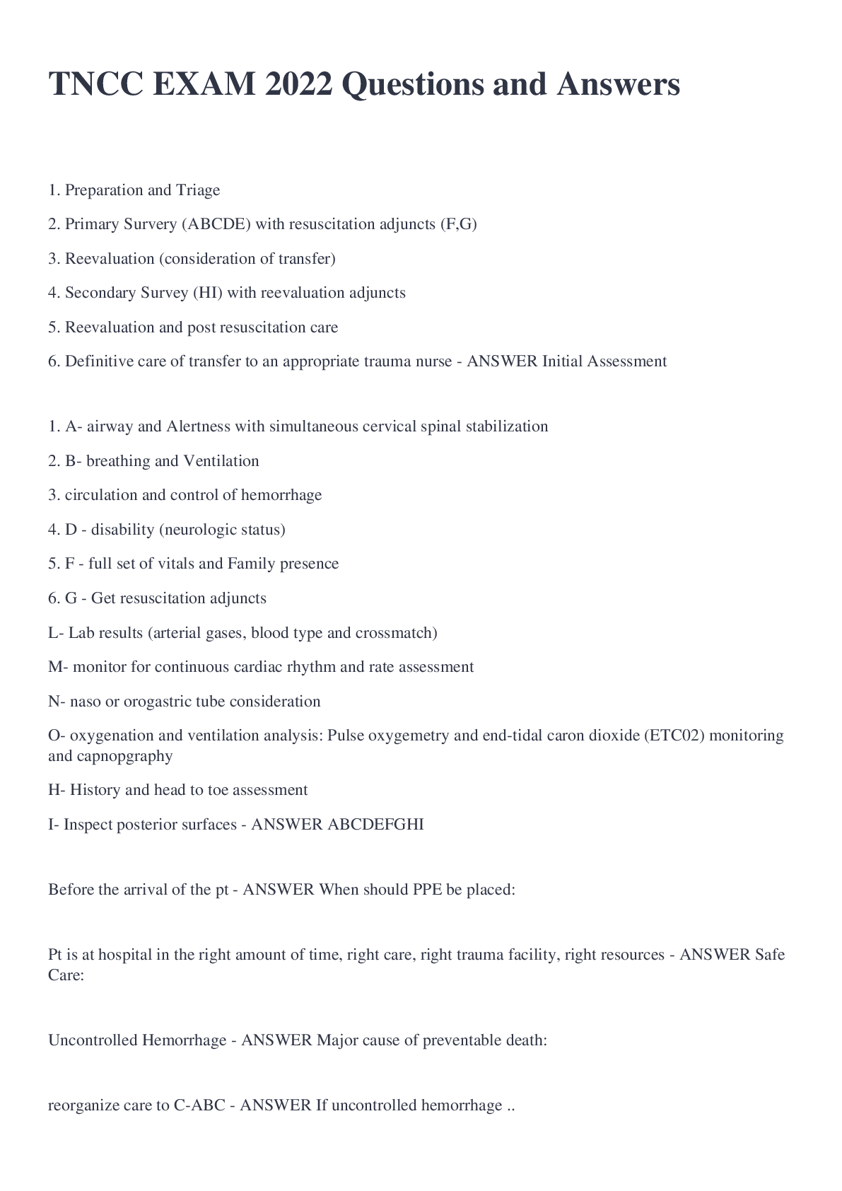
1. Preparation and Triage
2. Primary Survery (ABCDE) with resuscitation adjuncts (F,G)
3. Reevaluation (consideration of transfer)
4. Secondary Survey (HI) with reevaluation adjuncts
5. Reevaluation and post resuscit
...
1. Preparation and Triage
2. Primary Survery (ABCDE) with resuscitation adjuncts (F,G)
3. Reevaluation (consideration of transfer)
4. Secondary Survey (HI) with reevaluation adjuncts
5. Reevaluation and post resuscitation care
6. Definitive care of transfer to an appropriate trauma nurse - ANSWER Initial Assessment
1. A- airway and Alertness with simultaneous cervical spinal stabilization
2. B- breathing and Ventilation
3. circulation and control of hemorrhage
4. D - disability (neurologic status)
5. F - full set of vitals and Family presence
6. G - Get resuscitation adjuncts
L- Lab results (arterial gases, blood type and crossmatch)
M- monitor for continuous cardiac rhythm and rate assessment
N- naso or orogastric tube consideration
O- oxygenation and ventilation analysis: Pulse oxygemetry and end-tidal caron dioxide (ETC02) monitoring and capnopgraphy
H- History and head to toe assessment
I- Inspect posterior surfaces - ANSWER ABCDEFGHI
Before the arrival of the pt - ANSWER When should PPE be placed:
Pt is at hospital in the right amount of time, right care, right trauma facility, right resources - ANSWER Safe Care:
Uncontrolled Hemorrhage - ANSWER Major cause of preventable death:
reorganize care to C-ABC - ANSWER If uncontrolled hemorrhage ..
Used at the beginning of the initial assessment
1. A Alert. If the pt is alert he or she will be able to maintain his or her airway once it is clear.
2. V responds to verbal stimuli responds to pain. If the patient needs verbal stimulation to respond, an airway adjunct may be needed to keep the tongue from obstructing the airway.
3. P responds to pain. If the pt. responds only to pain, he or she may not be able to maintain his or her airway adjunct may need to be placed while further assessment is made to determine the need for intubation.
4. U Unresponsive. If the pt. is unresponsive, announce it loudly to the team and direct someone to chk in the pt is pulseless while assessing if the cause of the problem is the airway. - ANSWER Airway and AVPU:
ask pt to pen his or her mouth - ANSWER While assessing airway the patient is alert and responds to verbal stimuli you should..
jaw thrust maneuver to open airway and assess for obstruction. If pt has a suspected csi, the jaw thrust procedure should be done by two providers. One provider can maintain c-spine and the other can perform the jaw thrust maneuver. - ANSWER While assessing airway pt is unable to open mouth, responds only to pain, or is unresponsive you should..
1. The tongue obstructing the airway
2. loose or missing teeth
3. foreign objects
4. blood, vomit, or secretions'
5. edema
6. burns or evidence of inhalation injury
Auscultiate or listen for:
1. Obstructive airway sounds such as snoring or gurgling
2. Possible occlusive maxillofacial bony deformity
3. Subcutaneous emphysema - ANSWER Inspect the mouth for:
1. Check the presence of adequate rise and fall of the chest with assisted ventilation
2. Absence of gurgling on auscultation over the epigastrium
3. Bilateral breath sounds present on auscultation
4. Presence of carbon dioxide (CO2) verified by a CO2 device or monitor - ANSWER If the pt has a definitive airway in what should you do?
1. Suction the airway
2, Use care to avoid stimulating the gag reflex
3. If the airway is obstructed by blood or vomitus secretions, use a rigid suction device
If foreign body is noted, remove it carefully with forceps or another appropriate method - ANSWER If Airway is not patent
1. Apnea
2. GCS 8 or less
3. Maxillary fractures
4. Evidence of inhalation injury (facial burns)
5. Laryngeal or tracheal injury or neck hematoma
6. High risk of aspiration and patients inability to protect the airway
7. Compromised or ineffective ventilation - ANSWER Following conditions might require a definitive airway
Breathing: To assess breathing expose the chest:
1. Inspect for
a. spontaneous breathing
b. symmetrical rise and fall
c. depth, pattern, and rate of respiration
d. signs of difficulty breathing such as accessory muscle use
e. skin color (normal, pale, flushed, cyanotic)
f. contusions, abrasions, deformities (flail chest)
g. open pneumothoraces (sucking chest wounds)
h. JVD
i. signs of inhalation injury (singed nasal hairs, carbonaceous sputum) - ANSWER B
tracheal deviation and jvd - ANSWER Late signs of tension pneumo:
1. equal breath sounds bilaterally at the second intercostal space midclavicular line and the bases for fifth intercostal space at the axillary line - ANSWER Auscultate the chest for:
1. bony fractures and possible rib fractures, which may impact ventilation
2. palpate for crepitus
3. subcutaneous emphysema which may be a sign for a pneumothorax
4. soft tissue injury - ANSWER Palpate the chest for
1. open the airway, use jaw thrust
2. insert an oral airway
3. assist ventilations with a bag mask
4. prepare for definitive airway - ANSWER If breathing is absent..
trauma its need early supplemental oxygen, start with 15 mL O2 and titrate oxygen delivery. - ANSWER Oxygen on trauma patients
Circulation and Control of Hemorrhage
Inspect first for any uncontrolled bleeding
Skin color
palpate for central pulses - carotid and femoral - rate, rhythm, and strength
Skin temp: cool, diaphoretic, or warm and dry - ANSWER C
apply direct pressure to bleeding
elevate extremity
apply pressure over arterial sites
Consider a pelvic binder for pelvic fractures
consider a tourniquet
cannulate two veins with large caliber IV - if unable to gain assess consider IO
a. obtain labs, type and cross
b. infuse warm isotonic fluids
c. consider balanced resuscitation
d. use rapid infusion device - ANSWER C Interventions:
Disability - Neurologic Status
1. Assess pupils for equality, shape, and reactivity (PERRL)
2. Assess GCS (eye opening, verbal response, and motor response) - ANSWER D
1. Get a CT
2. Consider ABG 's if decreased LOC
3. Consider glucose check - ANSWER D Interventions
Exposure and Environmental Control
Remove all clothes and assess for any obvious injuries and uncontrolled bleeding - ANSWER E
IF clothing is needed for evidence preserve in paper bag.
Maintain body temp - cover the pt, turn up heat in room, administer warm fluids - ANSWER E Interventions:
Full set of vitals and family presence - ANSWER F
Get Resuscitation Adjuncts
L - Labs (maybe a lactic acid), a b g 's, blood type
M - monitors
N - naso or oro gastric tubes
O Oxygen and ETC02 monitors
P - pain assessment and management - ANSWER G
Reevaluation and Consider the need to Transfer - ANSWER Final step in primary survey
H,I - ANSWER Secondary Survery
History and Head to toe
MIST - prehospital report
MOI
Injuries sustained
S s/s in the field
T treatment in the field
if patients family present get a better hx on them - ANSWER H
Sample is part of history
S symptoms associated with injury
A allergies and tetanus status
M meds currently on including anticoagulant therapy
P past medical hx
L last oral intake
E Events and environment factors related to the injury - ANSWER SAMPLE
inspect for lacs, abrasions, asymmetry of facial expressions
palate for depressions and tenderness
look at ears for drainage - ANSWER Head to toe assessment: Head and face
immobilize cervical spine, tenderness, tracheal deviation - ANSWER Head to toe assessment: Neck and cervical spine
inspect, auscultate, palpate
any spontaneous breathing, rate, depth, and degree of effort, use of accessory muscles
lacs, contusions,
auscilate lung sounds and heart sounds - ANSWER Head to toe assessment: Chest
don't forget flanks!!!
inspect of lacs, puncture wounds, contusions,
auscultate then palpate:
bowel sounds?
any rigidity, guarding? begin with light palpation start to palpate with side that does not hurt
maybe do a fast scan? - ANSWER Head to toe assessment: Abdomen
any lacs? deformities? blood at the urtheral meatus
palpate pelvis with high pressure over the iliac wings downward and medially - ANSWER Head to toe assessment: pelvis and perineum
any deformities? bleeding? contusions, lacs? skin temp?? place splints on deformities, pulses - ANSWER Head to toe assessment: Extremities
inspect posterior surfaces
blogroll with at least 3 people. maintain c spine
take out backboard
Rectal tone per MD - ANSWER I
labs, wound care, tetanus, administer meds, prepare for transfer - ANSWER Secondary Reval Adjuncts
Vital signs
Interventions
Primary survey
Pain - ANSWER Post resuscitation care parameters that are continuously evaluated:
Capnography monitors numeric value, as well as continuous waveform, indicating real-time measurement and trending over time. - ANSWER Quantitative:
Colorimetric CO2 detectors provide info about the presence or absence of CO2. A chemically treated indicator strip changes color revealing the presence or absence of exhaled CO2 - ANSWER Qualitative
D displaced tube
O obstructed or kinked
P pneumothorax
E equipment failure , such as becoming detached from the equipment or loss of capnopgrahy - ANSWER DOPE
1. Preparation
2. Preoxygenation
3. Pretreatment
4. Paralysis and Induction
5. Protecting and positioning - v
6. Placement of proof - secure the tube
7. Post intubation - secure ETT Tube, get X-ray for placement - ANSWER Steps of Rapid Sequence Intubation
from hemorrhage is leading cause. Hypovolemia is caused by decrease in the amount of circulating volume. Goal is to replace volume. - ANSWER Hypovolemic Shock
results from hypo perfusion to the tissue due to an obstruction in either vasculature or heart. Goal is to relieve obstruction and improve perfusion.
Ex: tension pneumo or cardiac tamponade are two classic examples that may result from trauma. - ANSWER Obstructive Shock
Results from pump failure in the presence of adequate intravascular volume. There is a lack of cardiac output and end organ perfusion secondary to a decrease in myocardial contractility and/or valvular insufficiency.
Ex: MI's or dysrhythmia are common causes - ANSWER Cariogenic Shock
occurs as a result of maldistribution of an adequate circulating blood volume with the loss of vascular tone or increased permeability.
Ex: Anaphylactic - release of antihistamines
Septic Shock - systemic release of bacterial endotoxins, resulting in increased vascular permeability and vasodilation. Neurogenic shock - spinal cord injury results of loss in sympathetic nervous system control of vascular tone.
Goal: Volume replacement and vasoconstriction - ANSWER Distributive Shock
A breath every 5 to 6 seconds: 10-12 ventilations per minute - ANSWER Bag mask ventilation
Stroke Volume X HR - ANSWER Cardiac Output =
.. activation: .... are found in the carotid sinus and along the aortic arch, are sensitive to the degree of stretch in the arterial wall. When the receptors sense a decrease in stretch, they stimulate the sympathetic nervous system to release Epi, norepi, causing stimulation of cardiac activity and constriction of blood vessels, which causes a rise in heart rate and diastolic blood pressure - ANSWER Baroreceptors:
activation: consist of carotid and aortic bodies. ... detect changes in blood oxygen and Co2 and pH. When Co2 rises or oxygen level of pH falls these receptors are activated and information is relayed to the CNS and the cardiorespiratory centers in the medulla , which increases respiratory rage and depth and BP - ANSWER Chemoreceptors:
50 to 150 - ANSWER MAP Range
the decrease coagulopathy .. you will you bleed more - ANSWER The colder you are the more acidic you are..
in massive transfusion protocol... responsible for dissolving clots - ANSWER TXA
stabilized vital signs, improved mental status, improved urine output - ANSWER What are indicators of increased perfusion?
- ANSWER Prehospital shock index pg. 85
Flail chest - ANSWER Paradoxical chest wall movement
can be caused by blunt trauma. air escapes from injured lung to pleural space and negative intrapleural pressure is lost causing partial or collapsed lung - ANSWER Simple Pneumothorax
1. Dyspnea
2. Tachycardia
3. Decreased or absent breath sounds on the injured side
4. CP - ANSWER Simple Pneumo assessment:
Tx is based on size, presence of sx, and stability. For those are aysmpomatic and stable. Observation with or without oxygen. Larger pneumo who are unstable or likely to deteriorate a chest tube is placed. - ANSWER Simple pneumo interventions:
can result from penetrating wound through chest wall causing air to be trapped in to the intrapleural place. Might hear "sucking"
Tx: nonporous dressing tapes on 3 sided, then Chest tube and would closure surgical repair. - ANSWER Open Pneumo:
Air cannot escape intrapleural space.. can begin to compress heart. pt will have sever resp distress, hypotension, JVD. - ANSWER Tension pneumo
A 14 gauge needle that is inserted into the 2nd intercostal space in the midclavicular line on the affected side over the top of the rib to avoid neuromuscular bundle that runs under the rib.
Prepare for chest tube placement. - ANSWER Tension pneumo intervention
Caused by blood in the intrapleural space/ May also occur from lac to live or spleen combined with injury to the diaphragm.
Ensure two large bore IVS are placed.
Prepare for thoracentesis and chest tube insertion. If open thoracotomy is done chest tube is deferred. - ANSWER Hemothorax:
1. Hypotension
2. JVD
3. Muffled heart sounds - ANSWER Becks Triad:
Needle pericardiocentesis, but it is a temp solution. Requires surgical evaluation.
(Ultrasound guided) - ANSWER Cardiac Tamponade Intervention:
Aortic Dissection - ANSWER Unequal extremity pulse strength possibility of..
1. pain - hallmark sign, early sign
2. pressure - early sign
3. pallor, pules, paresthesia, paralysis - late sign - ANSWER Six P's of compartment syndrome:
Pediatric Assessment Triangle
1. General appearance - muscle tone, interactiveness, consoloability, poor or gaze, speech or cry
2. Work of breathing - inadequate or excessive, accessory muscle use, retractions, tripod position, abnormal upper airway sounds
3. Circulation of the skin - color, mottling or central or peripheral cyanosis, diaphoresis - ANSWER PAT
brachial pulse - ANSWER U
[Show More]