Bios255, Comprehensive Exam
This examination covers content learned in BIOS251, BIOS252, and BIOS255. This exam is worth 100 total points, consisting of 50 multiple-choice questions. You have 75 minutes to complete
...
Bios255, Comprehensive Exam
This examination covers content learned in BIOS251, BIOS252, and BIOS255. This exam is worth 100 total points, consisting of 50 multiple-choice questions. You have 75 minutes to complete the Exam.
Bios251
Describe a positive feedback system
A positive feedback system is mechanism in which the system responds to the perturbation in the same direction as the perturbation. Positive feedback is essentially a feedback mechanism that results in the amplification or growth of the output signal. An example of a positive is the amplification of labor contractions. The contractions are precipitated as the baby moves into position, stretching the cervix beyond its normal position. The positive feedback enhances the strength and frequency of the contractions until a child is born. After conception, the stretching halts, and the loop is interrupted.
Describe a negative feedback system
A negative feedback loop takes place when the result of a given reaction leads to a decrease in a given reaction. In this manner, a negative feedback mechanism brings a system to a target of stability or homeostasis.
Epithelial tissue
Vascular or avascular? Epithelial tissues are avascular.
Why might it be described as polar?
Epithelial tissues are described as avascular since no blood tissues cross the basement membrane to enter the tissue, and nutrients are mainly diffused or through absorption from underlying tissues.
What roles does it play in attachment and regeneration?
This study source was downloaded by 100000849840424 from CourseHero.com on 11-03-2022 07:37:03 GMT -05:00
Epithelial tissues depend on stem cells to replenish and replace the numerous cells that die from wear and tear.
Connective tissue
Describe the three types of protein fibers in connective tissue
Connective tissue fibers are grouped into three main groups: Collagen which is thick, strong, flexible, and resists stretch, reticular fibers are thin and form a supportive network; and elastin fibers which are mainly thick and elastic.
What is chondroitin sulfate and where is it located in the body?
Chondroitin sulfate is a chemical that is located in the cartilage around joints in the body. Skin
What is the function of keratin in the epidermis?
The core function of keratin is to adhere cells to each other and is critical in forming a protective layer on the outside of the skin.
Discuss the locations and functions of merocrine, Sebaceous, and apocrine glands Merocrine glands are mainly found in the palm of hands main function in the body is thermoregulation. Sebaceous glands located in the dermis middle layer of the skin secrete sebum which is critical in skin lubrication. Apocrine glands are found in the axilla and some cases in the skin of the scalp and face. Apocrine glands incessantly secrete fatty sweat into the gland tubule. Bones
Discuss the differences in the function of osteoclasts and osteoblasts
Osteoblast and osteoclast are the two main cells participating in that progress. Osteoclasts are responsible for aged bone resorption and osteoblasts are responsible for new bone formation. The resorption and formation are stable at physiological conditions.
Briefly describe the six steps of endochondral ossification
The first step is the enlargement of cartilages, where chondrocytes die, secondly, blood vessels
This study source was downloaded by 100000849840424 from CourseHero.com on 11-03-2022 07:37:03 GMT -05:00
grow into the perichondrium and cells convert into osteoblasts. Thirdly, more blood supply and
osteoblasts produce spongy bone tissue. Fourthly, Osteoclasts create a medullary cavity; appositional growth. In the fifth step, epiphysis centers calcify; blood and osteoblasts move in; secondary ossification centers and lastly epiphysis filled with spongy cartilage remains at joints.
Refer to Table 8.2, p. 226. Describe the appearances of condyles, facets, and heads Condyles are having a rounded protuberance at the end of some bones.
Facets are articular pillars and serve structural stability to the vertebral column. Heads in bones are around, and prominent extension of a bone forms part of a joint.
Examine Figure 8.5a, p. 229. Which bone constitutes most of the hard palate?
Cranial Bones
Define the terms diaphysis, metaphysis, and epiphysis
The diaphysis is the main or mid-section shaft of a long bone. It’s mainly composed of cortical and usually has bone marrow and adipose tissue.
Joints
Describe the anatomy of a typical synovial joint.
The synovial joint is characterized by the presence of a fluid-filled joint contained within a fibrous capsule. It is the most common type of joint found in the human body and contains several structures which are not seen in fibrous or cartilaginous joints. The three main features of
a synovial joint are (i) articular capsule, (ii) articular cartilage, (iii) synovial fluid.
What type of mobility is observed in synovial joints?
The mobility in the synovial joints is abduction, adduction, extension, flexion, and rotation.
Explain why the talocrural joint is classified as a hinge joint.
It is a hinge joint given that it allows dorsiflexion and plantarflexion of the foot.
Where is the sacroiliac joint located and which bones are found in this joint?
The sacroiliac joint is located between the iliac bones and the sacrum.
Bios252
This study source was downloaded by 100000849840424 from CourseHero.com on 11-03-2022 07:37:03 GMT -05:00
Muscles
What is a sarcomere?
A sarcomere is the basic contractile unit of muscle fiber. Each sarcomere is comprising of two main protein filaments—actin and myosin—which are the active structures responsible for muscular contraction.
Carefully, study steps 3 and 4 on p. 395. What is the effect of acetylcholine attaching to its
receptor?
The acetylcholine causes the receptors to open and allowing positively charged ions to enter the cell, eventually precipitating a downstream that leads to inhibition of muscle contraction.
Refer to p. 396, Briefly explain steps 6 through 13
Review Figure 11.15, p. 403. What are the differences between muscle twitches, incomplete tetanus, and complete tetanus?
Muscle twitches are precipitated by a myriad of reasons like stress, too many stimulants, poor eating habits, and as a side effect of some medications muscle twitches dissipate after a short period. On the other hand, incomplete tetanus occurs when the muscles undergo quick cycles of contraction and short relaxation phase of each. If the stimulus frequency is very high that the relaxation phase disappears totally, which is referred to as complete tetanus.
What are agonists, antagonists, and synergists?
Agonists- An agonist is a chemical that binds to a receptor and activates the receptor to produce a biological response.
Antagonists- A biological structure or chemical agent that interferes with the physiological action of another.
Synergists- the substance that enhances the effect of another substance, such as a drug Name the muscles that are part of the quadriceps group.
The quadriceps group of muscle comprises vastus lateralis, vastus medialis, vastus intermedius,
and the rectus femoris.
This study source was downloaded by 100000849840424 from CourseHero.com on 11-03-2022 07:37:03 GMT -05:00
Name the muscles that are part of the hamstring group.
The hamstring group of muscles comprises the long head of the biceps femoris, the short head of the biceps femoris, semitendinosus, and semimembranosus
Nervous system
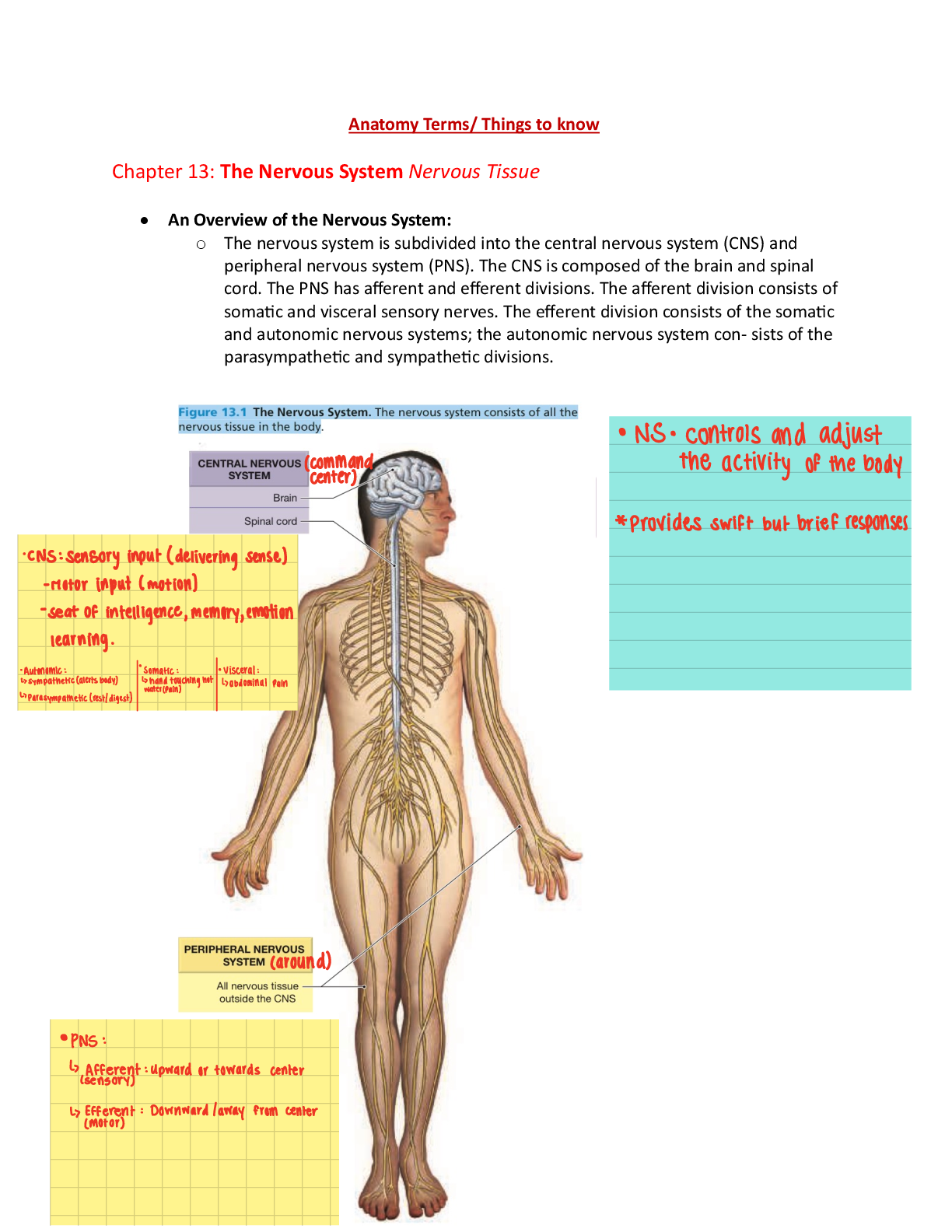
Describe the overall function of the nervous system
The nervous system is the main controlling, regulatory, and communicating system in the body Describe the events occurring in Figure 3.19, p. 92
What are some properties of graded/local potentials?
Refer to p. 434
Name the regions of the body innervated by the phrenic and sciatic nerves Phrenic nerves innervate the diaphragm, heart, lungs, and rib cage. the sciatic nerve innervates the muscles of the posterior thigh, entire leg, and entire foot.
What are the functions of cerebrospinal fluid?
The cerebrospinal fluid protects the brain and spinal cord from trauma. Supply nutrients to nervous system tissue. Remove waste products from cerebral metabolism can be tested for the diagnosis of a variety of neurological diseases through the use of a procedure called a lumbar puncture.
Discuss the location of the primary somatosensory cortex vs. the primary motor complex.
Is located in the anterior part of the parietal lobe, where it constitutes the postcentral gyrus. on Primary somatosensory is key in its key role of processing sensory information from various body parts.
Learn the mnemonic for cranial nerves on p. 533
Compare and contrast the effects of the parasympathetic and sympathetic nervous systems on the circulatory system.
The sympathetic nervous system releases norepinephrine (NE) while the parasympathetic nervous system releases acetylcholine (ACh). Sympathetic stimulation increases heart rate and
myocardial contractility. During exercise, emotional excitement, or under various pathological
This study source was downloaded by 100000849840424 from CourseHero.com on 11-03-2022 07:37:03 GMT -05:00
conditions (e.g., heart failure), the sympathetic nervous system is activated. The stimulation of the
sympathetic nervous system causes pupil dilatation, bronchiole dilatation, blood vessel constriction, sweat secretion, inhibits peristalsis, increases renin secretion by the kidneys, as well as can induce reproductive organ contraction and secretion. In contrast, parasympathetic stimulation decreases heart rate and constricts the pupils.
Use figures 16.26, p. 594 and 16.32, p. 597 to trace the path of light from the cornea to the choroid
Endocrine system
What are the differences between endocrine and exocrine glands?
Two main types of glands exist: exocrine and endocrine. The key difference between the two types is that, whereas exocrine glands secrete substances into a ductal system to an epithelial surface, endocrine glands secrete products directly into the bloodstream. Exocrine secretions form in the acinus, a small cluster of cells at the origination of glandular ducts. Exocrine glands sub classifies into subtypes based on the method of secretion, the compound produced, or the shape of the gland.
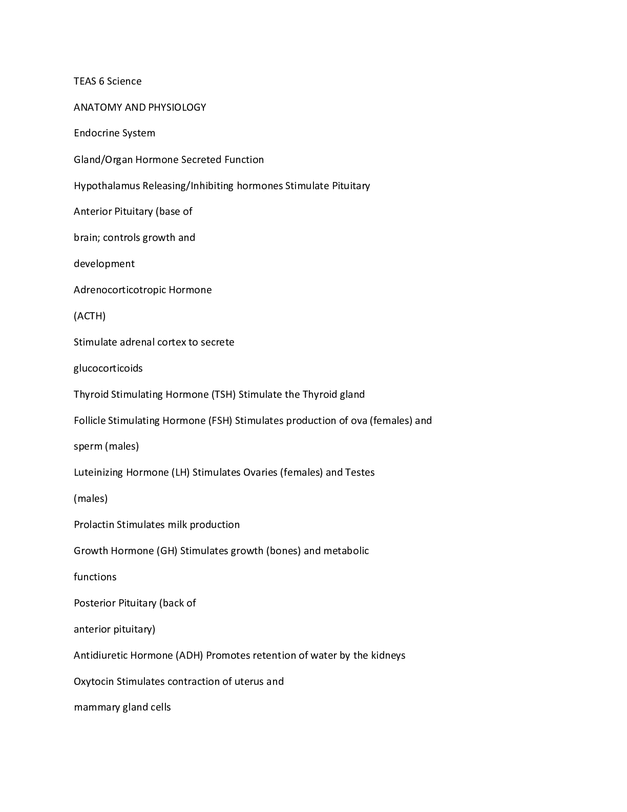
What is the function of the antidiuretic hormone?
ADH is a hormone produced by the hypothalamus in the brain and stored in the posterior pituitary gland. ADH dictates to the kidney how much water it ought to conserve. The antidiuretic hormone continuously regulates and balances the amount of water in the bloodstream.
Bios255
Cardiovascular system
Define the term hematocrit. How can hematocrit be increased?
The hematocrit (hct) is a measurement of how much an individual’s blood is made up of red blood cells. The test measures by volume but is then expressed as a percentage. If a person has
a hematocrit of 40%, this means that he or she has 40 milliliters of red blood cells in 100 milliliters of blood. Increasing the consumption of red meat (liver in particular), fish and shellfish
(oysters, clams, shrimp, and scallops), dried fruit (apricots, prunes, and peaches), green leafy
This study source was downloaded by 100000849840424 from CourseHero.com on 11-03-2022 07:37:03 GMT -05:00
vegetables, beans, iron-fortified bread and cereals, all rich in iron enhances hematocrit.
Concerning blood types, what does agglutination mean?
In hematology, red cell agglutination or auto agglutination is a phenomenon in which red blood cells clump together, forming aggregates. It is caused by the surface of the red cells being coated with antibodies.
With blood types, what do the positive and negative refer to?
Rhesus is a factor inherited protein found on the surface of a red blood cell. If a person’s blood has the rhesus protein, the individual is Rh-positive. If the blood does not have the rhesus protein, one is Rh-negative. Rh-positive is the most common blood type. Introducing the wrong blood type of blood into a person’s body can trigger potentially dangerous, even fatal immune reactions.
Explain what is occurring in Figure 18.15, p. 670
Name the tunics (layers) of veins and arteries. What is the main component of each layer? The walls of arteries and veins have three tunics. The inner layer is tunica intima, tunica media, and the outer layer is the tunica adventitia. Tunica intima comprises the endothelium, connective tissues, and a basal layer of elastic tissue called internal elastic lamina that separates the tunica intima from tunica media. Tunica media is chiefly comprised of circumferentially arranged smooth muscle cells. composed of loose connective tissue made up of fibroblasts and associated collagen fibers
What is the function of blood colloid osmotic pressure in net reabsorption pressure?
Colloid osmotic pressure (COP), the osmotic pressure exerted by large molecules, serves to hold water within the vascular space. It is normally created by plasma proteins, namely albumin,
that do not diffuse readily across the capillary membrane.
This study source was downloaded by 100000849840424 from CourseHero.com on 11-03-2022 07:37:03 GMT -05:00
[Show More]

.png)