NURS 6531 FINAL EXAM STUDY GUIDE
NURS 6531 FINAL EXAM STUDY GUIDE
PLEASE DO NOT SHARE IN THE GROUP AS A POST. EVERY MEMBER WHO HAS CONTRIBUTED HAS RECEIVED A COPY. THOSE WHO HAVE NOT CONTRIBUTED THEIR ASSIGNED
...
NURS 6531 FINAL EXAM STUDY GUIDE
NURS 6531 FINAL EXAM STUDY GUIDE
PLEASE DO NOT SHARE IN THE GROUP AS A POST. EVERY MEMBER WHO HAS CONTRIBUTED HAS RECEIVED A COPY. THOSE WHO HAVE NOT CONTRIBUTED THEIR ASSIGNED PORTIONS HAVE BEEN EXCLUDED.
Still waiting for:
79. Severe depression
80. GAD
When I receive these topics, I will post them in the group and you can just copy them and paste them into the study guide. If these topics are not received by next weekend, I will try to reassign and remove the members. Hate it is coming to that. These are grown people in graduate level masters classes. I have submitted to you everything I have now received.
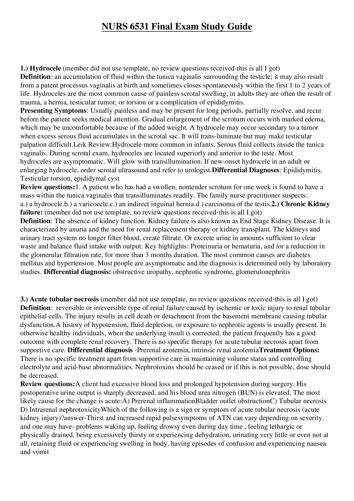
1.) Hydrocele (member did not use template, no review questions received-this is all I got)
Definition: an accumulation of fluid within the tunica vaginalis surrounding the testicle; it may also result from a patent processus vaginalis at birth and sometimes closes spontaneously within the first 1 to 2 years of life. Hydroceles are the most common cause of painless scrotal swelling.; in adults they are often the result of trauma, a hernia, testicular tumor, or torsion or a complication of epididymitis.
Presenting Symptoms: Usually painless and may be present for long periods, partially resolve, and recur before the patient seeks medical attention. Gradual enlargement of the scrotum occurs with marked edema, which may be uncomfortable because of the added weight. A hydrocele may occur secondary to a tumor when excess serous fluid accumulates in the scrotal sac. It will transluminate but may make testicular palpation difficult.
Leik Review:
Hydrocele more common in infants. Serous fluid collects inside the tunica vaginalis. During scrotal exam, hydroceles are located superiorly and anterior to the testes Most hydroceles are asymptomatic.
Will glow with transillumination. If new-onset hydrocele in an adult or enlarging hydrocele, order scrotal ultrasound and refer to urologist.
Differential Diagnoses: Epididymitis, Testicular torsion, epididymal cyst
Review questions:
1. A patient who has had a swollen, nontender scrotum for one week is found to have a mass within the tunica vaginalis that transilluminates readily. The family nurse practitioner suspects:
a.) a hydrocele.
b.) a varicocele.
c.) an indirect inguinal hernia.
d.) carcinoma of the testis.
2.) Chronic Kidney failure: (member did not use template, no review questions received-this is all I got)
Definition: The absence of kidney function. Kidney failure is also known as End Stage Kidney Disease. It is characterized by anuria and the need for renal replacement therapy or kidney transplant. The kidneys and urinary tract system no longer filter blood, create filtrate. Or excrete urine in amounts sufficient to clear waste and balance fluid intake with output. Key highlights: Proteinuria or hematuria, and /or a reduction in the glomerular filtration rate, for more than 3 months duration. The most common causes are diabetes mellitus and hypertension. Most people are asymptomatic and the diagnosis is determined only by laboratory studies.
Differential diagnosis: obstructive uropathy, nephrotic syndrome, glomerulonephritis
3.) Acute tubular necrosis (member did not use template, no review questions received-this is all I got)
Definition: reversible or irreversible type of renal failure caused by ischemic or toxic injury to renal tubular epithelial cells. The injury results in cell death or detachment from the basement membrane causing tubular dysfunction.
A history of hypotension, fluid depletion, or exposure to nephrotic agents is usually present. In otherwise healthy individuals, when the underlying insult is corrected, the patient frequently has a good outcome with complete renal recovery. There is no specific therapy for acute tubular necrosis apart from supportive care.
Differential diagnosis -Prerenal azotemia, intrinsic renal azotemia
Treatment Options: There is no specific treatment apart from supportive care in maintaining volume status and controlling electrolyte and acid-base abnormalities. Nephrotoxins should be ceased or if this is not possible, dose should be decreased.
Review questions:
A client had excessive blood loss and prolonged hypotension during surgery. His postoperative urine output is sharply decreased, and his blood urea nitrogen (BUN) is elevated. The most likely cause for the change is acute:
A) Prerenal inflammation
Bladder outlet obstruction
C) Tubular necrosis
D) Intrarenal nephrotoxicity
Which of the following is a sign or symptom of acute tubular necrosis (acute kidney injury)?
answer-Thirst and increased rapid pulse
symptoms of ATN can vary depending on severity. and one may have- problems waking up, feeling drowsy even during day time , feeling lethargic or physically drained, being excessively thirsty or experiencing dehydration, urinating very little or even not at all, retaining fluid or experiencing swelling in body, having episodes of confusion and experiencing nausea and vomit
4. Indirect inguinal hernia
Definition: Indirect inguinal hernia – Indirect inguinal hernia is caused by a birth defect in the abdominal wall that is present at birth. A scrotal-inguinal hernia results when a segment of the bowel slips through the internal inguinal ring, where it may remain in the inguinal canal or pass into the scrotal sac. An inguinal hernia may occur as a result of a defect in the anterior abdominal wall or because of a patent process vaginalis. Inguinal hernias predominantly affect men (9:1) and have the highest incidence in men aged 40 to 59. A hernia may move freely between the abdomen and the scrotum or can be spontaneously reduced by digital manipulation. When a hernia becomes strangulated or is unreducible, this compromises the blood supply and requires emergent surgical reduction. Strangulation should be suspected when a tender mass is palpated in the scrotum in addition to redness, nausea, and vomiting
Presenting Symptoms: Scrotal swelling, mild to moderate pain on straining, scrotal heaviness, and the possible presence of a bulge are common complaints. Increased edema after standing in an erect position but decreases when the patient is recumbent.
3 Differential Diagnoses: undescended testis, lymphadenopathy, femoral hernia
Pattern Recognition: Enlarged hemiscrotum or a bulge in the groin area that may spontaneously reduce when the patient is supine or with manual reduction. The provider will not be able to move the fingers above the mass, which should be soft and mushy but painless unless it is incarcerated and ischemic. Scrotal hernias do not transilluminate. Auscultation of bowel sounds over the mass is significant for the diagnosis of bowel in the scrotal sac.
Treatment options: If the herniated bowel is reducible, surgical referral for possible future repair is indicated. Difficulty in reducing a hernia is cause for urgent surgical intervention. However, pain may indicate incarceration of the bowel or complete inability to reduce the hernia, which is cause for immediate emergency department referral and surgical exploration.
Review questions:
1. Mr. S. comes to you with scrotal pain. The examinations of his scrotum, penis, and rectum are normal. Which of the following conditions outside of the scrotum may present as scrotal pain?
A. Inguinal herniation and peritonitis **
B. Renal colic and cardiac ischemia
C. Pancreatitis and Crohn ’ s disease
D. Polyarteritis nodosa and ulcerative colitis
Rationale: Conditions outside of the scrotum that may present with scrotal pain are abdominal aortic aneurysm, inguinal herniation, pancreatitis, renal colic, peritonitis, intraperitoneal hemorrhage, and polyarteritis nodosa. Keep in mind that any client with scrotal pain should be considered to have testicular torsion until proved otherwise, especially in the age groups of the neonate and adolescents.
2. The most common type of hernia is a(n):
A. indirect inguinal hernia. **
B. direct inguinal hernia.
C. femoral hernia.
D. umbilical hernia.
Rationale: An indirect inguinal hernia is the most common type of hernia affecting all ages and both genders and accounts for 50% of hernias treated. The point of origin is above the inguinal ligament and often travels into the scrotum. A direct inguinal hernia is less common (accounts for about 25% of hernias seen) and usually occurs in men older than age 40. The point of origin is above the inguinal ligament and rarely travels into the scrotum. The femoral hernia is the least common (about 10% of hernias seen) and occurs more often in women than in men. The point of origin is below the inguinal ligament and never travels into the scrotum in men. An umbilical hernia occurs more frequently in infants and is a protrusion of part of the intestine at the umbilicus.
3. Max, age 70, is obese. He is complaining of a bulge in his groin that has been there for months. He states that it is not painful, but it is annoying. You note that the origin of swelling is above the inguinal ligament directly behind and through the external ring. You diagnose this as a(n):
A. indirect inguinal hernia.
B. direct inguinal hernia. **
C. femoral hernia.
D. strangulated hernia.
Rationale: A direct inguinal hernia usually occurs in middle-aged to older men and is the result of an acquired weakness caused by heavy lifting, obesity, or chronic obstructive pulmonary disease (COPD). The origin of swelling is above the inguinal ligament directly behind and through the external ring. An indirect inguinal hernia is congenital or acquired and is more common in infants younger than 1 year of age and in men ages 16 – 25. The origin of swelling is above the inguinal ligament. The hernia sac enters the canal at the internal ring and exits at the external ring. A femoral hernia, which occurs more frequently in women, is acquired and results from an increase in abdominal pressure, as well as muscle weakness. The origin of swelling is below the inguinal ligament. Because Max is not having any pain and the condition has been this way for months, you know that the hernia is not strangulated. A strangulated hernia, which requires immediate referral to a surgeon, results in no blood supply to the affected bowel and causes nausea, vomiting, and tenderness.
5. Orchitis
Definition: Orchitis is a systemic, blood-borne infection that results in an acute inflammation of one or both testicles. It may coexist with infections of the prostate and epididymis; causes – viral infection (ex. Mumps), C. trachomatis and N. gonorrhoeae in adolescents, E. coli – men, complication of syphilis, mycobacterial, fungal; hydrocele and scrotal wall thickening may be seen as a complication of mumps
Presenting Symptoms: Gradual onset of acute or moderate pain, testicular swelling, and fever
3 Differential Diagnoses: epididymitis, testicular tumor, hernia, testicular torsion
Pattern Recognition: Testicular edema may be so pronounced that it is difficult to distinguish the testes from the epididymis. Palpation may reveal swollen, very tense testes that are painful, and the patient may be febrile. Inflammation of the testis usually involves systemic viral infections (commonly mumps) and includes unilateral or bilateral erythema, edema, and scrotal tenderness, which occurs 4 to 7 days after initial fever.
Treatment options: Anti-infective therapy is recommended, with guidance by local sensitivity reports. The following antibiotic regimens are effective against the most common causes of epididymitis: single-dose ceftriaxone given intramuscularly (IM), 250 to 500 mg, and doxycycline, 100 mg twice daily for 10 days for men younger than 35 years; in men older than 35 years, levofloxacin (given intravenously [IV] or orally [PO]), 500 to 750 mg/day, or ciprofloxacin, 500 mg (IV or PO), for 10 to 14 days. Antipyretics should be used to reduce discomfort and fever, and an anti-inflammatory agent should be prescribed. An antiemetic can also be prescribed for nausea and vomiting. Bed rest and scrotal elevation are also recommended for epididymitis. Hot or cold compresses may be helpful for orchitis.
Review questions:
A 35 year old sexually active man presents with a 1 week history of fever and pain over the left scrotum. It is accompanied by frequency and dysuria. The scrotum is edematous and tender to touch. He denies flank pain, nausea, and vomiting. He reports that eh pain is lessend when he uses scrotal-support briefs. The urinalysis shows 2 + blood and a large number of leukocytes. What is the most likely diagnosis?
A. Acute urinary tract infection
B. Acute pyelonephritis
C. Acute orthitis
D. Acute epididymitis **
Orchitis is caused by which of the following?
A. Mumps virus **
B. Measles virus
C. Chlamydia trachomatis
D. Chronic urinary tract infections that are not treated adequately
A 10 year old boy complains of sudden onset of scrotal pain upon awakening that morning. He is also complaining of severe nausea and vomiting. During the physical examination, the nurse practitioner finds a tender, warm, and swollen left scrotum. The cremastic reflex is negative and the urine dipstick is negative for leukocytes, nitrites, and blood. The most likely diagnosis is:
A. Acute epididymitis
B. Severe salmonella infection
C. Testicular torsion **
D. Acute orchitis
What type of follow up should this patient receive?
A. Refer to a urologist within 48 hours
B. Refer him to the emergency department as soon as possible **
C. Prescribe ibuprofen (advil) 600 mg QID for pain
D. Order a testicular ultrasound for further evaluation
6. Testicular torsion
Definition: Testicular torsion - obstruction of blood flow to the testes because of a twisting of the arteries and veins in the spermatic cord resulting in occlusion of blood flow. Occurs in 12-18 year olds. Usually unilateral, effecting the left testis. Two types: extravaginal and intravaginal. Extravaginal (rare, seen in neonates)- twisting of the spermatic cord, testis, and process vaginalis; intravaginal (seen in adolescents)- failure of the testis to adhere to the scrotal wall, creating a “bell clapper deformity.” Different from torsion of the appendix testis.
Presenting Symptoms: sudden in onset, extremely painful, and may awaken the patient from sleep or be trauma induced. Testicular pain, experience abdominal pain, nausea, and vomiting; 25% of patients have a fever. Clinical manifestations-testicle that rides high in the scrotum and an absent cremasteric reflex on examination
3 Differential Diagnoses: testicular appendix torsion, epididymitis, epididymo-orchitis, hydrocele
Pattern Recognition: Most common in the left hemiscrotum. Scrotal edema and erythema may be seen. The affected side may have a higher position as a result of rotation. The spermatic cord is swollen and extremely tender, the epididymis may be felt anteriorly, and the majority of patients will have an absent cremasteric reflex. In some instances a small area of cyanosis (blue dot sign) may be present on the scrotal skin and indicates torsion of the appendix testis.
Treatment options: Surgical consultation with surgical exploration – needs to occur in 6 hours.
Review questions:
1. A 24-year-old man presents with sudden onset of left-sided scrotal pain. He reports having intermittent unilateral testicular pain in the past but not as severe as this current episode. Confirmation of testicular torsion would include all of the following findings except:
A.unilateral loss of the cremasteric reflex.
B.the affected testicle held higher in the scrotum.
C.testicular swelling.
D.relief of pain with scrotal elevation. **
2. In assessing a man with testicular torsion, the NP is most likely to note:
A.elevated PSA level.
B.white blood cells reported in urinalysis.
C.left testicle most often affected. **
D.increased testicular blood flow by color-flow Doppler ultrasound.
3. Anticipated organ survival exceeds 85% with testicular decompression within how many hours of torsion?
A.1
B.6 **
C.16
D.24
4. To prevent a recurrence of testicular torsion, which of the following is recommended?
A.use of a scrotal support
B.avoidance of testicular trauma
C.orchiopexy **
D.limiting the number of sexual partners
7. Epididymitis
1 Definition: Inflammation or infection of the epididymis. Commonly occurs in men younger than 35 yrs. of age with chlamydia as the cause. Men older than 35 yrs. is likely as a result of bacterial ascension from bladder or bacteria introduced during cauterization/surgery. Diagnostic test STD testing, urine culture and scrotal ultrasound R/O testicular torsion.
2 Presenting Symptoms: pain, dysuria, urgency/frequency, low back pain/perineal pain, fever/chills/malaise, scrotal edema
3 Differential Diagnoses: testicular torsion, inguinal hernia, hydrocele, testicular tumor,
4 Pattern Recognition: Enlarged, tender epididymis, Urethral discharge may be evident, Positive prehns sign, Normal cremasteric reflex R/O testicular torsion.
5 Treatment options:
A Adult under 35yrs ceftriaxone 250mg IMx 1 PLUS Doxy 100mg BID a day or Azithromycin 1gm once.
B Adult over 35 yrs Bactrim DS 1 tab BID a day x 10 days or cipro 250 mg BID x 10 days.
C support/elevate scrotum
D Analgesic NSIADs,ice(early),heat (late),bed rest.
6 Review questions:
1. Jordan appears with a rapid onset of unilateral scrotal pain radiating up to the groin and flank. You are trying to differentiate between epididymitis and testicular torsion. Which test to determine whether swelling is in the testis or the epididymis should be your first choice?
A. X-ray
B. Ultrasound
C. Technetium scan
D. Physical examination
Answer B
If your client has a rapid onset of unilateral scrotal pain radiating up to the groin and flank and you are trying to differentiate between epididymitis and testicular torsion, an ultrasound test is useful to determine whether the swelling is in the testis or the epididymis and should be your first choice. Initially, before the swelling has reached its peak, a physical examination will probably differentiate, but within a few hours, when the testis also swells, it may not be possible to differentiate between epididymis and testis by palpation. A reactive hydrocele may also develop. A technetium scan will show an increased uptake in the case of epididymitis and decreased uptake in the case of torsion, but the least invasive and most inexpensive test is an ultrasound.
2. The nurse practitioner recognizes that the most common cause of epididymitis in a young man is:
A chlamydia
B E. coli
C mycoplasma
D Proteus species
Answer A
Chlamydia is a sexually transmitted infection which is the leading cause for epididymitis and nongonococcal urethritis in men less than 35 years old. Symptoms of epididymitis include unilateral testicular pain and tenderness, hydrocele and palpable swelling of the epididymis
3. Your 25-year-old male patient has had a fever, dysuria, low back pain, and scrotal edema. Which of the following is likely the diagnosis?
A acute bacteria prostatitis
B acute pyelonephritis
C epididymitis
D urinary tract infection
Answer C The combination of urinary tract infection symptoms and scrotal edema is often the case in epididymitis. Treatment for those aged < 35 years is ceftriaxone (Rocephin) or doxycycline because the most common causative agent is chlamydia. For those aged over 35 years, (Bactrim) Trimethoprim/sulfamethoxazole is the common treatment.
The proper treatment for urinary tract infection, acute pyelonephritis, and acute bacterial prostatitis is trimethoprim/sulfamethoxazole (Bactrim); however, scrotal edema is not present in these differential diagnoses
8. Benign prostatic hyperplasia
Definition: a non-cancerous enlargement of the prostate gland. Seen in 50% of men older than 50 and 90% alder than 80. BPH is related to elevations of androgen and estrogen that stimulate prostatic growth.
Presenting Symptoms: Symptoms are develop gradually. Obstructive BPH: urinary hesitancy, decreased caliber and force of the stream, and post void dribbling related to bladder outlet obstruction. Irritative symptoms include frequency, urgency, and nocturia and occur as a result of decreased functional bladder capacity and instability of infection. Hematuria may be present.
3 Differential Diagnoses: Bladder Calculi, Uretheral Stricture, Cancer of the Prostate, Bladder neck contracture, and UTI, and Bladder cancer
Pattern Recognition: UA should be ordered to exclude UTI. PSA is appropriate for men with a life-expectancy of more than 10 years in the presence of physical finding on DRE and if 5a-reductase inhibitor therapy (Proscar) is planned. DRE: Prostate that is enlarged but is symmetrical in texture and size (rubbery texture).
Treatment options: Lifestyle changes may decrease symptoms such as reduction of caffeine and alcohol intact, avoiding fluids before bedtime, and avoidance of diuretic medications.
Meds:
1. Alpha-adrenergic antagonist: (Terazosin, Tamsulosin, Doxazosin) relax smooth muscle in the bladder neck, prostate capsule, and prostatic urethra. Take at night. Watch for orthostatic hypotension. Tamsulosin may have less effect on BP than other alpha blockers.
2. 5-alpha-reductase inhibitors (blocks testosterone): Finasteride (Proscar): shrink the prostatic glandular hyperplasia by decreased tissue DHT levels, but it may take up to 6-12 months to see improvement in symptoms.
3. Phosphodiesterase-5 (PDE%): Sildenafil or Tadalafil: used for men with mild to moderate symptoms and erectile dysfunction
4. Saw Palmetto: herbal, may increase bleeding
Balloon dilation: reduces symptoms in the short term
TURP: effective for severe BPH, Gold standard treatment for bladder outlet obstruction, limited to prostates weighing less than 100g
Review questions:
1. Which statement is true about the use of alpha blockers in the treatment of symptomatic BPH?
They do not lower blood pressure in normotensive clients.
2. Milton, a 72 year old unmarried, sexually active white man presents to your clinic with complaints of hesitancy, urgency, and occasional uncontrolled dribbling. Although you suspect benign prostatic hypertrophy, what else should your differential diagnosis include? Urethral stricture (may develop as a result of sexually transmitted diseases and should be considered in a sexually active individual no matter what the age)
3. The action of a 5 alpha-reductace inhibitor in the treatment of BPH is to:
reduce action of androgens in the prostate.
4. Harry has BPH and complains of some incontinence. Your first step in diagnosing overflow incontinence would be to order a:
Post void residual urine measurement
5. Lower urinary tract symptoms in males can present as a constellation of storage or voiding symptoms. Storage symptoms include:
urgency and nocturia
6. A 63-year-old man presents to you with hematuria, hesitancy, and dribbling. DRE reveals a moderately enlarged prostate that is smooth. The client’s PSA is 1.2. What is the most appropriate management strategy for you to follow at this time?
Prescribe an alpha adrenergic blocker, which will relax bladder and prostate smooth muscle to improve flow and relieve symptoms.
7. In deciding whether to treat Morrison, who has BPH, you use the American Urological Association scale. No treatment is indicated if the AUA score is 7 or lower.
8. According to the AUA guideline on the management of BPH, when is referral for invasive surgery automatically warranted?
With the presence of refractory retention and bladder stones.
9. What differentiates prostate cancer symptoms from BPH?
Symptoms of prostate cancer in general tend to progress more rapidly than those of BPH.
9. Chronic prostatitis-
Definition : Inflammatory infection of the prostate. Usually caused by gram negative bacteria like E. coli. nonbacterial prostatitis mostly in young men caused by chlamydia, mycoplasma, Gardnerella.
Diagnostic test is urine culture.
Presenting Symptoms: fever /chills, low back pain, dysuria, urgency /frequency, nocturia
Differential Diagnoses: cystitis, BPH, Kidney stones, bladder ca, prostatic abscess, enterovesical fistula.
Pattern Recognition: Edematous prostate, may be warm and tender/boggy in palpitation, pain.
Treatment options:
Antibiotic choices : Bactrim,Levaquin,noroxin,ofloxacin
B sitz bath 3 times a day for 30mins
C no sexual intercourse until acute phase resolves
Review questions:
1 The most common gram-negative bacteria that causes both acute and chronic bacterial prostatitis is
A. Staphylococcus aureus .
B. Klebsiella .
C. Escherichia coli .
D. Enterobacteriaceae .
Answer C
The most common gram-negative bacterium that causes both acute and chronic bacterial prostatitis is Escherichia coli. The other aerobic gram-negative bacteria include Klebsiella, Pseudomonas, Enterobacteriaceae, Proteus mirabilis, and Neisseria gonorrhoeae. Occasionally other bacteria (Staphylococcus aureus and Streptococcus faecalis) are causes.
2 A history of urinary tract infections in males is often seen in men with chronic bacterial prostatitis. Other signs and symptoms of chronic bacterial prostatitis includes
A. irritative voiding symptoms, low back pain, and perineal pain.
B. nausea and vomiting, as well as fever.
C. loss of appetite and weight loss.
D. irritative voiding symptoms, inability to ambulate, and fever.
Answer A
Chronic bacterial prostatitis may have a variety of clinical presentations, but nausea and vomiting, loss of appetite and weight loss, as well as an inability to ambulate are rarely among the presenting symptoms of this disorder. Even fever is typically not present in chronic cases. Typically, there are irritative voiding symptoms that have persisted over time, low back pain, and perineal pain, although any one, or all, may be present. Sometimes clients are completely asymptomatic, although bacteria might be present on urinalysis, and expressed prostatic secretions usually demonstrate increased numbers of leukocytes. Physical examination may be unremarkable as well, although in some cases the prostate will feel boggy or indurated. There is often history of repeated urinary tract infections. Cystitis and/or chronic urethritis may be secondary or mimic prostatitis; however, cultures of the fractionated urine may localize the source of infection. Anal disease may share some of the symptoms of prostatitis, but physical examination should permit a distinction between the two.
3 When performing a prostate examination, you note a tender, warm prostate. What do you suspect?
A. Benign prostatic hypertrophy
B. Prostatic abscess
C. Prostate cancer
D. Bacterial prostatitis
Answer D
Bacterial prostatitis, in which the prostate feels very tender and warm, is usually caused by Escherichia coli. Clients with bacterial prostatitis usually also have a sudden onset of high fever, chills, malaise, myalgias, and arthralgias. In benign prostatic hypertrophy, the prostate gland would feel soft and nontender and would be enlarged. With prostatic abscess, the prostate feels like a firm, tender, or fluctuant mass. With prostate cancer, the prostate may have single or multiple nodules that are firm, hard, or indurated and are usually nontender.
10. Inguinal hernia
Definition: An inguinal hernia happens when contents of the abdomen—usually fat or part of the small intestine—bulge through a weak area in the lower abdominal wall. Inguinal hernias occur at the inguinal canal in the groin region. In males, the spermatic cords pass through the inguinal canals and connect to the testicles in the scrotum—the sac around the testicles. The spermatic cords contain blood vessels, nerves, and a duct, called the spermatic duct, that carries sperm from the testicles to the penis. In females, the round ligaments, which support the uterus, pass through the inguinal canals.
Presenting Symptoms: The first sign of an inguinal hernia is a small bulge on one or, rarely, on both sides of the groin—the area just above the groin crease between the lower abdomen and the thigh. The bulge may increase in size over time and usually disappears when lying down.
Other signs and symptoms can include
• discomfort or pain in the groin—especially when straining, lifting, coughing, or exercising—that improves when resting
• feelings such as weakness, heaviness, burning, or aching in the groin
• a swollen or an enlarged scrotum in men or boys
• Incarceration. An incarcerated hernia happens when part of the fat or small intestine from inside the abdomen becomes stuck in the groin or scrotum and cannot go back into the abdomen. A health care provider is unable to massage the hernia back into the abdomen.
• Strangulation. When an incarcerated hernia is not treated, the blood supply to the small intestine may become obstructed, causing “strangulation” of the small intestine. This lack of blood supply is an emergency situation and can cause the section of the intestine to die.
3 Differential Diagnoses: Ectopic testis, Femoral or inguinal adenitits, femoral hernia, sports hernia
Pattern Recognition: Physical exam: Feel for a bulge in the inguinal area when a patient strains or coughs. If the hernia is causing problems and it is not reducible then x-ray, CT scan or Ultrasound may be needed to see if it is incarcerated or strangulated.
Treatment options: Repair of an inguinal hernia via surgery is the only treatment for inguinal hernias and can prevent incarceration and strangulation. Health care providers recommend surgery for most people with inguinal hernias and especially for people with hernias that cause symptoms. Research suggests that men with hernias that cause few or no symptoms may be able to safely delay surgery until their symptoms increase.3, 6 Men who delay surgery should watch for symptoms and see a health care provider regularly. Health care providers usually recommend surgery for infants and children to prevent incarceration.1 Emergent, or immediate, surgery is necessary for incarcerated or strangulated hernias.
Review questions:
1. Gerard is complaining of a scrotal mass; however, the scrotum is so edematous that it is difficult to assess. How do you determine if it is a hernia or a hydrocele? Bowel sounds may be heard over a hernia.
2. Mr. S comes to you with scrotal pain. The examinations of his scrotum, penis, and rectum are normal. Which of the following conditions outside of the scrotum may present as scrotal pain? Inguinal hernia and peritonitis. Conditions outside of the scrotum that may present with scrotal pain are abdominal aortic aneurysm, inguinal herniation, pancreatitis, renal colic, peritonitis, intraperitoneal hemorrhage, and polyarteritis nodosa. Keep in mind that any client with scrotal pain should be considered to have testicular torsion until proved otherwise, especially In the age groups of the neonate and adolescents.
3. The most common type of hernia is an: indirect inguinal hernia
4. Max, age 70, is obese. He is complaining of a bulge in his groin that has been there for months. He states that is not painful, but it is annoying. You note that the origin of swelling is above the inguinal ligament directly behind and through the external ring. You diagnose this as a: direct inguinal hernia: which occurs in middle-aged to older men and is the result of an acquired weakness caused by heavy lifting, obesity, or COPD. An indirect inguinal hernia is congenital or acquired and is more common in infants younger than 1 and in men ages 16-25.
11. Varicocele
Definition: Enlarged pampiniform plexus veins within the scrotum. They form during puberty and can grow larger over time. More common on the left side.
Presenting Symptoms: Often asymptomatic. When there are symptoms they are: pain, infertility, and they may cause one testicle to grow slower or shrink.
3 Differential Diagnoses: testicular tumor, inguinal hernia, hermatocele or spermatocele
Pattern Recognition: Described as a “bag of worms” because of how they look and feel. Mass disappears when the patient lies down and reappears when the patient stands. Often found using the “Valsalva maneuver.” Scrotal Ultrasound can definitively diagnose. New onset varicocele can signal testicular tumor or mass that impedes venous drainage.
Treatment options: asymptomatic varicoceles do not need treatment. Surgical treatment is offered to those with fertility problems, pain, or asymmetrical growth of one testicle. Ibuprofen and Tylenol can help with pain. Least invasive, Percutaneous embolization, is done under general anesthetic and involves blocking blood flow to the pampiniform plexus veins and the varicocele shrink. If necessary, open or laparoscopic surgery is done.
Review questions:
1. A 17 year old boy reports feeling something on his left scrotum. On palpation, soft and movable blood vessels that feel like a “bag of worms” are noted underneath the scrotal skin. It is not swollen or reddened. The most likely diagnosis is (D)
A. Chronic orchitis
B. Chronic epididymitis
C. Testicular torsion
D. Varicocele **
Rationale: Varicose veins in the scrotal sac feels like “a bag of worms”
2. What risk factors contribute to varicocele? (D)
A. Younger age
B. Current cigarette smoker
C. Multiple sex partners
D. None of the above **
Rationale: There are no apparent significant risk factors for varicocele, but being overweight may slightly increase risk.
3. Treatment options for varicocele repair include all of the following except: (C)
A. Open surgery
B. Laparoscopic surgery
C. Treatment with a thrombolytic agent **
D. Percutaneous embolization
Rationale: Treatment for varicocele might not be necessary unless it causes pain, testicular atrophy, or infertility. Repair can involve open surgery, laparoscopic surgery or percutaneous embolization. A scrotal support may also provide relief of discomfort associated with this condition.
12. Balanitis
Definition: Inflammation of the glans penis.
Presenting Symptoms: Erythema, tenderness, edema, foul smelling discharge, ulceration, plaque.
3 Differential Diagnoses: psoriasis, Reiter syndrome, Lichen planus
Pattern Recognition: Most common in uncircumcised men, diabetics, and/or immunocompromised.
Treatment options: Local hygiene, warm compresses or sitz baths. Consider circumcision for prevention of recurrent balanitis. Most common pathogen is candida. Treat with antifungals (Clotrimazole, Nystatin, Fluconazole) or Abx (Bacitracin, Neosporin). If cellulitis present use po or IV cephalosporin or sulfa drug. Osmotic agents like granulated sugar placed on edematous tissue for several hours can reduce swelling. The puncture technique involves puncturing the edematous foreskin with 21 g needles allowing fluid to drain.
Review questions:
There is a higher risk of balanitis in which of the following conditions?
A. Renal Insufficiency
B. Diabetes Mellitus **
C. Graves’ disease
D. Asthma
Rationale: Balanitis is more common in uncircumcised, diabetic, and/or immunocompromised males.
2. Balanitis is caused by:
A. Staphylococcus aureus
B. Streptococcus pyogenes
C. Candida albicans **
D. Trichomonads
Rationale: Balanitis is most often caused by a Candidal infection of the glans penis.
3. Balanitis is a symptom of which one of the following diseases
A. Psoriatic arthritis
B. Reactive arthritis **
C. Alkylosing Spondylitis
D. Rheumatoid arthritis
Rationale: Circinate balanitis occurs in reactive arthritis (formerly known as Reiter Syndrome) presents as painless, asymptomatic, shallow, ulcerative lesions on the glans of the penis.
13. Prostate cancer screening
Prostate specific antigen (PSA) & Digital rectal exam (DRE)
Controversial and conflicting depending on your source. Latest info from epocrates says: Screening for prostate cancer using the PSA may prevent mortality from prostate CA in a very small number of men. Prostate screening put many men in danger of long-term harms such as urinary incontinence and erectile dysfunction. If men desire prostate screening, it should only be done after risks vs benefit is explained.
Routine screening for prostate cancer should not be done.
Men age 55-69 should be given the risks vs benefit information and then they decide if they want to be screened. Men age 70 and older should not be screened. African Americans, obese, and those with a family history are more at risk. These groups should be informed of their increased risks of PC and also risks of testing and then make an informed decision. DRE does not improve detection of prostate CA and should not be performed as part of a screening.
Review Questions:
The average American man has an approximately ____% lifetime risk of prostate cancer and an approximately ____% likelihood of clinical disease.
A. 15, 5
B. 25, 8
C. 40, 10 **
D. 60, 15
Rationale: The average American male has a 40% lifetime risk of latent prostate cancer, an approximate 10% risk of clinically significant disease, and an approximate 3% risk of dying of prostate cancer.
All of the following can cause an elevated PSA level except:
A. Current prostate infection
B. Recent cystoscopy
C. BPH
D. Prostatectomy **
Rationale: PSA levels can be transiently elevated in conditions other than prostate cancer, including prostatitis, or immediately after prostatic instrumentation such as cystoscopy. Levels often remain chronically elevated in patients with BPH. Serial increases even in the presence of a normal prostate examination should be evaluated further.
According to recent epidemiologic studies, prostate cancer is the number ____ cause of cancer death in men residing within the US. (B)
A. 1
B. 2 **
C. 3
D. 4
Rationale: Prostate cancer is the second leading cause of male cancer death in the US.
14. PSA and DRE
PSA (prostate-specific antigen) and DRE (Digital rectal exam)
Definition: The DRE can reveal a discrete, painless lesion or area of induration in the POSTERIOR lobe of the prostate. PSA is a blood test that test for presence of glycoprotein which is produced in benign and malignant prostate cells, while DRE can be uncomfortable due to examiner having to complete the exam for the patient. The benefits of PSA screening for cancer does not outweigh the harms.
Presenting Symptoms: Theses are exams done to look for prostate cancer, No presenting symptoms present for PSA and DRE.
3 Differential Diagnoses: BPH, prostate calculi, UTIs
Pattern Recognition: DRE is used to detect physical abnormalities of the prostate gland, a firm nodule on rectal exam, induration (deep thickening of the skin). Elevated PSA, evaluate for underlying cause.
Treatment options: Depends on the stage at diagnosis, patient’s age, tumor features, comorbidities.
Options include radical prostatectomy, cryotherapy, Hormonal therapy, androgen deprivation therapy (ADT). Pallative treatment with chem. And radiation therapy for pain.
Review questions:
1. You perform a DRE on a 72-year old man and find a lesion suspicious for prostate cancer. The findings are described as:
A. a rubbery, enlarged prostatic lobe. B. an area of prostatic induration**
C. a boggy gland. D. prostatic tenderness
Lesions are usually asymptomatic with DRE only done when suspicion is evident.
2. All of the following can cause a elevated PSA level except:
A. current prostate infection B. recent cystoscopy
C. BPH D. prostatectomy**
PSA level can be elevated in prostatitis, and immediately after prostatic instrumentation like cystoscopy. PSA level are chronically elevated in patients with BPH. Prostatectomy is removal of the prostate gland.
3. A 54 year old white man with no obvious risk for prostate cancer opted to undergo PSA screening and DRE testing. The DRE findings are normal and his PSA is 3..7ng/ml. You recommend:
A. Repeating the PSA test immediately. B. Repeat screening in 1 yr. **
C. Repeat screening in 2 yrs. D. Repeat screening in 5 yrs.
The American Cancer Society recommends conversation between the provider an the male patient 50 yr or older starts for men at average risk of prostate cancer and expect to live at least 10 more years. PSA level < 2.5ng/ml = testing every 2 years; PSA > 2.5ng/ml = testing every year.
15. Erectile Dysfunction
Definition: also known as impotence. Can be caused from reduced blood flow to the penis or nerve damage. Also, low self-esteem, anxiety, depression, stress and be associated with coronary artery disease.
Presenting Symptoms: The inability to achieve or maintain penile erection enough for satisfactory sexual performance
3 Differential Diagnoses: Decreased libido, anorgasmia and ejaculatory dysfunction
Pattern Recognition:
Treatment options: First line – phosphodiesterase (Viagra, Levitra, Cialis) use caution with nitrates
Review questions:
1. Which of the following mediations is associated with the highest incidence of erectile dysfunction?
a. Lamotrigine (Lamictal)
b. Clonazepam (Klonopin)
c. Paroxetine (Paxil) **
d. Doxepin (Sinequan)
Rationale: SSRI cause sexual adverse effects in up to 50% of men who take them. Paxil seems to have the highest risk of ED. It can also affect women achieving orgasm.
2. Which of the following is not a common risk factor for erectile dysfunction (ED)?
A. diabetes mellitus
B. hypertension
C. cigarette smoking
D. testosterone deficiency **
Rationale: Any disorder that causes injury to the nerves or impairs blood flow in the penis has the potential to cause ED
3. When taking a phosphodiesterase-5 (PDE-5) inhibitor, concomitant use of which medication must be avoided?
A. statins
B. sulfonylurea
C. angiotensin-converting enzyme (ACE) inhibitors
D. nitrates **
Rationale: The concomitant use of an ED medication with a nitrate is contraindicated because of the risk of profound hypotension
******THERE ARE MAY MORE QUESTIONS FOR REVIEW IN THE FITZGERALD FILE*****
16. HPV (Human PapillomaVirus)
Definition: most common STI in the US. Small percentage of women who have HPV develop cervical cancer with most lesions resolving spontaneously. Over 100 different types of HPV, 30-40 are genital subtypes. 13 are high risk types; especially types 17 and 18 which causes 70% of all cervical cancers. HPV-6 and 11 are associated with genital warts
Presenting Symptoms: Normally lesions are not visible on cervix during pelvic examination. HPV is detected through Pap test.
3 Differential Diagnoses: molluscum contagiosum, Marked cervicitis and erosion, Cervical polyp
Pattern Recognition: Found with Pap Smear and HPV testing during pelvic examines. Usually cannot visually see changes in cervical tissue. High grade lesions may appear as dark, brownish lesions around the cervix. In severe cases, patient make present with abnormal irregular vaginal bleeding. Can appear as genital warts in genitalia area.
Treatment options: HPV Vaccine (Gardasil) recommended for young girls and boys before becoming sexually active to prevent HPV strains 6, 11, 18, 31, 33, 45, 52, & 58, which are the strains that cause genital warts and cervical cancer. Cervarix is another vaccine only approved for girls. If HPV is detected, then biopsy has to be done as a definitive diagnostic test. CIN=no cervical dysplasia detected through biopsy. CIN 1= patient can manage conservatively with surveillance by Pap, or Pap and HPV test at particular intervals. CIN 2 and CIN 3 refer to OB/GYN
Genital Warts treatment- podophyllotoxin and podophyllum resin, trichloroacetic acid and bichloroacetic acid, Fluorouracil, Imiquimod, Sinecatechins, Interferons. ALL TOPICAL AGENTS
Surgeries include: Cryoblation with liquid nitrogen or nitrous oxide, Laser ablation, electrocautery, Ultrasonic aspiration, and excision biopsy
Review questions:
1. What percent of patients with genital warts have spontaneous regression of the lesions?
a. 10% B. 50% **
C. 25% D. 75%
About 50% of patients have spontaneous regression of genital warts without intervention.
2.Which HPV types are the most likely to cause genital warts or condyloma acuminatum?
A. 1,2, &3 B. 16 & 18
C. 6 & 11 ** D. 22 & 24
HPV types 6, 10, 40, 42, 43, 44, 54, 61, 70, 72, & 81 have low malignancy risks, however, types 6 & 11 most often cause genital warts. HPV 16 & 18 are most often associated with genital malignancies (cervical cancer).
3. Treatment for patients with condyloma acuminatum include all of the following except:
A. Topical acyclovir ** C. Podofilox
B. cryotherapy D. trichloroacetic acid
Topical Acyclovir is a antiviral medication used to slow the growth and spread of herpes infections. This cream is typically used for cold sore treatment whereas the ointment is used for genital herpes lesions.
17. UTI
Definition: UTI (cystitis) – Infection and inflammation of the kidney, bladder or urethra. Bacterial infection of the bladder mucosa is the most common type of urinary tract infection.
Presenting Symptoms: burning, frequency, and/or urgency during urination, pain during or after urination, sensation of incomplete bladder emptying, fever, chills, hematuria (gross or microscopic), lower abdominal and/or back pain, costovertebral angle tenderness, dribbling of urine in men, small volume and/or frequent voiding, foul-smelling urine
3 Differential Diagnoses: vaginitis, STD, PID, pregnancy, overactive bladder, enuresis, prostatitis, epididymitis
Pattern Recognition: E-coli 75-95% of cases, Proteus mirabilis, Klebsiella pneumoniae, Enterobacter, Staphylococcus saprophyticus
Treatment options: Consider Bactrim DS 1 tab PO daily fo 10-14 days; others to consider Macrobid (Nitrofurantoin) and Monurol (Fosfomycin
Alternate Treatment Options: Cipro, Levaquin, Beta-Lactams
Review questions:
1. What antibiotic would you expect NOT to give to a pregnant woman with a UTI? Bactrim – TMP avoid use during pregnancy secondary to possible risk of teratogenicity
2. What is the most common symptom of upper UTI in young children? Fever
3. What lab result will be altered if a patient has taken an antibiotic? Urine culture
4. What is the preferred method to collect a urine specimen in children who are not toilet-trained? catheterization
18. Pyelonephritis
Definition: Acute Pyelonephritis – Infection of the upper urinary tract and renal parenchyma. Usually uncomplicated, but when unmanaged can lead to bacteremia, scarring, and death.
Presenting Symptoms: Adolescents and adults: fever (could exceed 103), chills, costovertebral angle tenderness, flank pain (usually unilateral), abdominal tenderness malaise, myalgia, hematuria (occurs in 30-40% of women), nausea, vomiting, headache, dysuria, frequency, and urgency. Infants and children: fever (can progress to sepsis), failure to thrive, irritability, enuresis, nausea, and vomiting. Older adults: fever, mental status change, decompensation in another organ system, general deterioration
3 Differential Diagnoses: renal calculi, prostatitis, appendicitis, cholecystitis, urinary stones
Pattern Recognition: E-Coli responsible for 75% of cases; most common route of infection is movement of bacteria from the bladder to the upper urinary tract; in neonates, the most common route is hematogenous spread to the kidneys.
Treatment options: First line: Cipro 500mg PO BID for 7 days, Cipro XR 1000mg PO daily x 7 days, Levaquin 750mg PO daily for 5 days, Bactrim DS (160/800) 1 tab PO BID x 14 days
Review questions:
1. When would you consider a CT scan or renal ultrasound for patients with acute pyelonephritis? If the patient has not improved after 48-72 hours on an appropriate antibiotic.
2. What four reasons would you consider the infection complicated until proven otherwise? If the patient is a man, an older adult, or child, or has symptoms lasting more than 7 days
3. What medication would you expect not to use in acute pyelonephritis? Do not use nitrofurantoin because poor tissue concentrations are achieved in the renal parenchyma
19. Urinary calculi (Renal Calculi--Nephrolithiasis)
Definition: Commonly known as kidney stones. Renal, urologic, endocrine, and metabolic disorders can lead to the crystallization of material within the urinary system
Typically classified by composition:
Calcium Stones (most common approx 80%)
Calcium Oxalate (most common of calcium type stones)
Calcium Phosphate
Uric acid (common in warm dry climates-40% incidence)
Struvite Stones (result of infection-magnesium ammonium phosphate)
Cystine (caused by genetic disorder)
Mixed
Risk Factors: Influenced by urine composition, Increased incidence in men vs women (4:1)
Family History of Stones, Dehydration, Gout, Bariatric Surgery (increased oxalate excretion), DietaryHigh Protein Diet, High Vitamin C Diet (for men)
MedicationsTopiramate, Indinavir (HIV)
Presenting Symptoms: Hematuria, Dysuria, Abdominal, flank, or groin pain. Sudden onset, typically severe back and flank pain that waxes and wanes. Pain may radiate to groin, testes, suprapubic area, and labia. CVA tenderness
Differential Diagnoses: Acute peritonitis, pyelonephritis: flank pain, fever, and pyuria (Fever is unlikely in uncomplicated stone), Acute Appendicitis, Pancreatitis
Females Only: Salpingitis, Ectopic Pregnancy: occasionally mistaken (Clarified with renal/pelvic ultrasound)
Pattern Recognition: Classic Presentation: acute onset of unilateral severe colicky flank pain that waxes and wanes in waves where patient may become restless and unable to remain still at height of pain. Extreme pain is associated with N/V. Typically presenting with gross or microscopic hematura. 50% of patients pass stone within 48 hours.
Diagnosis: Urinalysis, Imaging (Xray or CT), Analysis of collected stone to assist in future prevention
Treatment options: Treatment varies based on size of stone as well as pain or obstruction. Analgesics, Alpha blocker, IV hydration in presence of nausea and vomiting, Strain Urine for Stone Retrieval, Shock wave lithotripsy (SWL), Ureteroscopy, Percutaneous nephrolithotomy and nephrostomy tube
Special Considerations: Urology referral for larger stones, inability to pass, or acute renal failure. Refer to ED for high fever (r/o urosepsis), extreme pain, acute renal failure
Pregnant women: rule out ectopic, abruptio placentae, preterm labor, and kidney stones
Prevention: Maintain adequate hydration,
Calcium (general) stones: Reduce sodium and animal protein, Adequate calcium intake. Calcium Oxalate- reduce oxalate rich foods (ie beets, swiss chard, sweet potatoes, nuts, tea, chocolate, soy).
Uric Acid Stones- limit animal protein
Review questions
1. To decrease the production of uric acid stones, the family nurse practitioner orders which medication?
A. allopurinol (zyloprim)
B. indomethacin (indocin)
C. bethanechol (urecholine)
D. phenazopyridine (pyridium)
Rationale: To decrease the formation of uric acid stones, a urinary alkylating agent, such as allopurinol is frequently used. As standard practice, the family nurse practitioner should check the patient’s serum uric acid level monthly for three months to ensure the levels are decreasing to normal ranges and then annually once serum uric acid levels are normalized.
2. You see a 58-year-old man diagnosed with a kidney stone who reports pain primarily during urination. You consider all of the following except:
A. improved hydration.
B. alpha blocker use.
C. prescribing a diuretic.
D. analgesia use
Rationale: Diuretics would deplete fluid volume and therefore contribute to stones.
3. Your 35-year-old patient is being worked up for microscopic hematuria. All of the following are differential diagnoses of microscopic hematuria except:
A) Kidney stones
B) Bladder cancer 527
C) Acute pyelonephritis
D) Renal artery stenosis
Rationale: Renal artery stenosis Renal artery stenosis refers to narrowing of the kidney arteries. It is commonly noted in individuals older than 50 years of age and is associated with atherosclerosis and hypertension. Hematuria is not associated with renal artery stenosis. Evidence of blood in the urine can be seen with kidney stones, bladder cancer, and acute pyelonephritis.
20. Chlamydia (member did not follow template, this is all I got)
Definition: Sexually Transmitted Infection caused by atypical bacterium; it is the most common bacterial cause of STI. Site of Infection: Urethritis Pharyngitis Proctitis
Females: Cervicitis Endometritis Salpingitis; can lead to Cervicitis, Dysuria-pyuria due to urethritis, PID, Perihepatitis (Fitzhugh-Curtis Syndrome)
Males: Epididymitis Prostatitis, can lead to Urethritis, epididymitis, prostatitis
Presenting Symptoms: Most often asymptomatic with highest prevalence among people age 25 years or younger
3 Differential Diagnoses: urogenital infection, Proctitis, Coinfection with N.Gonorrhoeae
Pattern Recognition: if left untreated in females can lead to PID that can lead to infertility, ectopic pregnancy, and chronic pelvic pain
Treatment options: Uncomplicated:
Azithromycin 1 g PO x 1 dose
Doxycycline 100 mg PO BID x 7days
No repeat test for cure necessary unless pregnant
Review questions:
1. what is the most common bacterial STI?
2. List one of the drugs of choice for Chlamydia
3. Cervical chlamydial infection can lead to PID if untreated, which can lead to _____
(infertility, ectopic, or chronic pelvic pain: all are correct)
21. Osteoarthritis:
Definition: Progressive destruction of the articular cartilage and subchondral bone accompanied by osteophyte formation and sclerosis. OA is confined to the joints & constitutional symptoms are absent.
Presenting Symptoms: Joint pain, usually asymmetrically, develops insidiously and accompanies or follows physical activity.
3 Differential Diagnoses: RA, Gout, Infective arthritis
Pattern Recognition: Morning stiffness lasting less than one hour; Joints are cool with possible crepitus and limited ROM; Overgrowth of osteocytes causes bony enlargement, especially bunions (MPT joint) Heberden’s nodules (DIP joints), and Bouchard’s nodes (PIP joints)
Commonly affected joints: DIP, PIP, CMC, MTP, hips, knees, cervical and lumbar spine
Treatment options: Weight loss, knee or elbow braces to stabilize joints during exercise; NSAIDS: Ibuprofen, Naproxen, Meloxicam, Celebrex, Diclofenac PO or topical
Review questions:
1. A 65-year-old carpenter complains of stiffness and pain in both hands and right knee shortly after waking and worsens in the afternoon. He feels some relief with rest. On, exam the nurse practitioner notices the presence of Heberden’s nodes. Which of the following is most likely?
a. Osteoporosis
b. Rheumatoid Arthritis
c. Osteoarthritis
d. Reiter’s syndrome
Answer C Rationale- Heberden’s nodes are classic sign of OA. They are bony overgrowths located at the distal interphalangeal joints.
2. Mary, age 72, has severe osteoarthritis of her right knee. She obtains much relief from corticosteroid injections. When she asks you how often she can have them, how do you respond?
A. Only once a year in the same joint
B. No more than twice a year in the same joint
C. No more than three to four times a year in the same joint
D. No more than five to six times a year in the same joint
Answer C. Rationale: Intra-articular corticosteroid injections provide much needed pain relief in weight-bearing joints of clients with osteoarthritis; however, they should be limited to no more than three to four in the same joint per year because of potential damage to the cartilage if given more frequently
3. Your 75-year-old client with osteoarthritis of the knee will be starting on a course of NSAIDs for pain management. The most important teaching point for your patient currently is:
A. You should start with a high dose first and taper down the dose as needed.
B. You should continue to take your Coumadin as you have been.
C. Report any excessive stomach upset or if you notice that your stools become dark or bloody.
D. At this point, it will not be helpful to lose weight.
Answer C When clients are taking NSAIDs, there is always a risk of gastrointestinal tract bleeding, so the clients should be advised to look for signs of this complication. The American Academy of Family Physicians (AAFP) recommends that the smallest possible doses of NSAIDs for the shortest possible duration are best to minimize the incidence of side effects. The AAFP cautions to avoid using NSAIDs in clients taking anticoagulant such as Coumadin, as there may be an additive effect and increased risk of bleeding. Obesity is one of the major contributors to the development of osteoarthritis of the knee. Any weight loss that can occur will remove stress from the client’ s joints and may improve overall cardiovascular health.
22. Rheumatoid Arthritis
Definition: chronic systemic autoimmune disease that primarily affects the lining of the synovial joints
Presenting Symptoms: Symmetric involvement; Morning stiffness and gelling; Hand and wrist (esp. MCP joints) involvement; Shaking hands is painful (withdraws hand on squeeze); Characteristics of onset
Onset over weeks to months; Prodromal symptoms of Anorexia, weakness, Fatigue. Usually starts in one joint
3 Differential Diagnoses: Osteoarthritis, Fibromyalgia, Systemic Lupus Erythematous
Pattern Recognition: ACR Rheumatoid Arthritis Diagnostic Criteria: Score of 6 or more is definitive for RA diagnosis
Joint Involvement
Large joints: Shoulders, elbows, hips, knees and ankles
Small joints: MTP, PIP, DIP and wrists
Points 0: One large joint involved
Points 1: Two to ten large joints involved
Points 2: One to three small joints involved (with or without large joint involvement)
Points 3: Four to ten small joints involved (with or without large joint involvement)
Points 5: More than 10 joints involved (with at least one small joint involved)
Serology (Rheumatoid Factor, Anti-Citrullinated Protein Antibody)
Points 0: RF negative and ACPA negative
Points 2: Low positive RF or ACPA
Points 3: High positive RF or ACPA
Acute Phase Reactants (C-Reactive Protein, Erythrocyte Sedimentation Rate)
Points 0: Normal C-RP and ESR
Points 1: Abnormal C-RP or ESR
Duration of symptoms
Points 0: Less than 6 weeks
Points 1: Six weeks or more
Treatment Options: DMARDS (Methotrexate first line Tx) and can use along with NSAIDs
Review Questions:
1. A 45-year-old woman in complaining of generalized stiffness, especially in both her wrist and hands. It is much worse in the morning and last a few hours. She also complains of fatigue and generalized body aches that have been present for the past few months. Which of the following is most likely?
a. Osteoporosis
b. Rheumatoid arthritis
c. Osteoarthritis
d. Gout
Answer B Rationale: When RA is active symptoms can include fatigue. Loss of energy, depression, low grade fever, muscle and joint aches, and stiffness. Muscle and joint stiffness are usually most notable in the morning and after periods of inactivity.
2. Ginny, age 48, has rheumatoid arthritis and gets achy and stiff after sitting through a long
movie. This is referred to as
A. longevity stiffness
B. gelling
C. intermittent arthritis
D. molding
Answer B. Rationale Gelling refers to the achiness and stiffness that occur in clients with rheumatoid arthritis after a period of inactivity.
3. Mrs. Matthews has rheumatoid arthritis. On reviewing an x-ray of her hip, you notice that there is a marked absence of articular cartilage. What mechanism is responsible for this?
A. Antigen-antibody formation
B. Lymphocyte response
C. Immune complex formation
D. Lysosomal degradation
Answer D. Rationale Lysosomal degradation results when leukocytes produce lysosomal enzymes that destroy articular cartilage in rheumatoid arthritis. The collagen fibers and the protein polysaccharides of articular cartilage are broken down by the enzymes. Immune complexes initiate the inflammatory process that brings leukocytes to the cartilage. Immune complexes are formed by the combination of immunoglobulin G with rheumatoid factors that are the result of antigen-antibody formation.
23. Myasthenia gravis
Definition: Myasthenia gravis is a chronic autoimmune neuromuscular disease characterized by varying degrees of weakness of the skeletal muscles of the body, which are responsble for breathing and moving parts of the body. In myasthenia gravis, the immune system--which normally protects the body from foreign organisms--mistakenly attacks itself.
Presenting Symptoms: Symptoms vary in type and intensity. The hallmark of myasthenia gravis is muscle weakness that increases during periods of activity and improves after periods of rest. Certain muscles that control eye and eyelid movements, facial expression, chewing, talking, and swallowing are often, but not always, involved. The muscles that control breathing and neck and limb movements may also be affected. Myasthenia gravis is caused by a defect in the transmission of nerve impulses to muscles. Normally when impulses travel down the nerve, the nerve endings release a neurotransmitter substance called acetylcholine. In myasthenia gravis, antibodies produced by the body's own immune system block, alter, or destroy the receptors for acetylcholine. The first noticeable symptoms include weakness in muscles, difficulty in swallowing, or slurred speech. Other symptoms may include blurred or double vision, drooping eyelid(s), and weakness in the arms, hands, fingers, legs, and neck. Myasthenia gravis is not directly inherited nor is it contagious.
3 Differential Diagnosis: Botulism, Motor Neuron Disease, Generalized Fatigue
Pattern Recognition: Myasthenia Gravis has the ability to affect ocular, bulbar, limb, and respiratory muscles. Transient worsening of myasthenic symptoms can be precipitated by concurrent infection, surgery, pregnancy, childbirth, certain medications. When severe, patients are at risk for life-threatening neuromuscular respiratory failure referred to as myasthenic crisis. Severe bulbar weakness that produces dysphagia and aspiration often complicates the respiratory failure. Patients in myasthenic crisis typically experience increasing generalized weakness as a warning, although occasionally a patient will present with respiratory insufficiency out of proportion to their limb or bulbar weakness.
Treatment Options: Symptomatic treatment (acetylcholinesterase inhibition) to increase the amount of acetylcholine (ACh) available at the neuromuscular junction; Oral pyridostigmine is the most widely used choice. Pyridostigmine provides marked improvement in some patients and little or none in others
Chronic immunosuppressive therapies (glucocorticoids and nonsteroidal immunosuppressive agents) to target the underlying immune dysregulation.
Rapid but short-acting immunomodulating treatments (therapeutic plasma exchange and intravenous IVIG Immune Globulin), Surgical treatment (thymectomy)
Review Questions:
1. Karina a client with myasthenia gravis is to receive immunosuppressive therapy. The nurse understands that this therapy is effective because it:
A. Stimulates the production of acetylcholine at the neuromuscular junction.
B. Promotes the removal of antibodies that impair the transmission of impulses
C. Inhibits the breakdown of acetylcholine at the neuromuscular junction.
D. Decreases the production of autoantibodies that attack the acetylcholine receptors. **
2. Treatment for Myasthenia Gravis Includes:
A. IVIG or Plasmapheresis **
B. Antibiotics
C. Analgesic’s
D. Sedation
3. What specific treatment is given patients diagnosed with Myasthenia Gravis that will focus on improving conduction?
A. Surgery
B. Physical Therapy
C. Mestinon (pyridostigmine) Neostigmine **
D. Fluroquinolones
24. Juvenile Rheumatoid arthritis
Definition: fever lasting at least 2 weeks and arthritis lasting at least 6 weeks.
Presenting Symptoms: high spiking fever (>101.3 – daily), rashes, markedly elevated WBC, anemia, lymphadenopathy diffuse joint pain. Patients younger than 16 years old, both sexes and can present as young as 1 year.
3 Differential Diagnoses: Infection (bacterial bone and joint), Viral and postinfectious arthritis, Inflammatory bowel disease, Kawasaki disease, Systemic lupus erythematosus
**The list can go on and on – UP TO DATE (systemic juvenile idiopathic arthritis: clinical manifestations and diagnosis – Author Yukiko Kimura MD**
Pattern Recognition: wrists, knees and ankles more typically. But hand, hips, cervical spine and temporomandibular joints are sometimes also affected. Salmon-pink colored rash (multiple round/oval macules, slightly raised, differing in size)
Treatment options: moderate – sever disease should be referred to pediatric rheumatologist for management.
NSAIDS (mild-moderate acute disease)
Anakinra, canakinumab or tocilizumab (moderate – severe acute disease)
Methotrexate
Glucocorticoids (DMARD)
**more information on UP TO DATE (systemic juvenile idiopathic arthritis: Treatment) – Author Yukiko Kimura MD
Review questions:
1. First-line treatment of SLE in a patient with mild symptoms is:
A. systemic corticosteroids.
B. hydroxychloroquine plus NSAIDs. **
C. anakinra.
D. methotrexate.
2. Common physical findings of SLE include all the following except:
A. weight gain. **
B. joint pain and swelling.
C. fatigue.
D. facial rash.
3. Which of the following is not characteristic of rheumatoid arthritis (RA)?
A. It is more common in women than in men at a 3:1 ratio.
B. Family history of autoimmune conditions often is reported.
C. Peak age for disease onset in individuals is age 50 to 70 years. **
D. Wrists, ankles, and toes often are involved.
25. Gout
Definition: Gout is a syndrome characterized by: hyperuricemia and deposition of urate crystals causing attacks of acute inflammatory arthritis; tophi around the joints and possible joint destruction; renal glomerular, tubular, and interstitial disease; and uric acid urolithiasis. The disease most commonly affects the first toe (podagra), foot, ankle, knee, fingers, wrist, and elbow; however, it can affect any joint.
Presenting symptoms: rapid-onset severe pain, joint stiffness, foot joint distribution, few affected joints, swelling and joint effusion, tenderness, tophi
Differential Diagnosis: Pseudogout (calcium pyrophosphate deposition disease), Septic arthritis, Trauma, Rheumatoid Arthritis
Pattern recognition: men aged between 40 and 60 years, use of gout-inducing medication, consumption of meat, seafood, or alcohol; hx of medical condition with high cell turnover rate
Treatment options: Acute gout, nonsteroidal anti-inflammatory drug (NSAID), colchicine, corticosteroid
Recurrent gout: 2-3 weeks post-acute episodeallopurinol, suppressive therapy, lesinurad, febuxostat suppressive therapy, lesinurad, probenecid, suppressive therapy, intravenous pegloticase, suppressive therapy.
Review questions:
1. The use of all of the following medications can trigger gout except:
A. aspirin.
B. statins. **
C. diuretics.
D. niacin.
The use of select medications, including thiazide diuretics, niacin, aspirin, and cyclosporine, can precipitate gout by causing hyperuricemia; alcohol use is also a possible precipitant.
2. Which of the following dietary supplements is associated with increased risk for gout?
A. vitamin A
B. gingko biloba
C. brewer’s yeast **
D. glucosamine
Dietary modification to avoid foods with high purine content is an important and often overlooked intervention to minimize the risk of future gout episodes. Examples of high-purine foods include certain seafood (scallops, mussels), organ and game meats, beans, spinach, asparagus, oatmeal, and baker’s and brewer’s yeasts when taken as dietary supplements.
26. Plantar fasciitis
Definition: Plantar fasciitis is an acute or chronic pain in the inferior heel at the attachment of the medial band of the plantar fascia to the medial calcaneal tubercle. It has been described as a chronic inflammatory process and may be an overuse injury. Pain is worst when taking the first few steps out of bed in the morning and after periods of rest. Although an aggravating condition, pain is self-limiting and usually resolves between 6 to 18 months without treatment.
Presenting symptoms: heel pain (stabbing or knife-like), pain relieved with rest, poststatic dyskinesia Differential diagnosis: Plantar fascia rupture, Inferior calcaneal bursitis, Calcaneal contusion, Abductor hallucis tendonitis
Pattern recognition: pain exacerbated by walking barefoot, pain improved with nonsteroidal anti-inflammatory drug (NSAID) use, no hx of acute injury to the heel, self-limiting pain, unilateral heel pain, positive dorsiflexion-eversion test, positive Windlass test, negative Tinel sign
Treatment options: all patients rest and management of precipitating factors, stretching, low-Dye taping or strapping, foot orthotics, night splint, nonsteroidal anti-inflammatory drugs (NSAIDs), corticosteroid injection, other physical therapies: cast immobilization, extracorporeal shockwave therapy (ESWT), surgery
Review questions:
1. Which of the following conditions places a patient at an increased risk of plantar fasciitis?
A. Diabetes**
B. Pregnancy
C. Alcoholism
D. Thyroid disease
There is a greater risk of plantar fasciitis with obesity, diabetes, aerobic exercise, flat feet, prolonged standing.
2. A 48-year old obese female presents to the clinic with complain of pain on the bottom of her feel with the first few steps in the morning that gets worse with prolonged walking. You would suspect:
A. Gout
B. Morton’s Neuroma
C. Rheumatoid Arthritis
D. Plantar fasciitis **
Classic case of plantar fasciitis: middle-aged adult complains of plantar foot pain either on one or both feet that is worsened by walking and weight bearing. Complains that foot pain is worse during the first few steps in the morning and continues to worsen with prolonged walking.
27. Cruciate ligament tear
Definition: Injured ligaments (ACL & PCL) are considered "sprains" and are graded on a severity scale.
Grade 1 Sprains. The ligament is mildly damaged in a Grade 1 Sprain. It has been slightly stretched, but is still able to help keep the knee joint stable.
Grade 2 Sprains. A Grade 2 Sprain stretches the ligament to the point where it becomes loose. This is often referred to as a partial tear of the ligament.
Grade 3 Sprains. This type of sprain is most commonly referred to as a complete tear of the ligament. The ligament has been split into two pieces, and the knee joint is unstable.
Partial tears of the anterior cruciate ligament are rare; most ACL injuries are complete or near complete tears.
Presenting Symptoms: When you injure your anterior cruciate ligament, you might hear a "popping" noise and you may feel your knee give out from under you. Other typical symptoms include:
Pain with swelling. Within 24 hours, your knee will swell. If ignored, the swelling and pain may resolve on its own. However, if you attempt to return to sports, your knee will probably be unstable and you risk causing further damage to the cushioning cartilage (meniscus) of your knee.
Loss of full range of motion, Tenderness along the joint line, Discomfort while walking
3 Differential Diagnoses:
Meniscal Tear -Main symptom is pain at affected joint line and stiffness. Physical exam maneuvers have poor accuracy, but joint line tenderness is common, as is pain with rotational maneuvers (e.g., positive McMurray test)
Posterior Capsular Sprain- Small effusion and stiffness common, along with painful extension. ACL and other ligaments are stable.
Patellar subluxation/dislocation - Severe pain around patella; may see kneecap off to the side. Hemarthrosis occurs, with difficulty in weight bearing, and a sensation of the kneecap being unstable. Exam usually reveals hemarthrosis, tenderness at patella and medial retinaculum, and apprehension when examiner attempts to displace patella laterally.
Treatment options: Surgery, Bracing, Physical Therapy
Review questions:
1. The Lackman maneuver is used to detect which of the following?
A. instability of the knee
B. nerve damage of the knee due to past knee injuries
C. integrity of the patellar tendon
D. tears on the meniscus of the knee
1. Which of the following is used to evaluate patients with medial collateral ligament injury?
a. Varus test
b. McMurray test
c. Lachman test
d. Valgus test
3. A positive drawer sign supports a diagnosis of:
a. sciatica
b. cruciate ligament injury
c. hip dislocation
d. patellar ligament injury
28. Osgood-Schlatter disease
Definition: Increased traction is placed on the patellar tendon at the site of the tibial tubercle causing inflammation and trauma to the apophysis.
Presenting Symptoms: Swelling, redness, and pain of the tibial tubercle at the site of the insertion of the patellar tendon. The patient will experience pain when participating in activity such as squatting, kneeling, running, jumping, or crouching and it will be relieved by rest. The pain will worsen when contraction of the quadriceps against resistance or prolonged sitting with knees flexed. This can occur on one or both sides.
3 Differential Diagnosis: Bursitis, Patellofemoral Syndrome, Fracture of the tibial plateau or proximal tibia
Pattern Recognition: Patients should be educated on risk factors such as increased rapid growth, repetitive jumping, and sports activities. In approximately 30% of cases, it will occur bilateral. Occurs in boys (13-15) usually more than girls (11-13). Usually resolves with skeletal maturation or growth plate closure.
Treatment Options: Rest, avoid activities that cause pain, Strength training of quadriceps, Ice for 20 minutes every 2-4 hours, Tylenol or Ibuprofen
Review Questions:
1. A 15-year-old high school athlete complains of a painful area below both knees. He tells you they feel bone-like and are tender to palpation. He denies any hot joints, fever, rash, or difficulties with weight-bearing. Which of the following conditions is most likely?
a) osteomyelitis
b) internal tibial torsion
c) Osgood-Schlatter Disease**
d) Calve-Perthe's Disease
2. When caring for the child with Osgood-Schlatter Disease, the nurse practitioner would know her treatment has been effective when:
a. The child no longer complains of pain at lower knee at rest
b. The child no longer complains of pain at the ankle at rest.
c. The child no longer complains of pain at the hip during exercise.
d. The child no longer complains of pain at lower knee during exercise.**
3. Common risk factors for Osgood Schlatter Disease include:
a. Highly active with open growth plates
b. Significant Growth Spurt
c. Typically asymmetrical but can occur bilaterally
d. Boys> Girls
e. All of the above **
29. Patellar fracture
Definition: A patellar fracture is a break in the patella, or kneecap, the small bone that sits at the front of your knee.
Presenting Symptoms: The most common symptoms of a patellar fracture are pain and swelling in the front of the knee. Other symptoms may include: Bruising, Inability to straighten the knee or keep it extended in a straight leg raise, Inability to walk
3 Differential Diagnoses: Soft tissue knee injury, ACL injury, Meniscus injury
Treatment options: Some simple patellar fractures can be treated by wearing a cast or splint until the bone heals. In most patellar fractures, however, the pieces of bone move out of place when the injury occurs. For these more complicated fractures, surgery is needed to restore and stabilize the kneecap and allow for the return of function.
Review questions:
1. What must you you do to diagnose a distal femur, patellar, and proximal tibia fracture?
Obtain imaging according to Ottawa knee rules
2. What are the key physical findings of patellar fractures?
palpable DEFECT on patella, HEMARTHROSIS, Failure to do STRAIGHT LEG RAISE = failure of EXTENSOR MECHANISM (retinaculum displaced)
3. What is the classification of patella fractures?
Just based on FRACTURE PATTERN
-Nondisplaced
-Transverse
-Pole/sleeve (upper/lower)
-Vertical
-Marginal
-Osteochondral
-Comminuted (Stellate)
30. Acute back pain -- Low Back Pain
Definition: Most common musculoskeletal problem worldwide. 2nd most common reason for visits to a physician’s office in the US. It is commonly classified by symptoms duration as acute, subacute, or chronic. Most people recover within 1 – 6 weeks. Reoccurrence rates are high, with 50% reporting reoccurrence within 6 months. It is usually transient, if it persists and becomes subacute with an increased chance for developing chronic low back pain. Acute back pain lasts less than 6 weeks and is usually mechanical. Subacute lasts 6 weeks to 3 months and can be mechanical or systemic medical (nonmechanical). Chronic is more than 3 months with symptoms more than half the days in the last 6 months. Risk factors are broad. They include jobs involving heavy lifting, pulling, pushing, prolonged standing or walking, and vehicular driving. Age, smoking, obesity, and genetic disposition.
Presenting Symptoms: Patients will present with axial or radicular pain. It is usually isolated to the back of the lumbar spine or lumbosacral junction with gluteal symptoms. It can be severe disrupting sleep, work, and activities of daily living. Patients may also experience thigh and leg pain.
3 Differential Diagnoses: Disk herniation, Muscle strain, Radicular pain from nerve root compression.
Treatment options: Physical therapy. Therapeutic exercises such as stretching, spinal manipulation therapy. Ice, heat. Ultrasound, a form of deep heat, can be helpful in treating muscle spasms. Transcutaneous electrical nerve stimulation (TENS) and traction. Pharmacological treatment: NSAIDS, acetaminophen are the first line recommended treatment. Skeletal muscle relaxants such as cyclobenzaprine are used for muscle spasms.
Review questions:
1. Heidi, age 29, is a nurse who has an acute episode of back pain. You have determined that it is a simple “mechanical” backache and order
A) Bedrest for 2 days
B) Muscle relaxants
C) “Let pain be your guide” and continue activities
D) Back-strengthening exercises
Answer: C
Rationale: Faster symptomatic recovery has been seen in clients with a simple “ mechanical ” backache who continue normal activities as much as they can with “ pain being their guide ” than in clients who use traditional medical treatments, such as bedrest and use of NSAIDs. Muscle relaxants should be ordered only if muscle spasms are actually present, although acetaminophen (Tylenol) and NSAIDs have also been shown to help the muscle spasms adequately. Back-strengthening exercises should be started
within 6 weeks of the onset of pain.
2. Beth, age 49, comes in with low back pain. An x-ray of the lumbar/sacral spine is within normal limits. Which of the following diagnoses do you explore further?
A. Scoliosis
B. Osteoarthritis
C. Spinal stenosis
D. Herniated nucleus pulposus
Answer: D
Rationale: A plain x-ray film will not show a herniated nucleus pulposus or a muscle strain. It will show spondylolisthesis, scoliosis, osteoarthritis, and spinal stenosis. Note that x-rays of the spine are not indicated in low back pain unless the cause of the pain is thought to have a bony origin or to be traumatic in nature or to rule out systemic disease
3. John, age 17, works as a stock boy at the local supermarket. He is in the office for a routine visit. You notice that he had an episode of low back pain 6 months ago from improperly lifting heavy boxes. In discussing proper body mechanics with him to prevent future injuries, you tell him,
A. “ Bend your knees and face the object straight on. ”
B. “ Hold boxes away from your body at arm ’s length. ”
C. “ Bend and twist simultaneously as you lift. ”
D. “ Keep your feet firmly together. ”
Answer: A
Rationale: In discussing proper body mechanics with John to prevent future injuries, you tell him to bend his knees and face the object straight on, to hold boxes close to his body and not at arm ’s length, and to spread his feet about shoulder-width apart. Using legs and arms, facing objects straight on, and keeping a wide stance provides a broad base of support and allows for use of supporting muscles, relieving stress on the back muscles. Never bend and twist simultaneously, but rather keep the spine straight to minimize injury
31. Spondylolithiasis-
Definition: spinal disorder in which a bone (vertebra) slips forward onto the bone below it
Presenting Symptoms: usually asymptomatic, occurs during growth spurt in late childhood/early adolescence, unlikely cause of back pain in adults after 40yo with no history of symptoms prior to age 30, if symptoms are present usually low back pain that is exacerbated by motion, especially twisting. Tenderness to deep palpation of the spinous process above slip (typically L4). Positive 1 leg hyperextension test (stork test), however not confirmatory test. Hamstring tightness occurs in 80%, muscle spasm and tenderness present.
3 Differential Diagnoses: osteomyelitis, Scheuermann disease, mechanical low back pain
Pattern Recognition: Imaging needed for diagnosis pursued in this specific order: standing lateral xrays, standing extension/flexion xrays, SPECT scan, CT, MRI
Treatment options: Rest, PT after pain with ADLs has subsided, OT, bracing. Surgery performed in skeletally immature patients w >30-50% slippage, progressive neurologic deficit, or pain persisting more than 6-12 months that is not relieved with rest and immobilization
Review questions:
1. With the straight-leg–raising test, the NP is evaluating tension on which of the following nerve roots? A. L1 and L2
B. L3 and L4
C. L5 and S1
D. S2 and S3
ANSWER: C
2. The most common sites for lumbar disk herniation are:
A. L1 to L2 and L2 to L3.
B. L2 to L3 and L4 to L5.
C. L4 to L5 and L5 to S1.
D. L5 to S1 and S1 to S2
ANSWER: C
3. Immediate diagnostic imaging for low back pain should be reserved for all of the following EXCEPT:
A. presence of signs of the cauda equina syndrome.
B. presence of severe neurological deficits.
C. presence of risk factors for cancer.
D. presence of moderate pain lasting at least 2 weeks.
ANSWER: D
32. Bursitis
Definition: acute or chronic inflammation of bursa (sac containing synovial fluid between tendon and either skin or bone). Thickening and proliferation of synovial lining, bursal adhesions, villus formation, tags and deposition of chalky deposits usually resulting from repetitive stress, infection, autoimmune disease, or trauma
Presenting Symptoms: localized pain and tenderness over a bursa and swelling if superficially sited
3 Differential Diagnoses: baker cyst, soft tissue infection, fracture
Pattern Recognition: pain, tenderness, decreased active range of motion, swelling, (warmth, erythema, low grade fever in septic bursitis)
Treatment options: 1st line rest, ice, modification in activity and lifestyle, NSAIDs. 2nd line corticosteroid injections. 3rd line surgery to remove bursa (septic bursitis: needle aspiration and antibiotic therapy)
Review questions:
1. First-line therapy for prepatellar bursitis should include:
A. bursal aspiration.
B. intrabursal corticosteroid injection.
C. acetaminophen.
D. knee splinting.
ANSWER: A
2. Clinical conditions with a presentation similar to acute bursitis include which of the following? (More than one can apply.)
A. rheumatoid arthritis
B. septic arthritis
C. joint trauma
D. pseudogout
ANSWER: all of the above
3. Patients with subscapular bursitis typically present with:
A. limited shoulder ROM.
B. heat over affected area.
C. localized tenderness under the superomedial angle of the scapula.
D. cervical nerve root irritation
ANSWER: C
33. Ulnar collateral ligament sprain -- (gamekeepers thumb):
Definition: Caused by forced abduction and hyperextension of the metacarpophalangeal joint, as might occur when a downhill skier falls and their thumb is thrust against a planted ski pole.
Presenting Symptoms: Patients complain of pain and swelling along the ulnar as aspect of the thumb joint initially. Weakness and instability may be the primary complaints of patients who present several weeks or months following the injury.
3 Differential Diagnoses: Arthritis, Carpal tunnel syndrome, de Quervain’s tendosynovitis.
Pattern Recognition: Tenderness and swelling along the ulnar aspect of the thumb (MCP) joint, weakness with pinch grip.
Treatment options: Ice to the thumb, immobilization with a spica splint for 6 weeks (start gentle passive ROM exercises after 3 weeks).
Review questions:
1. Which of the following is usually NOT part of treatment of a sprain?
A. immobilization
B. applying ice to the area
C. joint rest
D. local corticosteroid injection
Answer: D
Rationale: Rest, Ice, Compression, Elevation (RICE) for sprains
2. If any limitation or any increase in range of motion occurs when assessing the musculoskeletal system, the angles of the bones should be measured by using
A. Phalen ’ s test.
B. skeletometry.
C. the Thomas test.
D. a goniometer.
Answer: D
Rationale: If any limitation or increase in range of motion (ROM) occurs when assessing the musculoskeletal system, the angles of the bones should be measured by using a goniometer, which gives precise measurements of joint ROM. Phalen ’ s test is used to diagnose carpal tunnel syndrome; it is not a tool. Skeletometry does not exist. The Thomas test is for evaluation of hip ROM.
3. In assessing the skeletal muscles, you turn the forearm so that the palm is up. This is called
A. supination.
B. pronation.
C. abduction.
D. eversion.
Answer: A
Rationale: Turning the forearm so that the palm is up is supination. Turning the forearm so that the palm is down is pronation. Abduction is moving a limb away from the midline of the body. Eversion is moving the sole of the foot outward at the ankle.
34. Olecranon bursitis
Definition: The human body contains more than 150 bursae. These fluid filled sacs act as a cushion between tendons and bones. Bursitis develops when the synovial tissue that lines the sac becomes thickened and produces excessive fluid, leading to swelling and resulting pain. The bursae are lined by synovial tissue, which produces fluid that lubricates and reduces friction between these structures. The most commonly affected bursae are the subdeltoid, olecranon, ischial, trochanter, and prepatellar. In contrast to most forms of arthritis, bursitis typically presents with an abrupt onset with focal tenderness and swelling. The joint range of motion (ROM) is usually full but is often limited by pain. Risk factors include prolonged pressure or trauma to the elbow (also known as draftsman’s elbow).
Presenting Symptoms: Pain, swelling behind the elbow, swelling in the same area, often described as ball or sac hanging from elbow.
3 Differential Diagnoses: Rheumatoid arthritis, joint trauma, pseudogout.
Pattern Recognition: abrupt onset with focal tenderness and swelling. The joint range of motion (ROM) is usually full but is often limited by pain.
Treatment options: first-line therapy includes minimizing or eliminating the offending activity, applying ice to the affected area for 15 minutes at least four times per day, and taking nonsteroidal anti-inflammatory drugs (NSAIDs). If these conservative measures have not worked after approximately 4 to 8 weeks, intrabursal corticosteroid injection should be performed. Before injection, patients should be informed of the risks of this procedure, especially the most common problem, soreness at the injection site. After corticosteroid injection, infection, tissue atrophy, and inflammatory reaction are possible, but rarely encountered, complications.
Review questions:
1. The most common cause of acute bursitis is:
A. Inactivity
B. Joint overuse
C. Fibromyalgia
D. Bacterial infection
Answer: B
Rationale: Risk factors for acute bursitis include joint overuse, trauma, infection, or arthritis conditions such as rheumatoid arthritis or osteoarthritis. Because recurrence is common, prevention of further joint overuse and trauma should be emphasized.
2. First-line treatment options for bursitis usually include:
A. corticosteroid bursal injection.
B. heat to area.
C. weight-bearing exercises.
D. nonsteroidal anti-inflammatory drugs (NSAIDs).
Answer: D
Rationale: first-line therapy includes minimizing or eliminating the offending activity, applying ice to the affected area for 15 minutes at least four times per day, and taking nonsteroidal anti-inflammatory drugs (NSAIDs).
3. Likely sequelae of intrabursal corticosteroid injection include:
A. irreversible skin atrophy.
B. infection.
C. inflammatory reaction.
D. soreness at the site of injection.
Answer: D
Rationale: If conservative measures have not worked after approximately 4 to 8 weeks, intrabursal corticosteroid injection should be performed. Before injection, patients should be informed of
the risks of this procedure, especially the most common problem, soreness at the injection site. After corticosteroid injection, infection, tissue atrophy, and inflammatory reaction are possible, but rarely encountered, complications.
35. Lateral epicondylitis
Definition: also known as "Tennis Elbow", is the most common overuse syndrome in the elbow. It is a tendinopathy injury involving the extensor muscles of the forearm. These muscles originate on the lateral epicondylar region of the distal humerus.
Presenting Symptoms: Patients complain of pain over lateral epicondyle or outer aspect of lower humerus, which increases with resisted wrist extension, especially with elbow. Hand grip is often weak on affected side. Elbow ROM usually is normal. Often called tennis elbow, results from repetitive activity such as lifting, use of certain tool, playing sports involving a tight grip.
Prevent recurrence by avoiding precipitating causes, ensuring proper use of tools, using proper body mechanics, and developing of flexibility and strength of the involved musculature.
3 Differential Diagnoses:
1) Radial Tunnel Syndrome: assoc. w/ repetitive activities, hx. of acute trauma, pain over radial neck distal to the lateral epicondyle
2) Radial capitellar plica: differentiated from lateral epicondylitis by passive flexion of the elbow while pronating and supinating the arm,
3) Osteoarthritis: OA of elbow is usually assoc. w/ hx of traumatic injury, s/s include chronic pain, stiffness, mechanical symptoms and weakness.
Treatment options: Rest, applying ice15-20 minutes per day, avoidance of activity for up to several weeks is the 1st course of tx. PT is helpful in strengthening muscles in the arm, a counterforce brace centered over the back of the forearm can help relieve s symptoms. The use of a tennis elbow band can help prevent recurrence.
Review questions:
1. Patients with lateral epicondylitis typically present with:
a. electric-like pain elicited by tapping over the median nerve
b. reduced joint pain
c. pain that is worst with elbow flexion (elbow ROM is usually normal)
d. decreased hand grip strength** the pain is worse with resisted wrist extension
2. Risk factors for lateral epicondylitis include all of the following EXCEPT:
A. repetitive lifting
B. playing tennis
C. hammering
D. gout **
3. Up to what percentage of patients with medial epicondylitis recover without surgery?
A. 35%
B. 50%
C. 70%
D. 95% **
Rest, applying ice several times per day and avoiding of the activity for up to several weeks is the first course of treatment.
4. Initial treatment of lateral epicondylitis includes all of the following EXCEPT:
A. rest and activity modifications
B. corticosteroid injections**
C. topical or oral NSAIDS
D. counterforce bracing brace centered over the back of the forearm can help relieve symptoms
Rationale: corticosteroid injections can inhibit collagen repair
36. Carpal tunnel syndrome
Definition: a collection of symptoms and signs caused by compression of the median nerve in the carpal tunnel.
Presenting Symptoms: numbness and tingling of the thumb and radial fingers, aching and pain in the anterior wrist and forearm, weakness of hand, finger stiffness, cold sensitivity, and clumsiness in the hand.
3 Differential Diagnoses: Osteoarthritis , C6 radiculopathy, Ulnar neuropathy
Pattern Recognition: Women ages between 40 and 60 years are at highest risk. The symptoms are typically worse at night and characteristically awaken affected patients from sleep. CTS symptoms are often provoked by activities that involve flexing or extending the wrist or raising the arms. Risk factors for CTS include obesity, coexisting conditions (diabetes, pregnancy, rheumatoid arthritis, osteoarthritis of the hand, hypothyroidism, and connective tissue diseases. In addition, occupational biomechanical factors involving the hand and wrist, particularly repetition, forceful exertion, and vibration.
Treatment options:
a. Limiting the activity that caused the condition and elevating the affected extremity.
b. Application of a volar splint in a neutral position to help relieve the increase in intracanal pressure caused by wrist flexion and extension.
c. NSAIDS and acetaminophen provide pain relief.
d. Corticosteroid injection into the carpal tunnel to help reduce the swelling.
e. Surgery to release the transverse carpal ligament provides symptom relief in most patients whose CTS does not respond to conservative therapy.
Review questions:
1. Carpal tunnel syndrome is inflammation of the:
A) Ulnar nerve
B) Radial nerve
C) Brachial nerve
D) Median nerve **
Rationale: By compressing the median nerve within the carpal tunnel, characteristic symptoms—burning; tingling; numbness over the thumb, index, middle, and ring fingers—convey a positive test. The Phalen maneuver is a diagnostic test for carpal tunnel syndrome. The Phalen test is done by pushing the back of the hands together for 1 minute. Symptoms indicate a positive result for carpal tunnel.
2. The Phalen test is used to evaluate for:
A) Inflammation of the median nerve **
B) Rheumatoid arthritis
C) Degenerative joint changes
D) Chronic tenosynovitis
Rationale: The Phalen maneuver is a diagnostic test for carpal tunnel syndrome. The Phalen’s test is done by pushing the back of the hands together for 1 minute. By compressing the median nerve within the carpal tunnel, characteristic symptoms (burning, tingling, numbness over the thumb, index, middle and ring fingers) convey a positive test result.
3. Sandra, a computer programmer, has just been given a new diagnosis of carpal tunnel syndrome. Your next step is to
A. refer her to a hand surgeon.
B. take a more complete history.
C. try neutral position wrist splinting and order an oral NSAID. **
D. order a nerve conduction study such as an electromyography (EMG).
Rationale: For the client who has just been given a diagnosis of carpal tunnel syndrome, your next step is to try neutral position wrist splinting and order an oral NSAID. For symptoms of less than 10 months ’ duration, conservative treatment should be tried first. Taking a more complete history is not essential at this point because a diagnosis has already been made. Nerve conduction studies (i.e., electromyography [EMG]) confirm focal median nerve conduction delay within the carpal canal and also provide information about disease severity. For refractory cases, median nerve decompression may be accomplished by surgery, but complete recovery is not possible if atrophy is pronounced.
37. Ankle Sprain
Definition: The ankle joint is the most primitive joint in the body and important for walking, running, and the performance of sports. There are three major bones of the ankle: tibia, fibula, and talus. Ankle sprains happen at all ages and are a common problem seen by providers. A sprain is a ligamentous injury caused by an abnormal motion, sudden change in direction, or a misstep on an uneven surface. A minor ankle sprain can even jeopardize joint stability. The severity of the physical findings will determine the sprain category, and the sprain category will determine the treatment.
Grade 1: stretching or minor tearing of ligament fibers. Minimum pain, mild bruising, full ROM, mild joint tenderness, stable joint, and ability to bear weight.
Grade 2: Partial tearing of ligament and fibers. Mild to moderate pain, moderate swelling and bruising, painful, slightly limited motion and stability, tenderness over joint, mild joint laxity with stress, painful to bear weight and may be unable to do so.
Grade 3: Complete tearing of ligament fibers. Severe pain, Significant swelling and bruising, loss of motion and stability, severe pain, abnormal joint movement, inability to bear weight.
Presenting Symptoms: Most common is swollen and painful joint. Bruising and decreased motion usually present. It is important when obtaining history to determine whether the patient heard any audible sounds at the time of the injury. Immediate swelling or bruising causes concern for fracture.
3 Differential Diagnoses: Infection, facture, tendinitis, arthritis.
Treatment options: RICE are key first steps. NSAIDs can help with pain management.
Grade 1: RICE, active ROM exercises, non-weight bearing activity such as swimming, stationary bike, Return to sport in 2-3 weeks
Grade 2: RICE, active ROM exercise as tolerated, partial weight bearing as tolerated. May need crutches or cane. Gradual progress to full weight bearing. Return to sports in 4-8 weeks.
Grade 3: Referral to orthopedic surgeon (may need surgery), Cast for 10-14 days, non-weight bearing activity, gradual progression to full weight bearing, rehabilitation before returning to sports with semirigid ankle support.
Review questions:
1. Alexander, age 18, sprained his ankle playing ice hockey. He is confused as to whether to apply heat or cold. What do you tell him?
A. “ Use continuous heat for the first 12 hours, then use heat or cold to your own preference. ”
B. “ Use continuous cold for the first 12 hours, then use heat or cold to your own preference. ”
C. “ Apply cold for 20 minutes, then take it off for 30 – 45 minutes; repeat for the first 24 – 48 hours while awake. ”
D. “ Alternate between cold and heat for 20 minutes each for the first 24 – 48 hours. ”
Answer: C
Rationale: Tell a client who has sprained his ankle to apply cold for 20 minutes, then take it off for 30 – 45 minutes, and repeat that procedure for the first 24 – 48 hours while awake. Cold will cause vasoconstriction and decrease edema, preventing any further bleeding into the tissues. Ice has been proven to speed recovery in ankle sprains; however, ice should never be applied continuously because it
could hinder proper circulation and cause frostbite. Therefore, always recommend a protective padding
between the ice and the skin. Applying heat may increase swelling and subsequently slow recovery.
After any sprain, use the principles of RICE: R for rest, I for ice, C for compression, and E for elevation.
2. When Maxwell, age 12, slid into home plate while playing baseball, he injured his ankle. You are trying to differentiate between a sprain and a strain. You know that a sprain
A. is an injury to the ligaments that attach to bones in a joint.
B. is an injury to the tendons that attach to the muscles in a joint.
C. is an injury resulting in extensive tears of the muscles.
D. does not result in joint instability.
Answer: A
Rationale: A sprain is defined as an injury to the ligaments that connect bone to bone in a joint that results from a twisting motion and may cause joint instability. A strain is defined as an injury to the muscles and/or the tendons that attach muscles to bones
3. Which of the following is usually not a part of treatment of a sprain?
A. Immobilization
B. Applying ice to the area
C. Joint rest
D. Local corticosteroid injection
Answer: D
Rationale: Corticosteroids are not used to treat sprains.
38. Osteomyelitis
Definition: an acute or chronic bone infection with associated inflammation. It can occur as a result of hematogenous seeding, contagious spread of infection, or direct inoculation into intact bone via trauma or surgery.
Risk factors include: diabetes, recent trauma/injury, foreign body such as prosthetic implant, neuropathy or vascular insufficiency, immunosuppression, sickle cell disease, injection drug use, previous osteomyelitis
Presenting Symptoms: fever, chills, lethargy, pain, swelling, erythema in infected area. Restriction of movement in area involved
3 Differential Diagnoses: systemic infection from another source, neuropathic joint disease (charcot foot), localized inflammation or infection of overlying skin and soft tissues (gout)
Treatment options: direct empiric therapy toward probable organism and tailor according to culture results. 4-6 weeks of therapy is appropriate for acute osteomyelitis. >8 weeks for chronic osteomyelitis or MRSA infection.
Additional therapies include: hyperbaric oxygen therapy, negative pressure wound therapy, surgical drainage
Review questions:
1. Janice is recovering from osteomyelitis of her leg. She asks you for advice as to what she can do to promote healing. You tell her to:
A. put weight on the affected leg more frequently to promote increased circulation, oxygenation, and nutrition to the tissues of the wound area.
B. eat foods high in vitamins and calcium and increase her calorie and protein intake.
C. spend time in the fresh air and expose the wound to fresh air and sunlight.
D. be sure to use strict aseptic technique when changing the dressing, which should be kept wet at all times to improve wound healing.
Answer: B
Rationale: To help Janice recover from osteomyelitis and promote healing, advise her to do the following: Do range-of-motion and strengthening exercises; eat foods high in vitamins and calcium; and increase caloric and protein intake, as well as fluid intake, which helps minimize the risks of kidney damage, yeast infections, and adverse gastrointestinal effects. You do not have enough history in the stem of the question to determine if the client should bear weight. Likewise, it is unclear if exposing the wound to open air would be helpful or if wet dressings should be applied.
2. Dan, age 49, developed osteomyelitis of the femur after a motorcycle accident. Which of the following statements about the clinical manifestations of osteomyelitis is correct?
A. Integumentary effects include swelling, erythema, and warmth at the involved site.
B. There is a low-grade fever with intermittent chills
C. Musculoskeletal effects include tenderness of the entire leg.
D. Cardiovascular effects include bradycardia.
Answer: A
Rationale: The clinical manifestations of osteomyelitis include the integumentary effects of swelling, erythema, and warmth at the involved site, as well as drainage and ulceration through the skin and lymph node involvement, especially in the involved extremity. The client with osteomyelitis may also have tachycardia, localized tenderness, and a high fever with chills.
39. Muscular dystrophy
Definition: Group of more than 30 genetic diseases characterized by progressive weakness and degeneration of the skeletal muscles that control movement.
Duchenne MD – most common form affecting boys Some forms appear in childhood and infancy, while others may be seen until middle adulthood or later. Caused by the absence of dystrophin (protein involved in maintaining the integrity of muscles. Onset is between 3-5 years and the disorder progresses rapidly. Most boys are unable to walk by age 12 and later needs a respirator to breath. Girls in these families have 50% of inheriting and passing the defective gene to their child.
Becker MD – very similar but less severe than Duchenne md have faulty or not enough dystropin in their body
Facioscapulohumeral MD – usually begins in teenage years and causes progressive weakness in muscles of the face, arms, shoulders, and chest. It progresses slowly and can vary in symptoms from mild to disabling.
Myotonic MD – most common adult form. Typified by prolonged muscle spasms, cataracts, cardiac abnormalities, and endocrine disturbances. Individuals have long thin faces, drooping eyelids, and swan neck.
Cause of death from respiratory failure. Muscles are infiltrated by fatty and fibrous tissue causing progression of painless necrosis and degeneration of muscle fibers. X-chromosome gene is responsible for miscoding the protein dystrophin. Mental retardation is present in 30% of patients with Duchene muscular dystrophy. Elevated CK is due to the increased permeability of the sarcolemma and skeletal muscle necrosis. CK levels decreases when there are fewer muscles to be broken down. Waddling gait, clumsiness, and frequent falling are seen due to weakness of muscles of the pelvis and shoulders in young children. Gower’s sign when the patients walk up their legs to stand due to weak pelvic girdle.
Skeletal muscle atrophy and filled with fat and fibrous tissue infiltration appearing as muscle tissues in the calf. Calves hypertrophy. Ventilatory impairment is due to restrictive type. Pt presents with ineffective cough, impaired swallowing, and inability to mobilize secretions. The heart is a muscle, myocardial involvement is due to left ventricular fibrotic changes- 59% in children 6-10years have preclinical cardiac disease amd 25% with cardiac disease are 6 years old.
Digoxin is given to treat cardiac problems
Cardiac failure – common cause of death
Intestinal hypomotility, delayed gastric emptying, and gastric dilation are seen
Exon skipping meds – drisapersen, eteplirsen, skin exon 51
Differential Diagnosis: Congenital myopathy, Arthrogryposis, Polymyositis, Cerebral palsy
Treatments: No specific treatment to stop or reverse any form of MD. May include physical therapy, occupational, respiratory and speech therapy. Use of orthopedic appliances used for support, corrective surgeries. Corticosteroids to slow muscle degeneration, anticonvulsants to control seizures, some muscle activity, immunosuppressants to delay damage of the dying muscles, antibiotics to fight respiratory infections. And ventilatory assistance for some people.
Review Questions:
1. What is the protein lacking or absent in muscular dystrophy
a. coronin
b. tubulin
c. dystrophin
d. keratin
Ans: C
2. Type of muscular dystrophy that is most common in adulthood
a. Becker md
b. Duchenne md
c. facioscapulohumeral md
d. myotonic md.
Ans D
3. What type of muscular dystrophy do females get?
A. becker md
b. Duchenne md
c. myotonic md
d. none
Ans: D – none. Females are not affected but are carriers.
40. Scoliosis
Definition: Lateral curvature of the greater than 10 degrees frequently with rotatory component. Functional scoliosis – appearance of lateral curvature of the spine but without structural changes in the vertebral column.
Structural scoliosis – true deformity of the vertebrae rather than a postural problem
Etiology:
idiopathic (80% of all childhood scoliosis), diagnosis of exclusion with unknown cause
congenital – due to spinal congenital deformity and abnormal formation of the vertebrae that may be noted until several years of age.
Neuromuscular – may stem from myopathic cause, upper motor neuron disorder, or lower motor neuron disorder.
Infection, tumor, connective tissue disorder, or metabolic disease.
Differential diagnoses: Neurofibromatosis, cerebral palsy. Juvenile idiopathic scoliosis, multiple sclerosis, Rett syndrome
Pattern recognition: screen girls at 10 and 12 years old, boys only once bet 13-14 years old; unequal shoulder heights; unequal scapular prominences and heights; unequal waist angles; unequal rib prominences with forward flexion(ADAM’s Forward bending test); chest or back symmetry
* Functional scoliosis disappears when bending forward, accentuated with structural scoliosis
Scoliosis assessment:
0-9 degree convexity
10-19 mild scoliosis
20-29 moderate
30-39 marked scoliosis
Treatment Options: Serial casting or bracing for infants with greater than 25-degree curvature. “Growth friendly” spinal surgery when bracing is not tolerated or is ineffective. Thoracolumbar orthosis (TLSO) for skeletally immature patients with 25-45 degree curve. Spinal fusion - adolescents with 45- degree curvature or adults with greater that 50-60 curvature.
Neuromuscular scoliosis – wheelchair, varied bracing options, surgical approach for underlying cause
Review Questions:
1. The following are causes of scoliosis except:
a. unknown
b. spinal congenital deformity
c. arthritis
d. herniated disc.
Ans: D
2. It is defined as a true deformity of the spine
a. functional scoliosis
b. structural scoliosis
c. arthritic scoliosis
d. neurofibroma scoliosis
ans. B
3. Screening for females are recommended at what age?
A. 5 and 12
B. 7 and 11
3. 14 and 15
d. 10 and 12.
Ans: d
41. Septic Arthritis
Definition: infection due to bacterial invasion of the joint space. It affects the musculoskeletal system. Also referred to as infectious arthritis, pyarthrosis, pyogenic arthritis, bacterial arthritis. Risk factors include: age >80, low socioeconomic status, alcoholism, cellulitis and skin ulcers, violation of joint capsule, history of previous joint disease, and systemic illness.
Presenting Symptoms: typically presents with combination of joint pain, swelling, warmth, and decreased range of motion.
3 Differential Diagnoses: crystal arthritis, osteoarthritis, inflammatory arthritis (RA, SLE, sarcoidosis)
Pattern Recognition: swollen, tender joints, limited ROM
Treatment options: admit patient for parenteral abx and monitoring, drainage of purulent material. Consider referral to infectious disease and orthopedic
Review questions:
1. Bouchard’s nodule is found in which of the following?
A) Rheumatoid arthritis
B) Degenerative joint disease
C) Psoriatic arthritis
D) Septic arthritis
Answer: B) Degenerative joint disease
Rationale: Enlargement of the middle joint of a finger is called a Bouchard’s node. Signs of osteoarthritis (OA) include stiffness of joints, especially in the morning and after sitting for long periods. Visible signs of OA are an element in the diagnosis. (RA and gout often rely more heavily on lab tests.) Heberden nodes (bony overgrowths) are classic signs of OA. They are located at the distal interphalangeal joints. They are felt as hard, nontender nodules usually 2–3 cm in diameter but sometimes encompass the entire joint.
2. Podagra is associated with which of the following?
A) Rheumatoid arthritis
B) Gout
C) Osteoarthritis
D) Septic arthritis
Answer: B) Gout
Rationale: Gout (also known as podagra when it involves the big toe) is characterized by recurrent attacks of acute inflammatory arthritis—red, tender, hot, swollen joint. The metatarsal-phalangeal joint at the base of the big toe is the most commonly affected (approximately 50% of cases).
42. Fibromyalgia
Definition: chronic, widespread noninflammatory musculoskeletal pain syndrome with multisystem manifestations. Although the specific pathophysiology has not been elucidated, it is generally thought to be a disorder of altered central pain regulation.
Presenting Symptoms: patient presents `with array of complaints of widespread, nonspecific pain
3 Differential Diagnoses: RA, SLE, Sarcoidosis and other inflammatory connective tissue disorders, Diffuse/Advance OA, Polymyalgia rheumatica
Pattern Recognition: Universal symptoms include chronic widespread pain for greater than or equal to 3 months: bilateral limbs and in the axial skeleton; Fatigue and sleep disturbances; Often present: Mood disorders, including depression, anxiety and panic symptoms. Cognitive impairment, Headaches, Other regional pain syndromes like IBS, chronic pelvic pain, Paresthesia, Exercise intolerance, Sexual dysfunction, Ocular dryness, Multiple chemical sensitivity, Impaired social/occupational function. Symptoms can wax/wane on day to day basis, varying in quality, intensity and location
Treatment options:
Non-pharmacologic: Regular exercise, sleep and hygiene; Cognitive behavioral therapy; Aerobic exercise; Weight loss; Stop alcohol, smoking and substance abuse
Pharmacologic: Amitriptyline 10-50 mg PO AT BEDTIME; Duloxetine initially 30mg/day for 1 week then increase to 60 mg; Milnacipran; Pregabalin; cyclobenzaprine
Review questions:
1. Which of the following statements is most consistent with fibromyalgia?
A. It is predominantly diagnosed in African Americans.
B. It affects less than 1% of the general population. **
C. It is four to seven times more common in women than in men.
D. It is most often initially diagnosed in adults younger than 20 years old and older than 55 years old.
2. Fibromyalgia is more common in patients with:
A. type 2 diabetes.
B. rheumatoid arthritis and systemic lupus erythematosus. **
C. migraine headaches.
D. chronic obstructive pulmonary disorder (COPD).
3. Which of the following is inconsistent with the clinical presentation of fibromyalgia?
A. widespread body aches
B. joint swelling
C. fatigue **
D. cognitive changes
43. Syncope:
Definition: a temporary loss of consciousness associated with an increased rate of respiration, tachycardia, pallor, perspiration, and coolness of the skin. Although most causes of syncope are benign, this symptom presages a life-threatening event in a small subset of patients.
Presenting Symptoms: history and physical examination are the most specific and sensitive ways to evaluate syncope. The diagnosis is achieved with a thorough history and physical examination in 50-85% of patients. No single laboratory test has greater diagnostic efficacy. A detailed account of the event must be obtained from the patient. The account must include the circumstances surrounding the episode: the precipitant factors, the activity the patient was involved with prior to the event, and the patient's position when it occurred. Precipitating factors can include fatigue, sleep or food deprivation, warm ambient environment, alcohol consumption, pain, and strong emotions such as fear or apprehension.
Differential Diagnosis: Drop attacks, Coma, Vertigo, Seizure disorder, Stroke/TIA, Psychiatric
Pattern Recognition: Vasomotor/vascular conditions to consider in patients with suspected syncope include the following: Dehydration, Diabetic neuropathy, Diuresis, Drug-induced orthostasis, Dysautonomia, Ectopic pregnancy, Hemorrhage, Hypotension, Hypovolemia, Multisystem atrophy, Peripheral polyneuropathy, Postural hypotension, Subclavian steal, Vasodepressor/vasovagal response, Vasomotor insufficiency, Cardiac conditions to consider in patients with suspected syncope include the , following: Bradydysrhythmias, Cardiac myxoma, Cardiac outflow obstruction, Dysrhythmias, Hypertrophic subaortic stenosis, Paroxysmal supraventricular tachycardia, Paroxysmal ventricular, tachycardia, Primary pulmonary hypertension, Prolonged QT syndrome, Sick sinus syndrome, Sinoatrial block, Sinus pause (>3 s), Tachydysrhythmias, Tricuspid stenosis
Treatment options: General measures include reassurance, education, behavior modification. Meds are based on underlying cause, whether cardiac or neurologic. In cases with recurrent NMS, Mineralocorticoids a-adrenergic agonists are used; second line therapy: SSRIS, vagolytics
Review questions:
1. Jonas, age 62, experienced a temporary loss of consciousness that was associated with an increased rate of respiration, tachycardia, pallor, perspiration, and coolness of the skin. How would you describe this?
A. Lethargy
B. Delirium
C. Syncope**
D. A fugue state
2. The Hallpike maneuver is performed to elicit
A. a seizure.
B. vertigo.
C. syncope.**
D. a headache.
3. Common signs of high-grade aortic stenosis in an individual during exercise include all of the following except:
A. dyspnea.
B. angina.
C. seizure.
D. syncope.**
44. Migraine - Migraine Headaches
Definition: Migraine headache is a common headache disorder which can be disabling, It can be with or without an aura. Migraine headache with an aura occurs with preceding signs such as blind spots, flashing light or changes in visual fields, tunnel vision, halos preceding the headache. Aggravating factors include: foods high in triptans, teenage to middle age. Migraine without aura occurs with throbbing pain behind one eye, photophobia, phonophobia (noise), nausea/vomiting.
Aggravating factors include: Redwine, MSG, aspartame, menstruation, stress
Risk factors: Being female, or positive family history, excessive sleep, insomnia, long periods between eating, low weather pressure changes
Presenting Symptoms: Gradual onset, throbbing headache, sensitivity to light and noise, throbbing pain behind one eye which can be bilateral without treatment
3 Differential Diagnoses: Headache disorders, Secondary headaches, Giant cell arteritis, Mental health disorder, TIA, Syncope, Seizure
Pattern Recognition: Changes in mental state, aphasia, fatigue, anorexia, n/v, irritability, restlessness, aura, dizziness, tingling, numbness, weakness
Treatment options: Nonpharmacological: Rest in a quiet darkened room with an ice pack to forehead;
Ginger ale or dry toast or crackers for nausea, relaxation techniques. Avoid fatty foods, MSG, red wines, beer, caffeine, chocolate, nitrates/nitrites in hot dogs, sausage, luncheon meats, Sleep changes, stress, barometric weather changes, Avoid visual triggers (glares, sunlight, strobe lights); Avoid odor triggers, (tobacco smoke, perfumes, strong odors); Pharmacological: Abortive agents (Triptans or Ergotamine) or Prophylactic agents (beta-Blockers or Anticonvulsants); Sumatriptan (Imetrix)
NSAIDs, analgesics( extra strength Tylenol), Narcotics, ( codeine, hydrocodone), aspirin, Magnesium 600mg po daily or riboflavin 400mg po daily may be beneficial. Metoclopramide, prochlorperazine for nausea/vomiting
Review questions:
1. All of the following are possible etiologies for papilledema
a. Intracranial abscess
b. Ruptured aneurysm
c. Migraine headaches **
d. Cerebral edema
2. The following measures are used for treating various types of headache. Which is not considered to be effective therapy for migraine?
a. Propanolol prophylaxis
b. Resting in a quiet and darkened room
c. Trimethobenzamide suppositories for nausea
d. Sodium restriction to decrease water retention **
3.Which of the following is indicated in the prophylactic treatment of migraine headache
a. ibuprofen (Motrin)
b. Naproxen sodium (Anaprox)
c. Propanolol (Inderal) **
d. Sumatriptan (Imitrex)
4. An adult woman comes to the clinic presenting with gradual onset of throbbing headaches behind one eye worsening over several eyes is diagnosed as
a. Cluster headache
b. Migraine headache **
c. Trigeminal neuralgia
d. Temporal arteritis
45. Tension headaches
Definition: Tension headache can be primary or secondary in origin. Primary headache include migraine, tension headache, cluster headache, exercise headache.
Presenting Symptoms: Gradual onset, intermittent or constant, mild or severe, bilateral location
Pericranial tenderness, dull, throbbing pressure around the head, muscle tightness, muscle, stiffness in the neck, upper shoulders or scalp. No prodrome
3 Differential Diagnoses: Chronic migraines, Medication overuse headache, Sphenoid sinusitis, Meningitis, Brain or pituitary headaches, Sinus headache
Risk Factors: Emotional stress, anxiety, depression, SSRIs, overexertion, excessive use of alcohol, caffeine, high-nitrite foods, hormonal changes, poor posture, jaw clenching, teeth grinding
Treatment options:
Nonpharmacological: Application of ice pack to head, face, scalp or neck. Electrical stimulation or ultrasound. Darkened quiet room. Relaxation technique, deep tissue massage, acupuncture, spinal stimulation. CBT
Pharmacological: Aspirin or Tylenol (325 -1000mg po/pr max dose 4000mg/day); Ibuprofen (400—800mp po q 4-6hrs), naproxen (250- 500mg po bid max 1250mg/day); Off-label use: Amitriptyline, doxepin, venlafaxine, mirtazapine, Barbiturates: butalbital/acetaminophen/caffeine (50/300/40mg po 1-2 tab q 4hrs prn); Opioids not recommended for treatment of tension headache
Review questions:
1. In tension-type headache, which of the following statements is true?
a. Photophobia is seldom reported.
b. The pain is typically described as “pressing” in quality **
c. The headache is usually unilateral
d. Physical activity usually makes the discomfort worse
2. A first-line prophylactic treatment option for the prevention of tension-type headache is
a. Nortriptyline **
b. Verapamil
c. Carbamazepine
d. valproate
3. Migraine associated with muscle or neck pain, which is NOT an International Headache Society migraine diagnostic criterion, is often diagnosed as
a. Sinus headache
b. Cluster headache
c. Increased intracranial pressure
d. Tension-type headache **
46. Subarachnoid hemorrhage
Definition: Subarachnoid Hemorrhage are usually caused by a ruptured saccular aneurysm in the circle of Willis in the brain. Arteriovenous malformation or inherited bleeding disorders are another cause. A subarachnoid bleed occurs in the subarachnoid space, the area between the tissue that covers the brain and the brain. Risk factors are smoking, HTN, connective tissue disorders, polycystic kidney disease and family history of aneurysms. The mortality rate is 50% with 10-15 % of patients dying before they reach the hospital.
Presenting Symptoms: Sudden onset of severe headache described as the “worst headache of my life” accompanied by photophobia, nausea/vomiting, meningeal irritation (stiff neck, positive Brudzinski and Kernig sigh), rapid decline in level of consciousness. Some people experience a small leak called sentinel headache (sudden onset) resolved headache that then leads to a major hemorrhage. Sentinel headache can occur a few days before up to 20 days before the event. Seizures occur during the first 24 hours in less than 10% of the patients. SAH account for 5% of the total strokes (CVA 80%, 15% ICH)
Kernig’s Sign: Flex patients hips one at a time, then attempt to straighten the leg while keeping the hip flexed at 90 degree. Positive: resistance to leg raise, pain to back or neck pain with raising the leg.
Brudzinski Sign: Passively flex/bend the patients neck towards the chest. Positive: Patient reflexively flexes the hips and knee to relieve the pressure and pain.
Nuchal Rigidity: Neck flexed with the chin touching the chest. Inability to touch the chin to the chest due to pain is positive finding of nuchal rigidity (meningeal irritation)
Diagnostics: First- Non-contrast head CT (better than MRI at determining hemorrhage or ischemic stroke), lumbar puncture immediately if severe headache but CT is neg (LP: elevated opening pressure, RBC elevated in tubes 1-4, centrifugation of CSF can differentiate bleeding in SAH from traumatic LP), cerebral angiography if both are negative but still suspicious for SAH. Xanthochromia represents hemoglobin degradations and is highly suggestive of SAH. CT angiogram or MRA is done for preplanning of surgery.
3 Differential Diagnoses: Reversible cerebral vasoconstriction syndromes (RCVS), Cerebral infection (eg, meningitis, acute complicated sinusitis), Cerebral venous thrombosis, Cervical artery dissection
Pattern Recognition: cc sudden onset “worst headache of my life”
Treatment options: Prevention of vasospasm with highest risk day 3-8 (Nimitop 60 mg PO q4hr Treatment is continued for 21 days) all patients should receive nimodipine and euvolemia should be maintained , Transcranial Doppler (TCD) sonography is useful for detecting and monitoring vasospasm in SAH and performed daily, delayed cerebral ischemia, and prevention of rebleed (systolic BP < 160), Ventriculostomy to prevent hydrocephalus, prevention of seizure controversial, first-degree relatives of patients with SAH have a two- to five-fold increased risk of SAH compared with the general population, it may be reasonable to screen some family members for the presence of cerebral aneurysm.
Review questions:
1. Acute cerebral hemorrhage is best identified with which of the following imaging techniques?
a. transesophageal echocardiogram
b. CT Scan
c. Cerebral angiogram
d. MR angiography
Answer: B
47. Glaucoma
Definition: Glaucoma is a group of eye diseases traditionally characterized by elevated intraocular pressure. Glaucoma is defined as an optic neuropathy involving a characteristic atrophy of the optic nerve head, often accompanied with typical visual field defects.
Open-angle glaucoma is an optic neuropathy characterized by progressive peripheral visual field loss followed by central field loss in a typical pattern. It is usually but not always in the presence of elevated intraocular pressure (IOP). Increased aqueous production and/or decreased outflow are possible mechanisms for elevated intraocular pressure. The optic nerve or "disc" takes on a hollowed-out appearance on ophthalmoscopic examination, which is described as "cupping." Cupping is associated with the loss of ganglion cell axons.
Angle-closure glaucoma is a form of glaucoma characterized by narrowing or closure of the anterior chamber angle. The normal anterior chamber angle provides drainage for the aqueous humor, the fluid that fills the eyeball. When this drainage pathway is narrowed or closed, inadequate drainage of the aqueous humor leads to increased IOP and damage to the optic nerve. Normal IOP is 8 to 21 mmHg. In acute episodes of closed-angle glaucoma, pressures are often 30 mmHg or higher.
Risk factors for open-angle glaucoma include age (less than 1% of the population at age 55, 2% at 65, and 4% at 80), race (The estimated prevalence of open-angle glaucoma is approximately three times higher in blacks compared with whites), family history (relative risk of open-angle glaucoma increased 3.7- and 2.2-fold for individuals with an affected sibling or parent, respectively), having HTN or DM, and other factors (Other possible risk factors for developing open-angle glaucoma include myopia, pseudoexfoliation, low diastolic perfusion pressure, cardiovascular disease, a history of prior vitreoretinal surgery, and hypothyroidism).
Risk factors for angle-closure glaucoma Family history of angle-closure glaucoma Age >60 years, Female, Hyperopia (farsightedness), Certain medications, Pseudoexfoliation (a condition in which abnormal flaky deposits on eye surfaces can weaken the zonules that support the lens and cause it to shift forward), and Race.
Presenting Symptoms:
Open-angle - Individuals with open-angle glaucoma rarely experience symptoms. Thus, open-angle glaucoma is generally detected incidentally during comprehensive ophthalmic examination.
Angle-closure - The rapidity and degree of the intraocular pressure (IOP) elevation from angle-closure determines whether symptoms occur. If the IOP rises quickly, as is typical of acute primary angle-closure glaucoma, patients may experience some or all of the following symptoms - Decreased vision, Halos around lights, Headache, Severe eye pain, and Nausea and vomiting.
Pupillary dilation should be deferred in untreated cases of suspected angle-closure glaucoma, as this may exacerbate the condition
3 Differential Diagnoses:
Open-angle – other optic neuropathies (optic nerve pits, retinal vascular disorders, ocular hypertension), Physiologic cupping, ocular HTN.
Closed angle – optic nerve cupping, uveitis, keratitis
Pattern Recognition:
Open angle – *High IOP or large optic nerve cup. *Abnormal visual fields (with advanced glaucoma due to damage to the optic nerve). *Early has no symptoms – age > 55 needs yearly eye exams
Angle closure –* Can present with an acute painful crisis associated with blurred vision, more than 75% present with asymptomatic course with a progressive loss of the visual field. * Hazy cornea. *Narrow angle. *Pain may be present – supraorbital headache is typical. *Injection of conjunctiva, red eye (often severe limbal conjunctival injection with very high IOP). Shallow anterior chamber (one can shine a light from the side to perceive the forward position of the iris with narrow AC angles). *Thick cataract. *Pupil may be mid-dilated and nonreactive to light
Treatment options:
Open-angle:
*Prostaglandin analogues – commonly first line treatment. They lower intraocular pressure by 25-30% by increasing uveoscleral outflow and reducing aqueous production – latanoprost, bimatoprost, travoprost, ad tafluprost.
*Beta blockers – (timolol) – qd to bid depending on individual response to drug
*Carbonic anhydrase inhibitors – Diamox, 250mg qid or 500mg bid
*Alpha 2 agonists and cholinergic agonists
*Hyperosmotic agents (mannitol IV) in acute treatment
Angle closure:
*Laser iridotomy early in disease process
*Acute general trx –
IV mannitol, Pilocarpine, Beta blockers, Diamox, Anterior chamber paracentesis (as emergency treatment)
Review questions:
1. A 64-year-old woman presents to your clinic with a sudden right-sided headache that is worse in her right eye. She claims her vision seems blurred, and her right pupil is dilated and slow to react. The right conjunctiva is markedly injected, and the eyeball is firm. You screen her vision and find that she is 20/30 OS and 20/30 OD. She most likely has
A. open-angle glaucoma.
B. angle-closure glaucoma.
C. herpetic conjunctivitis.
D. diabetic retinopathy.
Answer B
Rationale: In angle closure glaucoma, the patient presents with a sudden onset of symptoms as described in this case. This client has a marked visual deficit and pain as well as a fullness of the eye. This is a medical emergency and should be referred immediately because blindness can occur within days without intervention. With open angle type, the onset is more insidious. Herpetic conjunctivitis is generally associated with a herpetic rash, and the pain is dull in character.
2. Sylvia has glaucoma and has started taking a medication that acts as a diuretic to reduce the intraocular pressure. Which medication is she taking?
A. A carbonicanhydrase inhibitor
B. A beta-adrenergic receptor blocker
C. A miotic
D. A mydriatic
Answer A
Rationale: Carbonic anhydrase inhibitors, such as acetazolamide (Diamox), act as diuretics to reduce the intraocular pressure in clients with glaucoma. A miotic causes contraction of the pupil and a mydriatic
dilates the pupil. Because of the effect of pupil dilation on aqueous outflow in angle-closure glaucoma, medications such as atropine and other anticholinergics that have a mydriatic effect should be avoided. Miotics such as pilocarpine (Pilocar) may be given to cause contraction of the sphincter of the iris and to contract the ciliary muscle, which promotes accommodation for near vision and facilitates aqueous humor outflow by increasing drainage through the trabecular meshwork in open-angle glaucoma. But the question is asked about diuretics, which pilocarpine is not. It is a cholinergic agent.
3. Signs and symptoms of acute angle-closure glaucoma include
A. painless redness of the eyes.
B. loss of peripheral vision.
C. translucent corneas.
D. halos around lights.
Answer D
Rationale: Signs and symptoms of acute angle-closure glaucoma include seeing halos around lights, severe eye pain and redness, nausea and vomiting, headache, blurred vision, conjunctival injection, cloudy cornea, mid- dilated pupil, and an increased intraocular pressure. Acute angle-closure glaucoma is less common than primary open-angle glaucoma, accounting for about 10% of all glaucoma cases in the United States. Emergency treatment is indicated, so a prompt referral is necessary when these signs and symptoms
48. Meningitis
Definition: Meningitis is an infection/inflammation of the meninges, CSF, and ventricles. It is caused by bacterial, viral, or fungal. Encephalitis is inflammation of the brain most commonly caused by a virus (Epstein Barr, herpes simplex: most common in the U.S, varicella-zoster, enterovirus, mosquito and tick bites), nuchal rigidity and fever usually absent. Viral is usually less severe and gradual onset of symptoms. EEG will be abnormal. Bacterial Meningitis: In adults usually caused by the bacteria Streptococcus pneumoniae, Neisseria meningitidis, Staphylococcus, and Haemophilus influence type b. Vaccines for pneumococcal (PCV 13 and PPV23) have prevented the infection of meningitis from that strain. Vaccine for meningococcal are MCV4 and MPSV4. MCV4 is recommended for all adolescents ages 11-12. A booster dose is given at age 16. Children never vaccinated can receive vaccine at age 13-18, ages 19-21 years of age not recommended but can receive booster dose if missed at age 16. MPSV4 is recommended for people older than 56 years who are at risk. Spread via respiratory tract, sinusitis, mastoiditis, otitis media.
Incubation period: 3-4 days average (range 1-10 days) period of being contagious, Bacteria can be found up to 24 hours after antibiotics started in the nose and pharynx. Public health should be contacted.
Diagnosis: Anyone with fever and altered neurological examination should be suspected of having meningitis. Head CT or MRI should be performed before a lumbar puncture.
CSF evaluation: Bacterial: WBC up to 100,000 with <80% neutrophils, reduced CSF glucose below the normal level of about 40% of the plasma level (<40), elevated CSF protein level (100-500), elevated opening pressure (bacterial and viral). Viral: elevated CSF WBC under 10,000 with mostly lymphocytes, normal glucose levels (20-40), normal to slightly elevated protein levels (50-100), and lymphocytosis.
Presenting Symptoms: classic traid of fever, headache, and nuchal rigidity. N. menigitidis causes purpura or petechial rash in 50% of the patients. Nausea, vomiting, photophobia, older patients may lack a fever or meningeal irritation but be confused or obtunded.
3 Differential Diagnoses: HIV infection, Syphilis, lymphomas and acute leukemias. DRUG-INDUCED MENINGITIS
Pattern Recognition: College students in dorms, microbiologists, patients with splenectomy, immune system disorder, HIV, people traveling to countries with high incidence of meningococcal disease are at risk and should be vaccinated.
Treatment options: Bacterial meningitis start antibiotics ASAP, increased mortality if not started within 8-10 hours. Ages 1-50 years old: Vancomycin 15-20 mg/kg every 8-12 hours plus third generation cephalosporin (cefotaxime 2g IV q4hr or ceftriaxone 2 g IV q12 hr, dexamethasone 10 mg IV q6 hours x 4 days. Older than 50: Ampicillin 2 g IV q4hr plus Ceftriaxone 2 g IV q12 hour OR Vancomycin 15-20 mg/kg q 6hr plus Cefotaxime 2 g IV q6 hours. Viral suspected of HSV or varicella zoster then start acyclovir 10mg/kg q8hr plus steroids.
Review questions:
1. When evaluating the person who has bacterial meningitis, the NP expects to find CSF results of:
a. low protein
b. predominance of lymphocytes
c. glucose at about 30% of serum levels
d. low opening pressure
Answer: C
2. An 18-year-old college freshman is brought to the student health center with a chief complaint of a 3-day history of progressive headache and intermittent fever. On physical examination he has a positive Kernig and Brudzinski sign. The most likely diagnosis is:
a. viral encephalitis
b. bacterial meningitis
c. acute subarachnoid hemorrhage
d. epidural hematoma
Answer: B
49. Giant cell arteritis --Temporal Arteritis
Definition: A systemic inflammatory disorder (vasculitis) of the medium and larage arteries of the body. Temporal arteries become inflamed and constricted or narrowed. Cause is unknown. Affects those age 70 to 80 yo, mean age is 72 years old, women more than men, common among white people of European/Scandinavian descent, family history.
Presenting Symptoms: Acute onset of headache that is located on one temple on an older patient. The affected temple has an indurated, reddened, and cord-like temporal artery (tender to touch) that is accompanied by scalp tenderness. Abrupt onset of visual disturbances and/or transient blindness of affected eye (amaurosis fugax). Some may complain of jaw pain or jaw claudication (caused by artery obstruction). Markedly elevated sedimentation rate (ESR) and C-reactive protein (CRP). Patients with polymyalgia rheumatica are at very high risk of developing temporal arteritis (up to 30%).
3 Differential Diagnoses: Malignant tumor, headache of other origin, vasculitis, polymyositis, non-arteritic anterior ischemic optic neuropathy, endocarditis
Pattern Recognition: Check ESR/sed rate (often reaches 100 mm/hr or more). Normal range is between 0 to 22 mm/hr (men) and 0 to 29 mm/hr (women). Check the C-reactive protein, which will be elevated. Headache on temple with transiet monocular loss of vision or partial visual field defect or blindness.
Treatment options: Sedimentation rate is screening test for temporal arteritis (elevated). Treatment with high-dosed prednisone for several weeks *(40 – 60 mg PO daily). Referral to rheumatology specialist for management. Permanent blindness may occur if not diagnosed early (ischemic optic neuropathy).
Review questions:
1. Which of the following diseases is associated with a high risk of giant cell arteritis?
A. history of transiet ischemia attacks (TIA)
B. Frequent migraine headaches with focal neurological findings
C. Polymalgia rheumatica (PMP)**
D. Systemic lupus erythematous (SLE)
Rationale: Giant cell arteritis (temporal arteritis) is more common among patient with PMR. It can cause blindness if not treated. PMR patients are taught how to recognize it. The quick onset of vision loss in one eye, accompanied by a tener indurated artery and scalp tenderness on the same side, are classic symptoms. Patients aare screened using the erythrocyte sedimentation rate (ESR) and C-reactive (CRP) test. Both will be markedly elevated.
Case: A 50-year-old man complains of marked scalp tenderness accompanied by a bad headache at his left temple. He reports a sudden loss of vision in the left eye for the past several hours. The neurological exam is normal except for the loss of vision in the left eye. (For Question 2 and 3)
2. Which of the following diagnostic tests would be most helpful in the diagnosis of this illness?
A. CT scan of the brain
B. Cranial nerve exam
C. Sedimentation rate **
D. CBC with differential
Rationale: Sedimentation rate is used to asasess for inflammation in the body, which is elevated in giant cell artertitis. GCA cana cause blindness if it is not treated with high dose steroids. Refer to ED ASAP. S/S include fever, fatigue, headache, jaw claudication, and transient visual loss (amarosis fugax) to permanent blindness. Patient with hx of polymyalgia rheumatica area at very high risk for GCA.
3. Which of the following conditions is most likely?
A. cluster headache
B. migraine headache with aura
C. migraine headache without aura
D. Giant cell arteritis **
Rationale: Loss of vision does not occur with headaches. Loss of vision, along with marked scalp tenderness and a bad headache at the left temple, are symptoms of GCA. Inflammation of the arteries of the head at the temple area causes thee symptoms.
50. Strokes
Definition: brain injury that happens when the blood supply to a part of the brain is interrupted. Without blood and oxygen, the brain tissue starts to die.
Presenting Symptoms: Sudden numbness facial weakness or asymmetry, aphasia, Altered LOC and coordination, Visual loss, severe headache. Vertigo, Diplopia, Unilateral hearing loss, N/V, Photophobia
Differential Diagnoses: 1-Migraine, 2-Seizure, 3-Subdural or epidural hematoma
Pattern Recognition: sudden-onset unilateral limb weakness and motor dysfunction > 24hours
-deficit in CN-III to XII, Babinski sign
-changes in hearing and vision, seizure, and head and neck pain
-potentially reversible condition if blood flow is reestablished
-ischemic strokes are most prevalent; hemorrhagic strokes are deadly
-CT scan is helpful in acute cerebral hemorrhage; MRI is a more sensitive test in the acute ischemic stroke
Treatment options:
-Thrombotic therapy (tPA): criteria: >18yrs old, within 4.5hrs from time of onset
-Carotid endarterectomy
-If subarachnoid hemorrhage, interventional radiology (introduce a coil into the aneurysm), time sensitive. -If intracerebral stroke, ventriculostomy to measure ICP then Sx to relief pressure
-PO oral anticoagulation agent if Hx of DM, previous stroke or > 80yrs old
-reducing atherosclerotic risk through control of hypertension, dyslipidemia, and diabetes mellitus
-post stroke: thiazide, ACEI, and ARBs
Review questions:
1. Of the four types of strokes, which one is the most common and has a gradual onset?
A. Thrombotic
B. Embolic
C. Lacunar
D. Hemorrhagic
Answer A Thrombotic strokes comprise 40% of all strokes, followed by embolic strokes (30%), lacunar strokes (20%), and hemorrhagic strokes (10%). Thrombotic and lacunar strokes usually have a gradual onset, whereas embolic and hemorrhagic strokes usually have a sudden onset.
2. Which of the following risk factors for a stroke can be eliminated?
A. Hypertension
B. Carotid artery stenosis
C. Smoking
D. Hyperlipidemia
-Answer C The best way to prevent a stroke is to identify at-risk clients and control as many risk factors as possible. Some risk factors, such as smoking, may be eliminated and others, such as hypertension, carotid artery stenosis, and hyperlipidemia, can be controlled or treated to reduce the risk of stroke
3. Which of the following complications is the leading cause of death shortly after a stroke?
A. Septicemia
B. Pneumonia
C. Pulmonary embolus
D. Ischemic heart disease
-Answer B The leading cause of death after a stroke is pneumonia as a complication. The second- and third-most common causes of death, respectively, are pulmonary embolus and ischemic heart disease. Pulmonary embolus results from immobilization, and ischemic heart disease is present because atherosclerosis affects the coronary arteries, as well as the cerebral vasculature. Septicemia does not usually occur after a stroke
51. TIA -- (transient ischemic attack) or “mini-stroke”
Definition: begins just like an ischemic stroke. In a TIA, the blockage is temporary and blood flow returns on its own.
Presenting Symptoms (same as stroke)
-nystagmus, flaccidity, ataxia, dysarthria
Differential Diagnoses (same as stroke)
-brain tumor
-syncope
-Transient global amnesia
Pattern Recognition:
-last less than an hour
-resolve usually within minutes, but certainly by 24 hours after onset
-Precede ischemic more than hemorrhagic
Treatment options:
-aggressive measures to reduce atherosclerotic risk
-prevention measures with antiplatelet therapy with aspirin or aspirin with extended-release dipyridamole (Aggrenox), if not effective, start clopidogrel (Plavix)
-if cardiac embolus related, PO warfarin therapy
Review questions:
1.Risk factors for transient ischemic attack (TIA) include all of the following except:
A. atrial fibrillation.
B. carotid artery disease.
C. combined oral contraceptive use.
D. pernicious anemia.
Answer D. TIA Risk factors include CAD and other form of atherosclerosis, structural cardiac problem, (valvular problems), hypercoagulable condition (antiphospholipid antibody and combined oral contraceptive.
2. A TIA is characterized as an episode of reversible neurological symptoms that can last:
A.1 hour. B
B.6 hours.
C.12 hours.
D.24 hours.
-answer D. TIA is a neurologic deficits that resolve completely within a few hours but no more than 24 hours after onset. If changes persist beyond 24 hours, the diagnosis of stroke should be considered. TIA should be considered a “stroke warning.”
3. When caring for a patient with a recent TIA, you consider that:
A. long-term antiplatelet therapy is likely indicated.
B. this person has a relatively low risk of future stroke.
C. women present with this disorder more often than men.
D. rehabilitation will be needed to minimize the effects of the resulting neurological insult.
Answer A. numerous studies have demonstrated a benefit of antiplatelet agent in reducing stroke risk in patient who have had TIA. Aspirin is the standard medical therapy for TIA and ischemic stroke therapy. Warfarin is indicated for patients at risk for cardiac embolism.
52. Trigeminal neuralgia
Definition: Pain disorder affecting the sensory branches of the trigeminal nerve (5th CN). AKA “tic douloureux” for painful spasm. Women affected more than men (3:2), and it affects older adults more than younger adults. Most cases occur between 50 and 70 years of age with mean age onset of 54 years. Unusual for individuals younger than 40 to be affected. The 5th CN contains motor and sensory nerves that originates in the brainstem. The sensory nerves conduct impulses from the face, head, cornea, conjunctiva, nose, and mouth and terminated in the thalamus. Motor portion of the nerve supplies the muscles of the jaw and sphenoid areas.
Two types of trigeminal neuralgia:
Primary – Thought to be caused by vascular compression and considered “classic trigeminal neuralgia”
Secondary – Differentiated by the ability to demonstrate a structural cause such as trauma, compression, or multiple sclerosis. The location of one of the cerebral arteries and its branches is thought to be a factor by creating compression on the nerve as it exits the brainstem.
Presenting Symptoms: Primary feature of this disorder is recurrent paroxysms of pain in the distribution of any branch of the trigeminal nerve. Pain described as burning, stabbing, sharp, penetrating, or electric shock-like and usually unilateral. Suspicion for MS should be raised if the pain is bilateral. The duration of each paroxysm varies from seconds to more than 15 minutes. Pain may recur once a month or several times per day. During an attack, the patient may cease talking, stop chewing, become still, rub or pinch the face, avoid making facial expressions during conversation, grimace, or make movements of the face and jaw. Between attacks the patient is free of symptoms except for fear of an impending attack.
3 Differential Diagnoses: Headache (migraine), acoustic neuroma, trigeminal neuroma, meningioma, aneurysms, acute polyneuropathy, chronic meningitis, other neuralgias, and dental abnormalities. Trigeminal neuralgia is a common cause of pain in multiple sclerosis.
Pattern Recognition: A characteristic feature is the trigger zone which is a small area of the skin or orobuccal mucosa that the patient can identify as the point that sets off an attack. Chewing, talking, facial movement, touch, drafts, or cool breezes may elicit paroxysm. The patient may be reluctant for the provider to examine their face for fear of triggering an attack. All CNs must be evaluated in detail and in secondary trigeminal neuralgia the corneal reflex may be abnormal. Diagnosis based primarily on the patient history and physical findings and requires no initial diagnostic laboratory or imaging studies unless history and physical findings suggest secondary trigeminal neuralgia.
Treatment options: A step-wise approach including co-management with a neurologist is warranted.
First-line treatment: Anticonvulsants—Provider should titrate to the maximum therapeutic dose necessary to provide pain relief and then titrate down to the lowest effective dose.
Carbamazepine – starting dose 100-200 mg daily, Maximum dose 200-400 mg 3 times per day
Adverse effects: aplastic anemia, agranulocytosis, ataxis, diplopia, Stevens-Johnson syndrome
Oxcarbazepine – starting dose 300 mg two times a day, Maximum dose 600-1200 mg two times per day.
Adverse effects: Nausea, vomiting, hyponatremia, Stevens-Johnson syndrome
Second Line Treatment:
Baclofen – Starting dose 5-10 mg 3 times a day, Maximum dose 30 mg 3 times per day.
Adverse effects: sedation, dizziness, dyspepsia, cognitive changes
Lamotrigine – Starting dose 25-50 mg daily Maximum dose 150-200 mg 2 times per day
Adverse effects: rash, Stevens-Johnson syndrome
Phenytoin – Starting dose 100-300 mg daily Maximum dose – 300-500 mg daily
Adverse effects: rash, dizziness, confusion, ataxia
MS patient treatment:
Gabapentin – Starting dose 100-300 mg daily, maximum dose 300-600 mg three times a day
Adverse effects: dizziness, sedation, decreased coordination
Misoprostol – Starting dose 200 mcg three times daily, Maximum dose 600 mcg daily
Adverse effects: Headache, nausea, diarrhea, contraindicated in pregnancy
Review questions:
1. A 56-year-old man complains of several episodes of severe lacerating pain that shoots up to his right cheek and is precipitated by drinking cold drinks or chewing. These episodes start suddenly and end spontaneously after a few seconds with several episodes per day. He denies any trauma, facial weakness, or difficulty swallowing. He has stopped drinking cold drinks because of the pain. Which of the following is most likely?
Trigeminal neuralgia
Cluster headache
Acute sinusitis
Sinus headache
Answer – A – Trigeminal neuralgia is a nerve disorder that causes a stabbing or electric-shock-like pain in parts of the face. Symptoms are very painful, sharp electric-like spasms that usually last a few seconds or minutes but can become constant. Pain is usually only on one side of the face, often around the eye, cheek, and lower part of the face. Pain may be triggered by touch or sounds. Painful attacks of trigeminal neuralgia can be triggered by common, everyday activities.
2. Which of the following cranial nerves is evaluated when a wisp of cotton is lightly brushed against the corner of the eye?
CN II
CN III
CN IV
CN V
Answer: D – Cranial nerve V is the trigeminal nerve and is used to test corneal reflex. The procedure to test cranial nerve V is inspection for muscle atrophy and tremors. Palpate the jaw muscles for tone and strength when the patient clenches teeth. Test superficial pain and touch sensation in each branch. Test temperature sensation if there are unexpected findings to pain or touch. Use a wisp of cotton to test the corneal reflex.
53. Bell’s palsy
Definition: Bell’s Palsy: acute, unilateral weakness or paralysis of the facial nerve (CN Vll) in the absence of brain dysfunction. Cause is unknown but believed to result from inflammation of the CN within the temporal bone from mechanical compression. Most common diagnosed peripheral facial nerve condition. Often linked to viral infections; herpes simplex virus, herpes zoster, Epstein-Barr, cytomegalovirus, adenovirus, rubella, and mumps virus. Usually BP is temporary and symptoms improve within a few wks. and complete recovery by 6 months. A small % will have permanent symptoms.
Presenting Symptoms: 1) Pain behind the ipsilateral ear (postauricular pain) preceding 1-2 days before the facial paralysis, 2) smooth forehead, wide palpebral fissure, inability to close the eye, flat nasolabial fold, asymmetric smile. 3) Tearing, drooling, postauricular pain, tinnitus, small hearing loss. 4) Dysgeusia (altered taste), increased sensitivity to sound (hyperacusis), hypoesthesia of the trigeminal nerve. Slow onset or slow progression suggests other another cause other than BP.
3 Differential Diagnoses: Lyme Disease, Stroke, Diabetes.
Pattern Recognition: 1) History of recent infections, usually viral (chickenpox, mumps, mononucleosis, HIV, flu); 2) BP causes a unilateral, full-face paralysis or paresis. Noted for lack of eyelid closure, no wrinkling of forehead.
Treatment options: Diagnostic approaches include;1) electromyography (measure the electrical activity of the facial muscle in response to stimulation); 2) Imaging scans (CT, MRI) but these are not ordered routinely, but may be used to rule out other causes. 3) Lyme Disease titer because BP is a rare finding during the secondary stage of Lyme disease. Treatment Options include; Corticosteroids. There is little benefit from antivirals. With ocular involvement, such as impaired eye closure and abnormal tear flow---consult ophthalmology. Tear substitutes, lubricants, and eye protection may be needed to reduce the risk of corneal drying/foreign body exposure.
Review questions:
1.You examine a 29-year-old woman who has a sudden onset of right sided facial asymmetry. She is unable to close her right eyelid tightly, frown, or smile on the affected side. Her exam is otherwise unremarkable. This presentation likely represents paralysis of CN:
A: lll
B. lV
C. Vll
D. Vlll
Correct answer is: C
2. Which represents the most appropriate diagnostic test for the patient in the previous question?
A. CBC with WBC Differential?
B. Lyme disease antibody titer?
C. CT scan of the head with contrast?
D. BUN and Creatine levels?
Correct answer is: B
3. In prescribing prednisone for the patient with Bells palsy, the NP considers that its use:
A. Has not been shown to be helpful in improving outcomes in the condition.
B. Should be initiated asap after the onset of facial paralysis.
C. Is likely to help minimize ocular symptoms.
D. May prolong the course of the disease.
Correct answer is: B
54. Epilepsy
Definition: Neurologic disorder that results in recurrent unprovoked seizures lasting at least 5 minutes. They are divided into syndromes. Seizures are classified as Partial or generalized. (Excessive and abnormal brain cell activity).
Presenting Symptoms and Pattern Recognition:
Partial seizures – Epileptic focus occurs in one hemisphere of the brain. Also known as focal seizures.
Simple partial seizures – awake state with abnormal motor, sensory, autonomic, or psychic behavior. Movement can affect any part of the body, localized or generalized. No loss of consciousness.
Motor: Tonic or clonic activity of one arm or leg
Sensory: Aura – such as auditory, olfactory, visual hallucinations
Autonomic: May have epigastric rising sensation
Psychic: Sense of déjà vu, fear, an indescribable feeling
Complex partial seizure – aura characterized by an unusual sense of smell or taste, visual or auditory hallucinations, stomach upset. Followed by vague stare and facial movements, muscle contraction and relaxation, and autonomic signs. Can lose consciousness.
May start out like a partial seizure or begin with immediate alteration in consciousness
Partial seizure evolving into generalized-Patient starts with a simple or complex partial seizure that becomes generalized into a tonic-clonic seizure.
Generalized - Epileptic focus involves both hemispheres of the brain simultaneously. Identified as nonconvulsive or convulsive.
Nonconvulsive – blank string lasting 3-50 seconds accompanied by impaired level of consciousness.
Absence – petit mal absence or petit mal seizures last 3-50 seconds and appear as if the patient is staring blankly in front of them. This generally follows with an impaired level of consciousness.
Atonic or loss of muscle tone resulting in “drop attacks” may occur
Convulsive – involves motor activity
Myocloinic – awake state or momentary loss of consciousness. Abrupt muscle twitches and jerks that can last seconds to minutes.
Tonic-clonic also known as grand mal seizures – rigid extension of arms and legs followed by sudden jerking movements with loss of consciousness. Bowel and bladder may be affected. A postictal confusion usually accompanies post seizure activity.
Tonic – involving increased muscle tone, rigidity
Clonic – muscle contraction and relaxation movements.
Causes:
Common causes include: head trauma, brain tumors, strokes, high fevers as a child. Conditions that cause damage to the brain. Stroke is the leading cause of epilepsy in adults.
3 Differential Diagnoses: Syncope, Transient ischemic attack (TIA), Toxic metabolic disturbance, Febrile seizure, Alcohol use or withdrawal, Psychogenic seizures
Treatment options: The treatment of epilepsy is determined by the severity and type. The goal is to control/prevent seizure activity through the use of a single agent. Epilepsy is not curable. Each drug is titrated slowly. First time seizures are generally not treated with AEDs. Diagnostics is completed through a sleep-deprived EEG, imaging and a thorough history.
Levetiracetam (Keppra) is a first line choice for both generalized and focal forms of epilepsy. Keppra does have psychiatric side effects. Valproate (Depakote) another first line drug is effective against generalized forms of epilepsy. Side effects of Depakote include weight gain, hair loss, PCOS, liver toxicity, and birth defects. Pregnant women should take Lamictal instead. Focal epilepsy and complex partial seizures are best treated with carbamazepine (Tegretol). Other medications for epilepsy area Primidone, Valproic acid, Gabapentin, Phenobarbital, Topiramate, Tiagabine, Felbamate, Zonisamide, Oxcarbazepine, and Pregabalin.
Review questions:
1. Treatment options for an adult with seizures include all of the following agents EXCEPT:
a. Carbamazepine
b. Phenytoin
c. Gabapentin
d. tamsulosin
Answer: D. Tamsulosin (Flomax)
Rationale: treatment for BPH. Seizure therapies include phenytoin, carbamazepine, clonazepam, ethosuximide, and valproic acid. Recently gabapentin, lamotrigine, and topiramate have become available for treatment as antiepileptic drugs (AEDs). Phenytoin and carbamazepine are considered to have a narrow therapeutic index meaning that a slightly higher dose from therapeutic level is considered toxic.
2. Which of the following best describes patient presentation during tonic-clonic (grand mal) seizure?
a. Blank staring lasting 3 to 50 seconds, accompanied by impaired level of consciousness
b. Awake state with abnormal motor behavior lasting seconds
c. Rigid extension of arms and legs, followed by sudden jerking movements with loss of consciousness
d. Abrupt muscle contraction with autonomic signs
Answer: C
Rationale: Rigid extension of arms and legs, followed by sudden jerking movements with loss of consciousness. Bowel and bladder incontinence can accompany. Many times patients will experience a postictal confusion afterwards.
3. A patient taking phenytoin can exhibit a drug interaction when concurrently taking:
a. Cyclosporine
b. Famotidine
c. Acetaminophen
d. Aspirin
Answer: A
Rationale: cyclosporine. Phenytoin (Dilantin) is a highly protein bound drug that can lead to displacement from the protein binding site, causing an increase in free phenytoin resulting in toxicity.
55. Parkinson’s disease
Definition: Idiopathic, progressive, chronic, incurable neurodegenative movement disorder characterized by bradykinesia in combination with rest tremor or rigidity. Symptoms include bradykinesia, ridigity, tremor, stiffness, dysarthria, dysphagia, gait freezing and tachyphemia. Occurs in about 1% of people older than 65, about 2.5% among people older than 80. Occurs in men > women. Family history is a risk factor.
Differential diagnoses: essential tremor, vascular parkinsonism, drug-inducated parkinsonism, dementia
Treatment options: improve motor function, maximize functional level, manage neuropsychariac complications, Dopamine agents – carbidopa/levodopa 25/100mg PO TID – avoid abrupt cessation
Pattern Recognition: Not considered genetic ; Ingestion of certain medications or toxins may produce secondary PD
Review Questions:
1. Dopamine or dopamine agonists used to treat Parkinson disease include all of the following except:
a. Levodopa
b. Chlorpromazine
c. Ropinirole
d. Pramipexole
Correct Answer: B
2. Parkinson disease is primarily caused by:
a. Degradation of myeline surrounding nerve fibers
b. Alterion in dopamine-containing neurons within the midbrain
c. Deterioration of neurons in the brainstem
d. Excessive production of acetylcholinesterase in the CSF
Correct Answer: B
3. The diagnosis of Parkinson disease relies on findings of:
a. Clinical evaluation of six cardinal features
b. Head MRI or CT scan
c. Pleocytosis in the CSF
d. A visual evoked potential test
Correct Answer: A – tremor at rest, rigidty, bradykinesia, flexed posture, loss of postural reflexed, mask-like facies
56. MS -- Multiple Sclerosis (MS)
Definition: A progressive autoimmune disorder characterized by a combination of inflammation and degeneration of the myelin of the brain’s white matter, leading to decreased brain mass and obstructed transmission of nerve impulses. Diagnosis is made by ruling out other conditions.
Presenting Symptoms: fatigue, urinary frequency, urgency, or hesitancy, sexual dysfunction, vertigo, weakness, numbness, blurred vision, diplopia, loss of vision, emotional changes, relapse symptoms develop rapidsly over hours or days and symptoms take weeks to recede
Differential diagnoses: Neuromyelitis Optica spectrum disorder, Sjogren’s syndrome, CNS lupus, Sarcoidosis, Lyme disease, Vitamin b12 deficiency, Stroke
Treatment Options:
a. no cure for MS
b. treatment focuses on speedy recovery, slowing the progression and managing symptoms
c. steroids
d. plasmapheresis
e. physical therapy
f. muscle relaxants
g. medications to reduce fatigue
Pattern Recognition: Affects nearly 1 million adults over the age of 18 in the U.S. Difficult to diagnose, most people are diagnosed between the ages of 20 to 50. Most common in areas farthest from the equator. More common in women than men. Genetic factors are thought to play a significant role in determining who develops MS
Review Questions:
1. The cause of multiple sclerosis is best described as:
a. A destructive process of the never fiber protecting myelin
b. An intracranial viral infection
c. Inflammation of the brain and/or spinal cord
d. An autoimmune disorder that destroys muscle fibers
Correct Answer: A
2. Common symptoms of MS include all of the following except:
a. Numbness or weakness in one or more limbs
b. Double vision or blurring vision
c. Facial weakness or numbness
d. Cold sensitivity
Correct Answer: D
3. Risk factors for MS include all of the following except:
a. Being older than 50 years of age
b. Female gender
c. Northern European ancestry
d. Autoimmune disease
Correct Answer: A
57. Delirium
Definition: Requires prompt recognition and treatment. Often the first and only indicator in older adults of underlying physical illness such as infection, MI, or drug toxicity, and is the leading complication of hospitalization for older adults. Delirium can develop from a general medical condition, substance intoxication or withdrawal, medications, or multiple causes. Delirium classified as hyperactive, hypoactive, and mixed variants.
Possible underlying causes of delirium:
Insufficiency of cerebral metabolism as demonstrated by diffuse slowing on an EEG in a patient with delirium.
Central abnormality caused by an imbalance of central cholinergic and adrenergic metabolism
Activation of cytokines
Stress reaction as evidenced by abnormally high circulating corticosteroid levels and an abnormality in brain network connectivity and changes in inhibitory tone
Presenting Symptoms: Hallmark sign – clouding of consciousness with an inability to focus, sustain, or shift attention, along with a change in cognition, including impairment in short-term memory, disorientation, and perceptual disturbances. Symptoms can last from weeks to months. Delirium has an acute onset. Patients may also exhibit signs of fear, anxiety, or anger. Symptoms may be worse in the late afternoon or evening.
3 Differential Diagnoses: Systemic causes—Infection (UTI, pneumonia, meningitis), MI, anemia
Primary cerebral disease – CVA, TIA, subdural hematoma, temporal arteritis, seizure
Metabolic disturbances – dehydration, hypo/hyper: sodium, calcium, magnesium, or potassium
Acid-base imbalance, hypoxia, hypoglycemia, hepatic or renal insufficiency, thyroid disfunction, vitamin deficiencies
Intoxication – alcohol, anticholinergics, narcotics, sedative-hypnotics, antidepressants, nonsteroidals, heavy metal poisons
Withdrawal – alcohol, benzodiazepines, sedatives, hypnotics, and narcotics
Pattern Recognition: Must be distinguished from other organic and psychiatric syndromes. Determine onset of symptoms, because delirium is acute unlike depressions and dementia. See above possible causes under differential diagnoses
Treatment options: Treatment is definitive and palliative.
Definitive – aimed at identification and treatment of the precipitating causes
Palliative – Directed at management of symptoms (agitation, restlessness, and hallucinations)
Nonessential medications should be tapered of dc’d. Regulate the sleep/wake cycle. Place patient in a setting that provides close behavioral monitoring and necessary medical interventions that maintains the patient safety. Medications used: antipsychotics (haloperidol and droperidol) for agitation and psychosis, and ST use of risperidone, quetiapine, and olanzapine may be used. Benzos for the treatment of alcohol and sedative withdrawal.
GOALS of TREATMENT: promote recovery, prevent additional complications, maintain the patient’s safety, and to maximize function
Review questions:
1. The most common trigger for delirium is:
a. alcohol withdrawal b. fecal impaction
c. head trauma d. acute infection
Answer: D – acute infection
The evaluation of a patient with delirium should be focused on defining the underlying cause. A thorough health history, including social and home assessment, and a physical exam should be conducted. Diagnostic testing should be focused to reveal the diagnosis of the underlying etiology with potentially reversible conditions.
2. Medications that commonly contribute to delirium include all of the following except:
a. first generation antihistamines b. cardioselective beta-adrenergic antagonists
c. opioids d. benzodiazepines
Answer: B – cardioselective beta-adrenergic antagonists
Problematic medications include: tricyclic antidepressants, first generation antihistamines, neuroleptics, opioids, long-acting benzodiazepines, and alcohol
3. Which of the following electrolyte disorders is commonly associated with delirium?
Hyponatremia b. hypernatremia
Hyperkalemia d. hypophosphatemia
Answer: A –hyponatremia – metabolic dysfunction is a common cause
4. Dave, age 76, is brought in by his wife, who states that within the past 2 days Dave has become agitated and restless, has had few lucid moments, slept very poorly last night, and can remember only recent events. Of the following differential diagnoses, which seems the most logical from this brief history?
a. Depression
b. Dementia
c. Delirium
d. Schizophrenia
Answer – C – The key phrase is the time of onset of the symptoms. Dave’s wife stated that her husband’s complaints occurred within the past few days, which is characteristic of delirium. In a depressive state, the onset may be weeks to months, whereas with dementia, the onset is usually insidious and gradual.
58. Neurofibromatosis
Definition: Neurofibromatosis is a genetic disorder of the nervous system. It mainly affects how nerve cells form and grow. It causes tumors to grow on nerves. You can get neurofibromatosis from your parents, or it can happen because of a mutation (change) in your genes. Once you have it, you can pass it along to your children. Usually the tumors are benign, but sometimes they can become cancerous.
There are three types of neurofibromatosis:
Type 1 (NF1) causes skin changes and deformed bones. It usually starts in childhood. Sometimes the symptoms are present at birth.
Type 2 (NF2) causes hearing loss, ringing in the ears, and poor balance. Symptoms often start in the teen years.
Schwannomatosis causes intense pain. It is the rarest type.
Presenting Symptoms: Flat, light brown spots on the skin (cafe au lait spots). Freckling in the armpits or groin area. Tiny bumps on the iris of the eye (Lisch nodules). Soft, pea-sized bumps on or under the skin (neurofibromas). Bone deformities. Tumor on the optic nerve (optic glioma). These tumors usually appear by age 3, rarely in late childhood and adolescence, and almost never in adults. Learning disabilities. Larger than average head size.
3 Differential Diagnoses:
Neurofibromatosis type 1-like syndrome
Familial cafe-au-lait spots
Segmental neurofibromatosis type 1.
Pattern Recognition:
Neurofibromatosis 1 is characterized by 2 of the following 7 criteria: 6 café-au-lait spots, skinfold freckles, 2 neurofibromas or 1 plexiform neurofibroma, 2 Lisch nodules, distinctive osseous lesion, optic pathway glioma, and/or a first-degree relative with neurofibromatosis 1. Ninety-five percent of neurofibromatosis 1 patients meet diagnostic criteria by the age of 8 years, and approximately 100% by the age of 20 years.
Treatment options:
Treatment for neurofibromatosis types 1 and 2 is clinical monitoring and medical intervention when appropriate. Cafe-au-lait spots and neurofibromas are benign and do not require treatment. Surgical excision can be performed on symptomatic lesions, but recurrence can occur. Plexiform neurofibromas have malignant potential. There is an 8% to 13% risk for plexiform neurofibromas to develop into malignant peripheral nerve sheath tumors. This should be suspected if there is a pain for more than one month, new neurologic deficits, change of the neurofibroma from soft to hard, or rapid increase in size. These malignancies are treated with wide local excision. Imatinib has been shown to decrease plexiform neurofibroma size.
Review questions:
1. Neurofibromatosis NF1 is also known as ________. It is caused by a gene mutation on chromosome 17 which encodes neurofibromin and affects around 1 in 4,000
Answer: von Recklinghausen's syndrome
2. What are common complications of neurofibromatosis type 1 (NF1)?
Answer: Short stature
Rationale: Short Stature is common in NF1; affected individuals are often shorter than their unaffected siblings. Macrocephaly is common in NF1 and should not cause undo alarm if present in affected infants or young children, unless serial head circumference measurements confirm the rapid crossing of percentiles. Chiari type 1 malformations are seen with increased frequency in the NF1 population.
Puberty usually occurs at a normal age, but precocious puberty with growth acceleration may occur in a small number of individuals. When precocious puberty is present, the patient must be evaluated for a chiasmal lesion causing disruption of the hypothalamic-pituitary axis.
3. What is the role of urine analysis in the workup of neurofibromatosis type 1 (NF1)?
Answer: Urinary free catecholamines (norepinephrine and epinephrine) as well as their metabolites (normetanephrine, metanephrine and vanillyl-mandelic acid) measured on a 24-hour urine collection are good biochemical screening tests for a suspected pheochromocytoma.
59. Alzheimer’s Disease
Definition: Alzheimer’s disease (AD) is a degenerative brain disease that causes a progressive decline in neurocognitive functioning and substantially interferes with social and/or economic welfare. Abnormal formation of beta-amyloid plaques and tau protein tangles results in severance of communication among neurons in the brain, leading to brain cell death and, eventually, substantial brain tissue loss in the cerebral cortex, hippocampus, and ventricles.
Presenting Symptoms: Most common initial symptoms is gradual worsening ability to remember new information. Insidious onset, gradual progression (possible brief plateaus). Decline in memory, cognition, language, personality, and mobility. Impaired judgement, abstract thinking, memory, reasoning, orientation, and attention. Difficulty with speech, and other forms of communication. Inability to interpret sounds, speech, and use of objects. Arousal disturbances: insomnia, daytime sleepiness. Hyperactivity, wandering, restlessness. Mood disturbances and emotional outbursts
Urinary and/or fecal incontinence. Paranoia, halluciantions, delusions (late findings)
3 Differential Diagnoses: Lewy body dementia, Brain tumor, Parkinson dementia
Pattern Recognition:
(1) The Theory of Template As the simplest theoretical hypothesis in pattern recognition, the Theory of Template mainly considers that people store various mini copies of exterior patterns formed in the past in the long-term memory. These copies, named templates, correspond with the exterior stimulation patterns one by one. When a simulation acts on people’s sense organs, the simulating information is first coded, compared and matched with pattern stored in brain, then identified as one certain pattern in brain which matches best. thus the pattern recognition effect is produced, otherwise the stimulation can not be distinguished and recognized. Because every template relates to a certain meanings and some other information, the pattern recognized then will be explained and processed in other ways. In daily life we can also find out some examples of template matching. Comparing with template, machine can recognize the seals on paychecks rapidly. Although it can explains some human pattern recognition, the Theory of Template, meanwhile, has some obvious restrictions. According to the Theory of Template, people have to store an appropriate template before recognize a pattern. Although pre-processing course is added, these templates are still numerous, not only bringing heavy burden to memory but also leading pattern recognition less flexible and stiffer. The Theory of Template doesn’t entirely explain the process of human pattern recognition, but the template and template matching cannot be entirely denied. As one aspect or link in the process of human pattern recognition, the template still works anyway. In some other models of pattern recognition, some mechanisms which are similar to template matching will also come out.
Treatment options: Cholinesterase Inhibitors (CEIs), Donepezil/Aricept, Galantamine/Razaydne
Rivastigmine/Exelon, NMDA Receptors Antagonists, Memantine/Nemenda
Review questions:
1. Which of the following is a false statement with regard to driving and Alzheimer’s-type dementia (AD)?
A. Patients with AD typically continue to drive for at least 3 years following the diagnosis.
B. Those with mild-to-moderate AD have an eightfold increase in the number of accidents.
C. Those at early stages of AD can continue to drive safely, though driving should be monitored regularly.
D. The National Transportation Safety Board recommends surrendering the driver’s license for all individuals with an AD diagnosis. **
2. The most common etiology of dementia is:
A. vascular disease.
B. Alzheimer’s disease. **
C. traumatic head injury.
D. drug–drug interaction induced.
3. When discussing the use of a cholinesterase inhibitor with a 72-year-old woman with a recent diagnosis of Alzheimer’s-type dementia and her family, you report that:
A. this medication will help return memory to her pre-illness baseline.
B. the risk associated with the use of this medication outweighs its benefits.
C. this medication will likely afford clear, although minor and time-limited, benefits. **
D. the medication should have been started earlier to help prevent any change in cognition.
4. When managing dementia, cholinesterase inhibitors offer the greatest benefit:
A. for prevention of Alzheimer’s-type dementia.
B. in patients with mild cognitive impairment.
C. in patients with mild-to-moderate AD. **
D. in patients with severe AD.
Cholinesterase inhibitors offer some relief from the symptoms of Alzheimer’s disease for some people for a limited period of time. These drugs are available on the Pharmaceutical Benefits Scheme (PBS) subject to certain conditions being met. The effect of these drugs varies for different people. Some will not notice any effect. Others may find that their symptoms improve slightly. And others will stay the same when they would have expected their symptoms to become gradually worse. There is no way to predict how an individual will respond.
Clinical trials suggest that cholinesterase inhibitors may provide limited benefits for people with mild to moderately severe Alzheimer’s disease and for people with dementia with Lewy bodies, vascular dementia or mixed dementia.
5. An 81-year-old man who was recently diagnosed with Alzheimer’s-type dementia is accompanied by his granddaughter for an office visit. The granddaughter reports that her grandfather often acts erratically with angry outbursts that can soon be followed by a more “normal” demeanor. She reports that the grandfather recently moved in with her, and she would like for this arrangement to continue as long as possible. In counseling the granddaughter, you consider all of the following except that: A.behavioral difficulties often arise in patients with AD if their usual routine is disrupted.
B.treatment with a cholinesterase inhibitor will likely improve his mental status to a point similar to his pre-dementia baseline. **
C.a home safety evaluation should be conducted, and appropriate modification performed.
D.any sudden change in mental status should be reported to the healthcare provider as soon as possible.
Rationale: Cognitive changes can decrease the attention span of older drivers, who often become more easily distracted. Those with Alzheimer’stype dementia (AD) can have a changed way of thinking and behaving. This can include forgetting familiar routes or how to drive safely and can lead to more driving mistakes or “close calls.” Though people at early stages of AD are often able to drive safely for a period of time, driving ability will be affected as the disease worsens and caregivers should monitor driving behavior. Patients with AD typically do not stop driving for at least 3 years after the initial diagnosis. There is an eightfold increase in accidents involving drivers with mild-to-moderate AD.
Dementia is defined by a chronic loss of intellectual or cognitive function of sufficient severity to interfere with social or occupational function; this condition is a symptom of an underlying diagnosis (see Table 16-7). In dementia, mental status changes can evolve insidiously over months or years with a gradually worsening course. The most common causes of dementia are Alzheimer’s-type and multi-infarct or vascular dementia. The evaluation of a person with suspected dementia is similar to assessment in delirium; the two conditions often overlap and can mimic each other (Table 16-8). Diagnosis of Alzheimer’s-type dementia (AD) requires a gradual onset of memory impairment plus one or more of the following: aphasia (language disturbance); apraxia (impairment of motor activities despite intact motor function); agnosia (failure to recognize objects despite intact sensory function); and executive functioning disturbance (planning, organizing, sequencing, abstracting). The deficits cause a significant impairment that represents a considerable decline from previous level of function.
60. Peripheral neuropathy
Definition: Peripheral neuropathy occurs when nerves are damaged or destroyed and can’t send messages from the brain and spinal cord to the muscles, skin and other parts of the body. Peripheral nerves go from the brain and spinal cord to the arms, hands, legs, and feet. When damage occurs, numbness and pain in these areas may occur. Peripheral neuropathy can affect multiple nerves (polyneuropathy) or only one nerve or nerve group (mononeuropathy) at a time. Mononeuropathy is usually the result of damage to a single nerve or nerve group by trauma, injury, local compression, prolonged pressure, or inflammation.
Presenting Symptoms: Gradual onset of numbness, prickling or tingling in your feet or hands, which can spread upward into your legs and arms. Sharp, jabbing, throbbing, freezing or burning pain. Extreme sensitivity to touch. Lack of coordination and falling. Muscle weakness or paralysis if motor nerves are affected. If autonomic nerves are affected, signs and symptoms might include: heat intolerance and altered sweating, Bowel, bladder or digestive problems; Changes in blood pressure, causing dizziness or lightheadedness
3 Differential Diagnoses: Diabetic neuropathy, Vitamin B12 deficiency, Amyloidosis
Pattern Recognition: Bilateral and symmetrical disturbances of function, usually begins in hands and feet, MOST COMMON CAUSE IS UNCONTROLLED DM
Treatment options:
Pain relievers – tramadol
Anti-seizure medications. Medications such as gabapentin (Gralise, Neurontin) and pregabalin (Lyrica), developed to treat epilepsy, may relieve nerve pain. Side effects can include drowsiness and dizziness.
Topical treatments. Capsaicin cream, lidocaine patches
Anti-depressants - tricyclic antidepressants, such as amitriptyline, doxepin and nortriptyline (Pamelor)
Serotonin and norepinephrine reuptake inhibitor duloxetine (Cymbalta) and the extended-release antidepressant venlafaxine (Effexor XR) also might ease the pain of peripheral neuropathy caused by diabetes.
Transcutaneous electrical nerve stimulation (TENS). Electrodes placed on the skin deliver a gentle electric current at varying frequencies
Physical Therapy
Alpha-lipoic acid
Lifestyle changes – diet, exercise, quit smoking, avoid alcohol
Acupuncture
Review questions:
1. A 51-year-old factory worker has noticed progressive weakness over the past year. Examination and testing reveal a painless largely motor peripheral neuropathy. Of the following agents, the one most likely to be etiologic in this case is
a. Lead
b. Manganese
c. Thallium
d. Cyanide
e. Mercury
Answer: MANGANESE
2. Lead poisoning, especially in adults, produces a painless neuropathy often targeting the radial nerve and resulting in a wrist drop. Lead poisoning in children is likely to produce increased intracranial pressure and cognitive dysfunction. Manganese is also a toxin, but long-term exposure to this metal may
produce parkinsonism rather than a sensory neuropathy. Cyanide was long regarded as the cause of an optic neuropathy, but this lethal toxin has probably been unjustly ascribed this capability. Mercury poisoning may produce a sensory neuropathy, but it is generally associated with paresthesias,
rather than dysesthesias.
3. What are the 4 major ways that peripheral neuropathy can present itself?
Answer: Traumatic, Systemic, Infectious/Autoimmune, Hereditary
4. What are the 2 major groups of medications used for Peripheral Neuropathy?
Answer: Neuroleptics & Antidepressants
61. Hyperthyroidism
Definition: Hyperthyroidism
Presenting Symptoms: Weight loss, tachycardia, fine tremor, moist skin, anxiety, new onset of atrial fibrillation
3 Differential Diagnoses:
Pattern Recognition: Graves disease
Treatment options: high dose of antithyroid drugs, corticosteroids, iodine solution, beta-blockers, supportive care
Review questions:
1. The best screening test for detecting hyperthyroidism and hypothyroidism is: TSH.
A TSH level is the best screening test for detecting hypothyroidism. A normal TSH rules out primary hypothyroidism in asymptomatic patients
2. A middle-aged woman is complaining of feeling anxious and irritable for several months. She reports weight loss and a fine tremor in her hands that bothers her even when she is at rest. She has lost weight without changing her usual diet or activity. The patient reports that her appetite has increased. Her menstrual cycles have become irregular. Which of the following conditions is best described? Hyperthyroidism.
Symptoms of hyperthyroidism are nervousness, irritability, increased perspiration, heart racing, hand tremors, anxiety, difficulty sleeping, thinning of the skin, fine brittle hair and muscular weakness—especially in the upper arms and thighs. More frequent bowel movements may occur but diarrhea us uncommon. Weight loss sometimes significant, may occur despite a good appetite, vomiting may occur and for women, menstrual flow may lighten, and menstrual periods may occur less often
3.
62. Cushing’s Syndrome
Definition: Cushing’s syndrome results from an overexposure of tissues to corticosteroids from exogenous (medications) or endogenous sources (pituitary, adrenal, tumor, etc.). Cushing’s disease, the most common cause of Cushing’s syndrome, is glucocorticoid excess related to excessive production of adrenocorticotropic hormone (ACTH) by the pituitary gland.
Etiology:The most common cause is adrenocorticotropic home dependent etiologies: pituitary adenoma, nonpituitary adrenocorticotropic hormone-producing tumors. ACTH-independent etiology: autonomous cortisol production from adrenal tissue. Exposure to exogenous sources of corticosteroids. Occurs between 25-40 years of age. Women > Men 3:1
Risk factors: Long term use of corticosteroids, adrenal tumor, Pituitary tumor, Family history of Cushing’s syndrome
Presenting Symptoms: Truncal obesity, dorsal cervical fat pad (buffalo hump), Amenorrhea, clitoral hypertrophy, Central weight gain, Edema, moon face, Abdominal striae, thin skin with poor wound healing, ecchymosis, Hirsutism, Hypertension, Weakness and fatigue, Glucosuria, polyuria, polydipsia, Osteoporosis, Personality changes, mood changes, Slow growth in children, Hyperpigmentation, Acne, 3 Differential Diagnoses: Syndrome X, PCOS, Diabetes Type 1 and 2
Pattern Recognition:
The analyses using pattern recognition methods of these 8 characteristics before endocrine workups were found to be useful to discriminate the patients with CS who needed to undergo surgery. These results indicate that we could screen the patients with CS using routine clinical test parameters, before performing the initial test for CS. In conclusion this efficient and versatile system is clinically very useful, and would improve patients’ QOL.
Treatment options:
NONPHARMACOLOGIC MANAGEMENT: Surgery if tumor is present; High protein diet, potassium supplements; Educate: early treatment of infection, daily weights, emotional lability. Reduce ACTH production, Transsphenoidal resection , Radiation. Reduce adrenocortical cortisol secretion: Bilateral adrenalectomy.
Pharmacologic Management: If taking exogenous glucocorticoids, wean off; Medical therapy alone is not usually appropriate, but may be required if surgery must be delayed or is contraindicated; Medications to control cortisol secretion should be prescribed and managed by an endocrinologist
Review questions:
1. The nurse practitioner who suspects that one of her hypertensive patient’s has Cushing’s syndrome would expect to find which of the following laboratory results?
a. hyponatremia
b. hypoglycemia
c. elevated serum cortisol levels
d. decreased urine 17-ketosteroids
Answer is C.
2. ______ Disorder in Cushing’s is due to excessive ACTH production and ACTH dependent
Answer is Pituitary (60-70)
3. Several conditions can cause Cushing syndrome. What is the most common cause?
Iatrogenic administration of exogenous corticosteroids (predisone)
4. A 46-year-old woman complains of fatigue, weakness, lethargy, decreased concentration and memory, and increased facial hair over the past 12 months. She also reports gaining over 30 pounds (13.6 kg) in the past 2 months. She has a history of asthma with repeated flares during the past 6 months requiring multiple courses of prednisone therapy. A likely diagnosis for this patient is:
A. type 2 diabetes.
B. Cushing’s syndrome. **
C. Cushing’s disease.
D. central obesity.
5. Cushing’s syndrome results from an excess of:
A. luteinizing hormone.
B. follicle-stimulating hormone.
C. cortisol. **
D. aldosterone.
6. A first-line approach to treating Cushing’s syndrome in a 56-year-old woman who has been taking oral corticosteroids to treat rheumatoid arthritis for the past 2 years is:
A. gradually tapering corticosteroid use. **
B. referral for surgery.
C. consideration of radiation therapy.
D. prescribing mifepristone.
7. Untreated Cushing’s syndrome can lead to all of the following except:
A. rheumatoid arthritis. **
B. hypertension.
C. type 2 diabetes.
D. osteoporosis.
8. Cushing’s disease is the specific type of Cushing’s syndrome that is caused by:
A. long-term exposure to corticosteroids.
B. a benign tumor of the adrenal gland.
C. a benign pituitary tumor. **
D.an ectopic tumor that produces ACTH.
9. A first-line approach to treating Cushing’s syndrome in a 56-year-old woman who has been taking oral corticosteroids to treat rheumatoid arthritis for the past 2 years is:
A. gradually tapering corticosteroid use.
B. referral for surgery.
C. consideration of radiation therapy.
D. prescribing mifepristone. **
Rationale- Cushing’s syndrome occurs when the body is exposed to elevated levels of cortisol for an extended period of time. The most common cause of Cushing’s syndrome is the result of long-term use of high-dose systemic corticosteroids, though the body can also produce an excessive amount of cortisol from the adrenal gland. Cortisol plays various roles in the body, including regulation of blood pressure, helping the body cope with stress, and regulating the metabolism of proteins, carbohydrates, and fats.
The hallmark signs of Cushing’s syndrome include progressive weight gain and fatty tissue deposits, particularly around the midsection and upper back, in the face (moon face), and between the shoulders (buffalo hump). Other signs include pink or purple stretch marks (striae) on the abdomen, thighs, breasts, and arms; thinning, fragile skin that bruises easily; slow healing of cuts, insect bites, and infections; and acne. Those with Cushing’s syndrome often experience fatigue; muscle weakness or myopathy; depression, anxiety, and irritability; new or worsened high blood pressure; glucose intolerance that can lead to diabetes; headache; and bone loss. Women with this condition report thicker and more visible body and facial hair (hirsutism), as well as irregular or absent menstrual periods. An exogenous cause of Cushing’s syndrome is the longterm use of high doses of systemic corticosteroids, such as prednisone, for the treatment of inflammatory conditions (e.g., rheumatoid arthritis, systemic lupus, and asthma). Corticosteroid exposure can result from the use of oral or parenteral medications, and, less commonly, inhalers, nasal sprays, and skin creams. Repeated injections of cortico- steroids for joint or back pain can also lead to development of this condition. The most common reason for Cushing’s syndrome is protracted (greater than 2 weeks) use of higherdose systemic corticosteroids. These drugs have the same effect as cortisol and are often prescribed at doses that attain supraphysiological levels in the body. Endogenous causes of Cushing’s syndrome can also occur. Overproduction of cortisol can occur from one or both adrenal glands, or can result from overproduction of adrenocorticotropic hormone (ACTH), which is produced by the pituitary gland and regulates cortisol production. Overproduction of ACTH can occur because of a pituitary gland tumor (pituitary adenoma) or an ectopic ACTH-secreting tumor (such as in the lung). Cushing’s disease is a specific form of Cushing’s syndrome caused by a benign tumor on the pituitary gland that overproduces ACTH. Overproduction of cortisol can also occur from a benign tumor of the adrenal cortex (adrenal adenoma) or, more rarely, an adrenocortical carcinoma. Diagnosis of Cushing’s syndrome can be difficult, particularly when endogenous in origin. For patients with longterm use of high-dose corticosteroids, Cushing’s syndrome is usually suspected as a result of this exposure. For patients without a history of long-term corticosteroid use, urine, blood, and saliva tests can evaluate cortisol levels. MRI or CT scans can be used to detect abnormalities of the pituitary or adrenal glands. These tests can also help rule out other medical conditions with similar signs and symptoms, such as polycystic ovary syndrome, depression, eating disorders, and alcoholism. Expert consultation is advised. Without intervention, Cushing’s syndrome is associated with increased rates of cardiovascular events (heart failure or myocardial infarction) and infection. In addition, this condition can lead to osteoporosis, hypertension, type 2 diabetes, frequent and unusual infections, and loss of muscle mass. Treatment is designed to reduce the level of cortisol in the body. For patients taking long-term treatment of corticosteroids, tapering the dose as soon as possible is recommended. Abrupt discontinuation of long-term corticosteroid therapy can lead to adrenal crisis; therefore, reducing its use to a low or maintenance dose is the safest initial intervention. For endogenous Cushing’s syndrome, surgical resection is the primary treatment of choice to remove a tumor of the adrenal gland overproducing cortisol, or to remove a tumor of the pituitary gland or other sites that are overproducing ACTH. For those with Cushing’s disease, first-line treatment is transsphenoidal surgery, which results in an approximately 80% cure rate. Often surgery is curative, though radiation therapy may be needed in conjunction with surgery. Radiation therapy is an option for those who are not suitable candidates for surgery. Cortisol replacement therapy is often used following surgery to provide a normal physiological level of cortisol. In most cases, this treatment can be tapered over time as the body returns to normal adrenal hormone production. Certain medications can be used to control cortisol production. These include mitotane (Lysodren®) and metyrapone (Metopirone®). Mifepristone (Korlym®) has been approved for use in patients with endogenous Cushing’s syndrome and type 2 diabetes or glucose intolerance and have failed surgery or cannot have surgery. This agent does not impact the production of cortisol but blocks the effects of cortisol on tissues.
63. Diabetes (no definition received-member did not provide answers)
Definition: Diabetes – type 1, 2
Presenting Symptoms: Dka, patients may present with few days or weeks of polyuria, polydipsia, polyphagia, weight loss, weakness
3 Differential Diagnoses:
Type 1: neonatal diabetes latent autoimmune diabetes in adults, maturity onset diabetes of the young
Type 2: prediabetes, DM1, monogenic diabetes, ketosis-prone diabetes, gestational diabetes
Pattern Recognition: Type 1: too much water, too much pee and weight loss
Treatment options:
Type 1: glucose monitoring, diet exercise, insulin
Type 2: lifestyle changes, blood pressure management, lipid management
Review questions:
1. Mrs. Allen is a 67-year-old woman with type 2 diabetes who complains of seeing flashing lights and floaters, decreased visual acuity, and metamorphopsia in her left eye. The most likely diagnosis is:
open-angle glaucoma, central retinal artery occlusion anterior uveitis, retinal detachment.
2. Risk factors for POAG include all of the following except: African ancestry, type 2 diabetes mellitus. advanced age, blue eye color.
3. You see a 38-year-old African American male with hypertension who is currently being treated with thiazide-type diuretic. His current blood pressure reading is 156/94 mm Hg and he has no history of diabetes mellitus or chronic kidney disease. Following current best evidence, you consider adding which of the following medications? ACEI, angiotensin receptor blocker (ARBs), beta-adrenergic receptor antagonist, calcium channel blocker
64. Metabolic Syndrome (no definition received-member did not provide answers)
Definition: Metabolic syndrome
Presenting Symptoms: Lipid abnormalities, insulin resistance, abdominal obesity, high blood pressure; may result in diabetes, heart disease, stroke, colon cancer, nonalcoholic steatohepatitis (NASH), polycystic ovary disease, chronic renal failure
3 Differential Diagnoses: Chronic Liver disease, Cushing Syndrome, Congenital Adrenal Hyperplasia
Pattern Recognition: metabolic syndrome is a cluster of risk factors that increase one’s chance of heart disease stroke and diabetes. Practice good diet and lifestyle changes (i.e. regular exercise)
Treatment options: Diet changes (increase fiber, reduce fats and carbs), lifestyle modification
Review questions:
1. Which of the following is included in the National Cholesterol Education Program (NCEP) Adult Treatment Panel (ATP) III definition of metabolic syndrome?
A) Elevated triglycerides (150 mg/dL or greater)
B) Decreased high-density lipoprotein (HDL) (<40 mg/dL in men, <50 mg/dL in women)
C) Abdominal obesity/central adiposity (waist circumference >102 cm [40 inches] in men, >88 cm [35 inches] in women)
D) All of the above
2 . Which of the following is a criterion outlined by the American College of Endocrinology's definition of insulin resistance syndrome?
A) HDL greater than 50 mg/dL
B) Fasting glucose 80–100 mg/dL
C) Triglyceride level less than 150 mg/dL
D) Blood pressure greater than 130/85 mm Hg
3 . Which of the following statements regarding the epidemiology of metabolic syndrome is TRUE?
A) Metabolic syndrome is roughly the same among non-Hispanic white women and men.
B) Metabolic syndrome is more prevalent among non-Hispanic black men than non-Hispanic black women.
C) Metabolic syndrome is more prevalent among Mexican American men than Mexican American women.
D) All of the above
65. Anemias – iron deficiency
Definition: Iron deficiency anemia- anemia with a decrease in serum iron level
Presenting Symptoms: Daily fatigue, exertional dyspnea. Craving for nonfood items such as ice or dirt (PICA).
3 Differential Diagnoses: Thalassemia minor, folate deficiency, vitamin B12 deficiency
Pattern Recognition: pallor of the skin, conjunctiva, nail beds.
Treatment options: Ferrous sulfate 325mg PO TID between meals with vitamin C or orange juice for 3-6 months. Increase fluids and fiber. Increase intake of red meat and green leafy vegetables
Review questions:
1. Stu, age 49, has slightly reduced hemoglobin and hematocrit readings. What is your next action after you ask him about his diet?
a. Repeat the laboratory tests
b. Perform a fecal occult blood test
c. Start him on an iron preparation
d. Start him on folic acid
Answer B
Rationale: Test for fecal occult in the stools should be done on all clients suspected of having iron-deficiency anemia. In the early stages of iron-deficiency anemia both the hemoglobin and hematocrit measures are normal to slightly reduced. It is necessary to whether the iron deficiency is related solely to inadequate dietary intake, decreased absorption, or chronic blood loss.
2. Tina, age 2, had a complete blood count (CBC) drawn at her last visit. It indicates that she has microcytic hypochromic anemia. What should you do now at this visit?
a. Obtain a lead level
b. Instruct Tina’s parents to increase the amount of milk in her diet
c. Start Tina on ferrous sulfate (FeoSol) and check the CBC in 6 weeks
d. Recheck the CBC on this visit
Answer A
Rationale: The provider should always check a lead level before starting iron supplementation in children because an elevated lead level will cause anemia despite a normal iron level. Supplementation can cause iron overload. Regular milk (cow’s milk) is often the cause of anemia in children; thus, a thorough diet history must be obtained. Children younger than 1 year are usually on iron-fortified infant formulas, and when they switch to cow’s milk, they do not receive sufficient iron.
3. Which is the best serum test to perform to spot an iron-deficiency anemia early before it progresses to full-blown anemia?
a. Hemoglobin
b. Hematocrit
c. Ferritin
d. Reticulocytes
Answer C
Rationale: A serum measurement of ferritin, the body’s iron-storing protein, can tell exactly how much iron is on hand in the body. It is the best way to spot an iron deficiency early before it progresses to full-blown anemia. If ferritin level is borderline, a dietary and supplementation regimen of iron will rebuild the iron stores. Hemoglobin is the iron-containing pigment of the red blood cells that carries oxygen from the lungs to the tissues. Hematocrit is the volume of erythrocytes packed in a given volume of blood. The H & H values give the values only at a given time without regard for the body’s stores. Reticulocytes are the last immature stage of red blood cells.
66. Hemolytic Anemia
Definition: Thalassemia Minor-genetic disorder in which the bone marrow produces abnormal hemoglobin
Presenting Symptoms: Typically, asymptomatic. Discovered incidentally because of abnormal CBC results. Ethnic background Asian and Mediterranean.
3 Differential Diagnoses: Iron deficiency anemia, Folate deficiency, Vitamin B12 deficiency
Pattern Recognition:
Treatment options: Gold-standard test: Hemoglobin electrophoresis. Genetic counseling: Educate about the possibility of having a child with the disease if partner has the trait. Thalassemia does not require treatment (asymptomatic genetic disease).
Review questions:
1.Thalassemia is caused by
a. Blood loss
b. Impaired production of all blood-forming elements
c. Increased destruction of red blood cells
d. Autoimmune antibodies
Answer: C
Rationale: Thalassemia is caused by a decreased synthesis of hemoglobin and malformation of red blood cells (RBCs). It is an inherited disorder that occurs primarily in Asians or persons of Mediterranean ancestry. Aplastic anemia is a depression or cessation of all blood-forming elements. An acquired hemolytic disorder is most often drug induced or autoimmune; in such cases antibodies that cause premature destruction of RBCs are produced.
2. Beta thalassemia minor is considered a:
a. Macrocytic anemia
b. Normocytic anemia
c. Microcytic anemia
d. Hemolytic anemia
Answer: C
Rationale: Beta thalassemia minor is a genetic disorder in which the bone marrow produces small pale, red blood cells in which hypochromic microcytic anemia occurs.
3. What is the best diagnostic test for thalassemia?
a. CBC
b. MCV
c. Hemoglobin electrophoresis
d. Bone marrow biopsy
Answer: C
Rationale: The gold standard for diagnosis of thalassemia (or sickle cell anemia) is the hemoglobin electrophoresis
67. Sickle Cell Anemia
Definition: Chronic hemolytic anemia that is genetically (autosomal recessive) transmitted and characterized by sickle shaped RBCs
Presenting Symptoms: Signs and symptoms develop in infancy or childhood. Delayed growth or development, increased susceptibility to infection. In Crisis, patients experience- sudden onset of severe pain in extremities, back, chest, and abdomen, achy joints weakness, and dyspnea.
Labs/diagnostics-Hemoglobin will be decreased; peripheral smear will show distorted sickle-shaped RBCs; cellulose acetate and citrate agar gel electrophoresis to confirm HGB genotype-The diagnostic or gold standard test for sickle cell anemia, G6PD anemia, and alpha or beta thalassemia is the hemoglobin electrophoresis.
3 Differential Diagnoses: Gout, Septic Arthritis, Avascular necrosis, Osteomyelitits, Other anemia-hemoglobinopathies
Treatment options: Treat both acute and chronic complications of the disease. Acute- fluids, analgesics and oxygen. Hydroxyurea for acute chest syndrome. Chelation deferasirox, Referral to hematologist
Review questions:
1. An 18-year-old male with a history of sickle cell anemia calls the nurse practitioner on the phone complaining that he woke up with a painful penile erection 4 hours ago. The nurse practitioner would follow which of the following treatment plans?
Answer- Recommend immediate referral to the emergency department Priapism is considered a medical emergency. The cause is complex neurological and vascular factors. Priapism is associated with hematological disorders, especially sickle cell disease.
2. The most sensitive and specifi c diagnostic test for both sickle cell anemia and thalassemia is:
Answer-Hemoglobin electrophoresis Review the complete blood count and reticulocyte count for clues to the diagnosis or complicating diseases. Hemoglobin electrophoresis and isoelectric focusing are the most commonly used tests for the diagnosis of sickle hemoglobinopathies. Although the defi nitive diagnosis of thalassemia syndromes requires direct DNA-based techniques, hemoglobin electrophoresis is a useful ancillary test to differentiate thalassemias from the sickle hemoglobinopathies and to help differentiate among the thalassemias.
3. When advising a patient about injectable infuenza immunization, the nurse practitioner (NP) considers the following about the use of this vaccine:
Answer- Its use is not recommended in sickle cell anemia.
68. Aplastic Anemia
Definition: Aplastic anemia is a syndrome of bone marrow failure characterized by peripheral pancytopenia and marrow hypoplasia. Although often normocytic, mild macrocytosis can also be observed in association with stress erythropoiesis and elevated fetal hemoglobin levels.
The condition occurs when your body stops producing enough new blood cells. It is a rare and serious condition that can develop at any age. It may occur suddenly, or it can occur slowly and get worse over a long period of time.
Presenting Symptoms: Fatigue, Shortness of breath with exertion, Rapid or irregular heart rate, Pale skin, Frequent or prolonged infections, Unexplained or easy bruising, Nosebleeds and bleeding gums, Prolonged bleeding from cuts, Skin rash, Dizziness, Headache
3 Differential Diagnosis: Acute Lymphoblastic Leukemia (ALL), Acute Myeloid Leukemia (AML), Megaloblastic Anemia
Pattern Recognition: People who have aplastic anemia have low reticulocyte levels. EPO level. EPO, or erythropoietin, is a protein made by your kidneys. It is created in response to low oxygen levels in the body, typically caused by low red cell counts and anemia. EPO causes your bone marrow to make more red blood cells.
Treatment Options: Treatment course is dictated by the underlying etiology of the aplastic anemia. Removal of the offending agent is appropriate if exposure to a toxic agent or drug is thought to be the underlying cause. Treatment for aplastic anemia may include medications, blood transfusions or a stem cell transplant, also known as a bone marrow transplant.
Patients with acquired aplastic anemias tend to respond favorably to immunosuppressive therapy, e.g. antithymocyte globulin, cyclosporine, corticosteroids. The only curative treatment option for patients in bone marrow failure is hematopoetic cell transplantation. Outcomes are improved in younger patients (decreased incidence of complications related to the AA) with HLA-identical sibling donors or from autologous transplantation from stored cord-blood.
Patients for whom transplant is not an option may also be considered for androgen therapy to alleviate the pancytopenia, though this treatment seems to be most effective at improving the anemia, then the thrombocytopenia, and last the neutropenia. Hematopoetic growth factors such as granulocyte colony-stimulating factor or granulocyte-macrophage colony stimulating factor may be useful for raising the neutrophil count but are less effective at treating the thrombocytopenia or anemia.
Review Questions:
1.Select treatment options for aplastic anemia
A. IV equine ATG
B. Bone marrow transplant **
C. Splenectomy
D. Immune suppression
2. How would you diagnose aplastic anemia?
A. Blood smear
B. Bone marrow biopsy **
C. Spleen biopsy
D. CBC
E. Magic 8-ball consultation
3. Aplastic anemia can be acquired (more common) and inherited. What are some of the ways it can be acquired? (select all that apply)
A. Post viral infection **
B. Pregnancy **
C. Ionizing radiation **
D. Drugs and chemicals **
E. Who knows? (idiopathic) **
Feedback: yes, all. Also: paroxysmal nocturnal hemoglobinuria. Chemotherapy drugs have a dose-response severity. The inherited disorder is called Fanconi's anemia.
4. Aside from the gradual onset signs of anemia, what other clinical presentations would you see with aplastic anemia?
1. Koilonychias, "spoon nails"
2. Associated thrombocytopenia, e.g. history of bleeding from the gums **
3. Neutropenia, e.g. repeat bacterial infections **
4. Purpura **
5. Pica
69. VITAMIN DEFICIENCY ANEMIA
Definition: the lack of healthy RBC due to the lack of vitamins B12, folate (B9), and vitamin C resulting in macrocytic anemia
Presenting Symptoms: fatigue, SOB, dizziness, decreased vibration sensation, peripheral neuropathy, ataxia, loss of proprioception, irritability, headaches. neurologic symptoms in Vitamin B12 only.
3 Differential Diagnoses: identify cause of vitamin deficiency, identify macrocytic anemia from drug or alcohol abuse, hypothyroidism, chemo, hemolysis. Vitamin B12 deficiency, folic acid deficiency, myelodysplastic syndrome, liver disease, hypothyroidism
Pattern Recognition: diagnoses are often incidental from CBC results. macrocytic MCV >100 FL, sore beefy red tongue, brittle nails
Treatment options: identify underlying cause; don’t treat asymptomatic anemia. Oral vitamin supplementation, B12 - nuerological symptoms more than 3 months old are permanent, symptoms can resolve in 48 hours. Folate- 4-6 weeks; Diet modification /vitamin supplements. Folate: legumes, asparagus, eggs, leafy greens, citrus, fruits, vegetables. B12: meat protein, dairy, eggs
Review questions:
1. Which of the following is not associated with B12-deficiency anemia?
A) Spoon-shaped nails and pica
B) A red and swollen tongue (glossitis) and a vegan diet
C) Macrocytes and multisegmented neutrophils
D) Tingling and numbness in both feet
Answer:
A) Spoon-shaped nails and pica Symptoms of iron-deficiency anemia include tiredness, lethargy, shortness of breath, palpitations; less common symptoms include a smooth tongue (atrophic glosssitis), altered sense of taste, pica (desire to eat non- food items), and spoon-shaped nails (koilonychia).
2. A nurse practitioner is giving dietary counseling to a 30-year-old male alcoholic who has recently been diagnosed with folic acid deficiency anemia. Which of the following foods should the nurse practitioner recommend to this patient?
A) Tomatoes, oranges, and bananas
B) Cheese, yogurt, and milk
C) Lettuce, beef, and dairy products
D) Spinach, liver, and whole wheat bread
Answer:
D) Spinach, liver, and whole wheat bread Folic acid deficiency is associated with excessive alcohol intake and malnutrition. Folate occurs naturally in the fol- lowing foods: beans and legumes, citrus fruits and juices, dark leafy vegetables (spinach), liver, poultry, pork, shellfish, wheat bran, and whole grains.
3. Which of the following findings are seen in a patient with folate-deficiency anemia?
A) Microcytic and hypochromic red blood cells
B) Microcytic and normochromic red blood cells
C) Normal size and color of the red blood cells
D) Macrocytic and normocytic red blood cells
Answer:
D) Macrocytic and normocytic red blood cells Anemia resulting from Vitamin B12 or folate deficiency is sometimes referred to as “macrocytic” or “megalo- blastic” anemia because red blood cells are larger than normal. A diagnosis of pernicious anemia first requires demonstration of megaloblastic anemia with a complete blood count (CBC) with differential that evaluates the mean corpuscular volume (MCV), as well the mean corpuscular hemoglobin concentration (MCHC). Pernicious anemia is identified with a high MCV (macrocytic) and a normal MCHC (normochromic).
4. The red blood cell (RBC) peripheral smear in pernicious anemia will show:
A) Microcytic and hypochromic cells
B) Microcytic and normochromic cells
C) Macrocytic and normochromic cells
D) Macrocytic and hypochromic cells
Answer:
C) Macrocytic and normochromic cells Anemias resulting from Vitamin B12 or folate deficiency are sometimes referred to as “macrocytic” or “megaloblastic” anemia because red blood cells are larger than normal. A diagnosis of pernicious anemia first requires demonstration of megaloblastic anemia with a complete blood count (CBC) with differential that evaluates the mean corpuscular vol- ume (MCV), as well the mean corpuscular hemoglobin concentration (MCHC). Pernicious anemia is identified with a high MCV (macrocytic) and a normal MCHC (normochromic) anemia.
70. Adrenal insufficiency
Definition: Primary Adrenal Insufficiency, Addison Disease – adrenal glands don’t produce enough cortisol (and sometimes aldosterone), typically from autoimmune disorder (80%), TB infection, or rarely cancer.
Presenting Symptoms: Chronic malaise, dizziness, nausea, chronic abdominal pain, skin darkening, muscle cramps, decreased libido, loss of appetite leading to weight loss, Low BP, low blood sugar, dizziness, salt craving; Decreased pubic and axilla hair in women with altered menses. Depression, impaired memory, and agitation in 20-40%. Addisonian crisis (rapid onset of Low blood glucose, extreme fatigue, Low BP, fainting, low NA+, High K+, Lower body pain, vomiting, diarrhea)
3 Differential Diagnoses: Eating disorder, alcoholism, malnutrition, Hyperthyroidism, Diabetes
Pattern Recognition: Can be confused with eating disorders, Hyperpigmentation (often in hand creases, lips, elbows and knees, mucosal membranes). Orthostatic hypotension, Could be triggered by TB
Chronic steroid use followed by abrupt discontinuation. Elevated ACTH, hyponatremia, hyperkalemia
Treatment options:
Oral Hydrocortisone (20-30 mg daily given in multiple doses; Longer acting are better: dexamethasone, prednisolone, and SQ hydrocortisone; Mineralcorticosteroid replacement via Fludrocortisone (0.05 – 0.2 mg daily); For renal disturbance and hypotension; For resistant hyponatremia, liberal salt use (with no other contraindications); Addisonian crisis (emergency referral). IV hydrocortisone, 100 mg q6h for 24 hours. Fluid resuscitation
Review questions:
1. All of the following are considered target organs that are more likely to become damaged due to chronic hypertension except:
A) Eyes
B) Heart
C) Adrenal glands
D) Kidneys
Answer:
C. Adrenal glands Over time, the effects of hypertension can include a heart attack, kidney failure, and congestive heart failure. The body structures most vul- nerable to high blood pressure include the blood vessels (including eyes), heart, brain, and kidneys. Catecholamines are a class of hormones secreted by the adrenal glands. They include adrenaline and noradrenaline, which regulate heart rate, blood pressure, sweating, and other reactions that prepare the body for physical activ- ity. Corticosteroids, like cortisol, and mineralocorticoids, such as aldosterone, are also secreted by the adrenal glands. Cortisol helps the body cope with stress, while aldosterone regulates blood pressure and blood levels of sodium and potassium.
2. A 34-year-old woman complains of progressive weakness, fatigue, poor appetite, and weight loss. She has also noticed the development of hyperpigmentation, mainly on the knuckles, elbows, and knees. All of the following blood tests can be used to help confirm a diagnosis of Addison’s disease except:
A. sodium.
B. potassium.
C. cortisol.
D. folate. **
3. The hormone cortisol plays a role in all of the following processes except:
A. maintaining glucose control.
B. maintaining thyroid function. **
C. suppressing the immune response.
D. helping the body respond to stress.
4. Which of the following is a mineralocorticoid?
A. cortisol
B. aldosterone **
C. insulin
D. hydrocortisone
5. Secondary adrenal insufficiency can occur with the presence of a diseased or malfunctioning:
A. pituitary gland.**
B. thyroid.
C. pancreatic beta cells.
D. hypothalamus.
6. A 43-year-old man is experiencing an acute adrenal crisis and presents with prominent nausea, vomiting, and low blood pressure. He appears cyanotic and confused. The most appropriate treatment is an injection of:
A. epinephrine.
B. insulin.
C. adrenaline.
D. hydrocortisone.**
7. A 24-year-old female runner is diagnosed with Addison’s disease. In counseling her about exercise,
you recommend:
A. tapering her running to only 10 minutes per day for 2 to 3 days per week.
B. ceasing any prolonged strenuous exercise.
C. ensuring an ample amount of sodium is ingested. **
D. switching to a nonimpact exercise.
Addison’s disease is a disorder that occurs when there is an inadequate amount of hormones produced by the adrenal glands. The condition occurs in all age groups and has a similar prevalence in male and female genders. The adrenal glands, located on top of each kidney, are responsible for producing a variety of hormones. Glucocorticoid hormones, such as cortisol, play a role in maintaining glucose control, suppressing the immune response, and helping the body respond to stress. Mineralocorticoid hormones, such as aldosterone, regulate sodium and potassium balance. Sex hormones in males (androgens) and females (estrogens) are involved in sexual development and sex drive. Patients with Addison’s disease (also called adrenal insufficiency or hypocortisolism) do not produce enough cortisol and, in some cases, aldosterone.
71. Pituitary Gland Disorders
Definition: The pituitary gland is a tiny organ, the size of a pea, found at the base of the brain. As the “master gland” of the body, it produces many hormones that travel throughout the body, directing certain processes or stimulating other glands to produce other hormones. Frequent type of pituitary disorder is a pituitary gland tumor.
Presenting Symptoms: Loss of peripheral or blurred vision, Experience menstrual irregularities and sexual dysfunction, Enlargement of face, fingers, and forehead, Bruise easily without an injury, and Weakening of muscles, fatigue, weight loss/gain, Sensitivity to cold or difficulty staying warm, decreased appetite, infertility, hot flashes
In children-Abnormal growth rate, Early signs of puberty, girls before 7 and boys before 8 years, and Excessive drinking of water.
3 Differential Diagnoses: Infection, Thyroid disorders, Traumatic Injuries
Pattern Recognition: Most common in children-short stature. In adults-intolerance to cold, anemia
Treatment options: MRI to detect tumors, prolactin, T4, T3, TSH, CBC, prolactin, LH, FSH, ACTH, serum insulin, CMP. For tumors-radiation, surgery. Hormone replacement for deficiencies, Corticosteroids. Referral to endocrinology.
Review questions:
1. Secondary adrenal insufficiency can occur with the presence of a diseased or malfunctioning:
A. pituitary gland.
B. thyroid.
C. pancreatic beta cells.
D. hypothalamus.
Answer-A
2. Cushing’s disease is the specific type of Cushing’s syndrome that is caused by:
A. long-term exposure to corticosteroids.
B. a benign tumor of the adrenal gland.
C. a benign pituitary tumor.
D. an ectopic tumor that produces ACTH.
Answer-C
3. TSH is released by the:
A. thyroid follicles.
B. adrenal cortex.
C. hypothalamus.
D. anterior lobe of the pituitary.
Answer-D
72. Lupus -- Lupus (Systemic Lupus Erythematosus)
Definition: A chronic inflammatory rheumatic disease that affects multiple systems: joints, skin, kidneys, blood cells, brain, heart, lungs. Most common in women of childbearing age; pregnancy risks are increased for mother and baby; increases risk for preeclampsia and preterm birth. African Americans have more severe disease and poorer prognosis. No known cause. Suspected reaction between genetics and environmental trigger such as viruses, physical trauma, emotional stress; lupus-like syndromes known to be triggered by some drugs. Characterized by relapse and remission stages. Signs and symptoms are often non-specific symptoms of fatigue, arthralgia, oral ulcers, and rashes and mimic other conditions. Often misdiagnosed because symptoms are non-specific. Accompanied by Raynaud phenomenon in 1/3 of patients. Patient often has iron deficiency anemia, anemia of chronic disease, leukopenia, lymphopenia, thrombocytopenia.
Presenting Symptoms: Fatigue, fever, weight loss, joint pain, rashes, seizures, edema, headaches, stiffness, edema, chest pain.
3 Differential Diagnoses: Fibromyalgia, rheumatoid arthritis, multiple sclerosis.
Pattern Recognition: Characteristic sign is the malar rash on the cheeks and bridge of the nose, better known as the butterfly rash; only appears in 35% of patients with the illness. Labs: elevated ESR, CRP, proteinuria, and serum gamma globulins. Renal function should be monitored with BUN, Creatinine, 24 hour urine protein, and creatinine clearance to assess for nephritis. ANA is most sensitive but not a specific tool for diagnosing lupus; the most specific tools to diagnose lupus are anti-Sm and Anti-dsDNA. Chest xray shows inflammation in lungs; echo shows changes in heart; kidney biopsy shows damage to renal function.
Treatment options: Avoid prolonged sun exposure and use SPF-30 sunscreen due to photosensitivity. Moderate physical exercise, healthy diet, low-dose Omega-3, supplemental vitamin D and calcium, NSAIDs for pain, fever; corticosteroids with the dose based on severity; antimalarial drug (hydroxychloroquine) has been effective for long-term treatment; immunosuppressive agents (methotrexate and others) are used when symptoms do not respond to initial therapy. These increase patient’s risk for infection, cancer, liver damage. Patient should get annual flu vaccine, control blood pressure to control cardiovascular and renal damage.
Review questions:
1. A positive ANA test is a sensitive marker for the presence of:
A. hyperparathyroidism.
B. systemic lupus erythematosus (SLE). **
C. Kawasaki disease.
D. leukocytosis.
2. Long-term effects of SLE can include all of the following EXCEPT:
A. birth defects when occurring in a pregnant female. **
B. kidney failure.
C. avascular necrosis.
D. pericarditis.
3. Common physical findings of SLE include all of the following EXCEPT:
A. weight gain. **
B. joint pain and swelling.
C. fatigue.
D. facial rash.
4. All of the following diagnostic findings are expected in a patient with SLE EXCEPT:
A. elevated ESR.
B. anemia.
C. negative ANA test. **
D. proteinuria.
5. First-line treatment of SLE in a patient with mild symptoms is:
A. systemic corticosteroids.
B. hydroxychloroquine plus NSAIDs. **
C. anakinra.
D. methotrexate.
73. Hashimoto’s syndrome (aka chronic autoimmune thyroiditis and chronic lymphocytic thyroiditis)
Definition: An autoimmune disease and most common cause of hypothyroidism in America. Difficult to diagnose in early stages as patient may appear to have hyperthyroidism or have normal labs. Women are affected by the illness more than men, usually between 30-50 years old. Often associated with gastric disorders and other autoimmune disorders: SLE, RA, DM, pernicious anemia.
Presenting Symptoms: Early symptoms include: weight gain, fatigue, constipation, dry skin. Later symptoms include: depression, dementia, memory loss, muscle cramps, joint pain, hair loss, apnea, cold intolerance, peripheral neuropathy, goiter, fatigue, cold dry skin, periorbital edema, brittle nails, hypertension, slow speech, ataxia. Hashimoto’s Goiter is diffusely enlarged, firm, has fine nodules, neck pain and tightness. Most severe form of Hashimoto’s is Myxoedema coma. Treatment is with T4 replacement with Levothyroxine.
3 Differential Diagnoses: Grave’s Disease, Hypopituitarism, and thyroid cancer.
Pattern Recognition: Labs: elevated TSH, low T4 and increased anti-thyroid peroxidase (TPO) antibodies. Patients may have anemia, low GFR, hyponatremia; elevated creatine kinase, prolactin, total cholesterol, LDL, and triglycerides.
Treatment options: Levothyroxinee 1.6 to 1.8 mcg/kg/day. Dose should be monitored frequently as an excess of the hormone can cause arrhythmias and osteoporosis. Should be taken early in the morning on an empty stomach. The elderly and patients with CV disease require lower doses. Pregnant women require 30% increase in dose.
Review questions:
1. Hypothyroidism most often develops as a result of:
A. primary pituitary failure.
B. thyroid neoplasia.
C. autoimmune thyroiditis. **
D. radioactive iodine exposure
2. Which is following is the least helpful test for the assessment of thyroid disease?
A. total T4
B. thyroid-stimulating hormone (TSH)
C. free T4
D. thyroid peroxidase (TPO) antibodies **
3. Periodic routine screening for hypothyroidism is indicated in the presence of which of the following clinical conditions?
A. digoxin use
B. male gender
C. Down syndrome **
D. alcoholism
74. Grave’s disease
Definition: Graves Disease-is an autoimmune disease in which thyroid-stimulating antibodies cause increased thyroid function; most common cause of hyperthyroidism.
Presenting Symptoms: Tachycardia, palpations, tremor, restlessness, hyperactivity, anxiety, emotional lability, insomnia, sweating, heat intolerance, pruritus, skin changes, Wt. loss, increased appetite, fatigue, dyspnea, oligo-/amenorrhea (woman), loss of libido, erectile dysfunction (Men), gynecomastia, loose frequent stools, blurred vision or diplopia, lacrimation, photophobia, ocular dryness, retro-orbital discomfort, loss of color vision or visual acuity.
3 Differential Diagnoses: 1) Toxic multinodular goiter 2) Toxic Adenoma 3) Thyroiditis.
Pattern Recognition: Risk factors: female gender, postpartum period, stressful life events, medications: iodine, amiodarone, lithium, highly active antiretroviral (HAART); rarely, immune-modulating medications (e.g. interferon therapy), smoking (higher risk of developing ophthalmopathy). Patterns: thyroid enlargement (goiter), fine hair, warm skin, palmar erythema, resting tacycardia, fine tremor, hyperreflexia, proximal myopathy.
Treatment options: 1) Propylthiouracil (PTU) shrinks thyroid gland/ decreases hormone production 2) Methimazole (tapazole) shrinks thyroid gland/ decreases hormone production. Side effects: skin rash, granulocytopenia/aplastic anemia, thrombocytopenia (check CBC with platelets), hepatic necrosis ( monitor CBC, LFTs).
Review questions:
1. Physical exam findings in patients with grave’s disease include
A. Muscle Tenderness
B. Coarse, dry skin
C. Eyelid retraction
D. Delayed relaxation phase of the patellar reflex
Answer: C
2. The mechanism of action of radioactive iodine in the treatment of grave’s disease is to:
A. destroy the overactive thyroid tissue
B. Reduce production of TSH
C. Alter thyroid metabolic rate
D. Relieve distress caused by increased thyroid size
Answer: A
3. Which of the following medications is a helpful treatment option for the relief of tremor and tachycardia seen with untreated hyperthyroidism?
A. Propranolol
B. Diazepam
C. Carbamazepine
D. Verapamil
Answer: A
75. PCOS
Definition: PCOS (Polycystic Ovarian Syndrome): is a common endocrine disorder with heterogeneous manifestations that affects up to 7% of the U.S. population
Presenting Symptoms: Individual symptoms may vary. Menstrual dysfunction, infertility, hirsutism, acne, obesity, HTN, insulin resistance or DM, acanthosis nigricans, clitoromegaly, ovarian enlargement.
3 Differential Diagnoses: 1) Cushing syndrome 2) HAIR-AN syndrome 3) Testosterone-producing ovarian or adrenal tumor
Pattern Recognition: Risk factors: cause and effect are difficult to extricate in this disorder. Pattern:
Treatment options: The goal of treatment in PCOS depends on symptoms and patient’s goals for fertility. Therapy must be individualized according to the needs and desires of each patient. Medications may include: low-dose oral contraceptives, metformin, clomiphene, spironolactone, Eflornithine hydrochloride 13.9%
Review questions:
1. In woman with PCOS who are not attempting to conceive, what is the best medical maintenance therapy to treat menstrual disorder?
A. Metformin
B. Combination hormonal contraceptive
C. Progestins
D. Leuprolide Acetate
Answer: B
2. What is the first-line treatment for hirsutism in Patients with PCOS who are already taking an oral contraceptive pill?
A. Flutimide
B. Finasteride
C. Spironolactone
D. Drospirenone
Answer: C
3. Which of the following should be the first-line treatment for overweight women with PCOS and infertility to improve ovulation and pregnancy rates?
A. Clomiphene citrate
B. In vitro fertilization
C. Weight reduction
D. Carbohydrate restriction
Answer: C
76. Hodgkin’s Lymphoma
Definition: A cancer of the beta lymphocytes (B cells), which are a type of white blood cell that primarily makes up the lymphatic system. There are 2 types: Classic Hodgkin lymphoma (95% of cases) versus Nodular lymphocyte-predominant Hodgkin lymphoma (5%).
Presenting Symptoms: night sweats, fevers, and pain with ingestion of alcoholic drinks; generalized pruritis with painless enlarged lymph nodes (primarily in the neck); anorexia and weight loss; mediastinal involvement (mediastinal mass)
Differential Diagnoses: lymphadenopathy may be a primary or secondary sign of numerous disorders – infectious process, viral syndromes, mononucleosis, autoimmune disease, other malignancies
Pattern Recognition: higher incidence among young adults (20-40 years) or older adults (>60 years), males, and Whites; diagnosis made by microscopic evaluation of the involved tissue, typically taken from a lymph node biopsy (excisional biopsies preferred – fine needle aspiration does not provide enough tissue for accurate diagnosis); Reed-Sternberg cells will be found on pathologic examination of the excised lymph node
Treatment options: curable in about 75% of patients, but treatment-related toxicities have become a competing cause of mortality; cornerstone of HL treatment: combination chemotherapy (ABVD [Adriamycin, Bleomycin, Vinblastine, Dacarbazine] has the best overall efficacy with the least toxicities); selection of initial treatment based on presenting stage and prognostic factors; radiation may be used in combination with chemo; high-dose chemo with autologous hematopoietic stem cell transplant is the cornerstone of salvage therapy for patients with recurrent HL
Review questions:
1. Your client, George, age 60, presents with pruritus and complains of lymphadenopathy in his neck. He also complains of night sweats and has noticed a low-grade fever. He has not lost any weight and otherwise feels well. He is widowed and has been dating a new woman recently. On physical exam, you find enlarged supraclavicular nodes. You suspect:
A. Hodgkin’s lymphoma
B. lung cancer
C. a lingering viral infection from a bout of flu he had 6 weeks ago
D. non-Hodgkin ’ s lymphoma.
Answer – A. Hodgkin’s lymphoma
Rationale: This presentation is classic for HL. These symptoms are not as commonly seen in cases of non-Hodgkin ’s lymphoma. Although supraclavicular nodes may be present in lung cancer, one does not typically see these other symptoms.
2. Your client, Jackson, has decreased lymphocytes. You suspect:
A. bacterial infection
B. viral infection
C. immunodeficiency
D. parasitic infections
Answer – C
Rationale: A decrease in lymphocytes would be most consistent with immunodeficiency disorders, long-term corticosteroid therapy, or debilitating diseases such as Hodgkin ’s lymphoma or lupus erythematosus. Lymphocytes are increased primarily in viral infections (hepatitis, infectious mononucleosis, CMV, herpes zoster) and only occasionally in bacterial infections (pertussis, brucellosis). Eosinophils are elevated in parasitic infections such as malaria, trichinosis, and ascariasis.
3. One of the most common causes of involuntary weight loss is:
A. pulmonary disease
B. malignancy
C. endocrine disturbances
D. substance abuse
Answer – B
Rationale: Malignancies (lymphoma, lung, gastrointestinal tract), along with gastrointestinal diseases and psychiatric disorders, are the most common causes of involuntary weight loss.
77. Idiopathic thrombocytopenic purpura (ITP)
Definition: A disorder characterized by an abnormal decrease in the number of platelets in the blood (which help stop bleeding). A decrease in platelets can results in easy bruising and bleeding. “Idiopathic” means the cause is unknown
“Thrombocytopenia” means a decreased number of platelets in the blood
“Purpura” refers to the purple discoloring of the skin, as with a bruise
*In most cases, the cause of ITP is unknown. Children often develop IT after a viral infection and usually recover fully without treatment. In adults, ITP is often long term. In ITP, the bone marrow produces a normal number of platelets. However, antibodies produced by the immune system attach to the platelets, marking the platelets for destruction. The spleen, which helps the body fight infection, recognizes the antibodies and removes the platelets from the system, decreasing the number of circulating platelets.*
Presenting Symptoms: easy bruising, petechiae, purpura, epistaxis, gingival bleeding, and blood in urine or stools (combined with a low platelet count)
Differential Diagnoses: pseudothromboyctopenia (platelet clumping in the presence of EDTA), malignancy (myelodysplastic syndrome, leukemia), drug-induced thrombocytopenia, autoimmune or infectious diseases
Pattern Recognition: a complete medical history and physical exam should be obtained first; diagnostics includes a CBC (a normal platelet count is between 150,000-450,000 but people with ITP often have platelet counts below 20,000; red and white blood cells will be normal with ITP), blood smear (used to confirm number of platelets in a CBC; blood is observed under a microscope), and a bone marrow exam (if ITP, the bone marrow will be normal)
Treatment options: initial treatment is with glucocorticoids (prednisone); if low platelet count persists, then a low-dose steroid is continued and IV immunoglobulins are administered (IV anti-D, IVIG) over a 3-12 month period; if platelets still low, then immunize and perform splenectomy; once stable platelet count is reached, no therapy is required and the individual is observed
Review questions:
1. Marissa is going to have a splenectomy for her idiopathic thrombocytopenic purpura (ITP). Which of the following statements about this is true?:
A. splenectomy produces permanent remission in 70% – 90% of people with ITP
B. splenectomy is a first-line therapy
C. vitamin B 12 supplementation is essential
D. anticoagulant therapy is indicated postoperatively when the platelet count rises.
Answer – D
Rationale: A splenectomy is not the first-line therapy for ITP, it is usually the last option. Vitamin B12 supplementation is not necessary with a splenectomy. Anticoagulation after a splenectomy helps prevent portal or splenic vein thrombosis. Anticoagulants should only be given once the platelet count rises to prevent bleeding.
2. What is the initial treatment for a patient with ITP?
A. immunoglobulin therapy
B. treatment with steroids
C. prompt splenectomy
D. platelet transfusion.
Answer – B.
Rationale: Steroids are first-line therapy for ITP. Immunoglobulin therapy and splenectomy are other treatment options that can be considered once steroids fail. Platelet transfusions are not helpful with ITP long-term; they are only beneficial short-term for emergencies.
3. Which of the following is not a laboratory finding in ITP?
A. normal leukocytes
B. normal platelet morphology
C. neutropenia
D. normal hematocrit. Answer – C.
Rationale: WBCs and RBCs are normal with ITP; if they are abnormal, ITP is not the diagnosis and malignancy may be the issue. Platelet morphology is normal with ITP; the bone marrow is producing normal platelets, but the immune system is attacking the platelets.
78. Thrombosis
Definition: formation or presence of a blood clot in a blood vessel. The vessel may be any vein or artery as, for example, in a deep vein thrombosis or a coronary (artery) thrombosis. The clot itself is termed a thrombus. If the clot breaks loose and travels through the bloodstream, it is a thromboembolism. Thrombi can form in superficial and deep veins.
Causes can be-oncological, protein C or protein S deficiency, antithrombin deficiency, homocystinuria, factor XIII polymorphisms, increased plasma coagulation, Prothrombin gene variant, Factor V Leiden
Presenting Symptoms: Superficial-tenderness at the site, redness, a feeling of warmth and possible swelling of the affected limb. Because the vein is close to the surface may feel hard or rope like.
Deep vein-swelling, pain, erythema. The Homan sign (pain with dorsiflexion of the foot) may also be present but does not exclude a DVT.
Pulmonary Embolism (PE)- maybe asymptomatic when accompanying a DVT. Alone a PE may manifest with chest pain, shortness of breath, palpitations, syncope, or a vague feeling of doom.
3 Differential Diagnoses: Superficial-Infections or injury, DVT-injury, cellulitis, superficial phlebitis
PE-pneumonia, tumor, MI
Pattern Recognition: Superficial- rope like vein is palpable, pain and redness
DVT-unilateral swelling of one limb, unilateral pain, tender, may see change in color, positive Homan’s in <30%. PE- Sudden shortness of breath, chest pressure, hemoptysis
Treatment options: Superficial- Doppler U/S to diagnosis and treat with moist heat, elevation, stockings 30-40 mmHG, NSAIDS, short term anticoagulation, weight loss, smoking cessation, and increased mobility.
DVT- Doppler Ultrasound, Contrast Venography is Gold Standard. If extensive or present with a PE-send to ER. May treat DVT outpatient with Lovenox then coumadin after 72 hours for 3-12 months for first DVT, lifetime for recurrent DVT or Xarelto (maybe lifetime). Smoking cessation don’t cross legs, wear stockings, no massaging.
PE-VQscan, hospital observation
Review questions:
1. Due to an increased risk of blood clots, an alternative to the contraceptive ring (NuvaRing®) or patch (Ortho Evra®) is preferred in all of the following women except:
A. a 42-year-old nulliparous woman with type 2 diabetes mellitus and high LDL.
B. a 31-year-old woman with a history of naturally occurring multiple gestation pregnancy.
C. a 28-year-old who smokes >15 cigarettes per day.
D. a 33-year-old with a family history of venous thrombosis.
Answer-B
2. More common etiologies of acute lower-extremity atherosclerotic arterial disease include:
A. arterial embolism with underlying atrial fibrillation.
B. chronic venous insufficiency.
C. extension of venous thrombosis.
D. vessel trauma
Answer-A
3. At what point after childbirth can a combined oral contraceptive be started without other risk factors for venous thrombosis in a woman who is not breastfeeding?
A. 1 day
B. 1 week
C. 3 weeks
D. 6 weeks
Answer-C
79. Severe depression Mariah Long
80. GAD Mariah Long
81. Alcohol abuse disorder
Definition: Alcohol withdrawal syndrome, also known as the “shakes” occurs in patients with alcohol dependence when their alcohol consumption is decreased of stopped (Kim & Connors, 2019). Patients will start presenting with change in mental status, seizures, hallucinations, delusions, nausea, vomiting, tremor, tachycardia, hyperthermia, and hypertension (Kim & Connors, 2019). Testing that needs to be completed will include; BUN and serum creatinine, liver function tests, ethanol, electrolyte panel, CBC, CT head, CXR (Kim & Connors, 2019). Alcohol level greater than 0.08%, standard drink Beer: 12 oz, Wine 5 oz, Liquor 1.5oz.
Presenting Symptoms: Patients will start presenting with change in mental status, seizures, hallucinations, delusions, nausea, vomiting, tremor, tachycardia, hyperthermia, and hypertension (Kim & Connors, 2019).
3 Differential Diagnoses: Dehydration, Alcohol ketoacidosis, Mental health disorders such as; Anxiety disorder, Bipolar disorder, depression.
Pattern Recognition:
Treatment options: Acute treatment includes; benzodiazepines, supportive care, phenobarbital, vitamin supplementation. Alcohol withdrawal syndrome typically starts 6 hours after alcohol cessation, peaks 2-3 days, and persist up to 7 days after cessation, it is recommended to start on CIWA protocol, assess symptoms every 4 hours, PRN and follow protocol at facility for scaling of medications.
Review questions:
1. Which of the following is not a component of the CAGE questionnaire?
A. Have you ever felt you should cut down on your drinking?
B. Have you been annoyed by people criticizing your drinking
C. Have you ever felt guilty about your drinking?
D. Have you ever engaged in a violent act while drinking?
Answer: D. Have you ever engaged in a violent act while drinking?
Rationale: Have you ever felt you ought to Cut down on drinking? Have people Annoyed you by criticizing your drinking? Have you ever felt bad or Guilty about your drinking? Have you ever had a drink first in the morning to steady your nerves or get rid of a hangover? (Eye-opener).
2. During an office visit, a 38-year-old woman states, “I drink way too much but do not know what to do to stop.” According to Prochasaka’s change framework, her statement is most consistent with a person at the stage of:
A. Precontemplation
B. Contemplation
C. Preparation
D. Action
Answer: B. Contemplation.
In providing care, the NP must maintain an attitude that, as with any substance abuse, the patient is capable of changing and achieving sobriety. Change occurs dynamically and often unpredictably. A commonly used change framework is based on the work of James Prochaska, who notes five stages of preparation for change:
• Precontemplation: The patient is not interested in change and might be unaware that the problem exists or minimizes the problem’s impact.
• Contemplation: The patient is considering change and looking at its positive and negative aspects. The person often reports feeling “stuck” with the problem.
• Preparation: The patient exhibits some change behaviors or thoughts and often reports feeling that he or she does not have the tools to proceed.
• Action: The patient is ready to go forth with change, often takes concrete steps to change, but is inconsistent with carrying through.
• Maintenance/relapse: The patient learns to continue the change and has adopted and embraced the healthy habit. Relapse can occur, however, and the person learns to deal with backsliding.
3. Lorazepam or oxazepam is the preferred benzodiazepine for treating alcohol withdrawal symptoms when there is a concomitant history of?
A. Seizure disorder
B. Folate deficiency anemia
C. Multiple substance abuse
D. Hepatic dysfunction
Answer: D. Hepatic dysfunction.
Rationale: Benzodiazepines have long been used to treat alcohol withdrawal symptoms. Chlordiazepoxide (Librium) and diazepam (Valium), therapeutic agents with a long half-life, are reasonable treatment options for a patient with adequate hepatic function, but agents with shorter half-lives (e.g.,lorazepam) or agents that have an absence of active metabolites (e.g., oxazepam) should be used in patients with hepatic dysfunction to prevent prolonged effects. Providing a higher dose, long-acting benzodiazepine, such as diazepam 20 mg on day one, followed by a dosing schedule reduced by 5 mg daily (increased if symptoms are particularly severe), is often effective and is currently favored over a fixed-dosed dosing schedule. If benzodiazepine allergy or intolerance is an issue. Carbamazepine offers a therapeutic alternative; atypical or standard antipsychotics play no role in managing alcohol withdrawal symptoms. Alpha-adrenergic agonists (e.g., clonidine) or beta-adrenergic antagonists (i.e., propranolol) are helpful in managing the distressing physical manifestations. of alcohol withdrawal such as tachycardia and tremor. Adjunctive therapy with an anticonvulsant can be used to treat or prevent withdrawal seizures. The use of these medications does not prevent the progression of alcohol withdrawal, and these should not be used as monotherapy but only with appropriate use of a benzodiazepine. Attention must be focused on treating alcohol-induced nutritional deficiencies, in particular with high-dose vitamin B supplementation, including thiamine, pyridoxine, and folic acid, and vitamin C. Magnesium deficiency is a common correctable problem in alcohol abuse. The recommended dietary allowance for magnesium in men is 400 to 420 mg/day, whereas for nonpregnant and nonlactating women it is 310 to 320 mg/day. Supportive care, including sufficient fluid intake and frequent clinical reassessment, including vital signs, is important.
82. Common substance abuse:
Definition: a maladaptive pattern of substance use that leads to impairment or distress. Commonly abused substances include: alcohol, caffeine, cannabis, hallucinogens, inhalants, opioids, sedatives, hypnotics and anxiolytics, stimulants (cocaine, amphetamines), tobacco, and anabolic steroids. Manifestations: impairment or distress involve school, work, family, legal, physical, or social difficulties.
Substances taken in larger amounts or for longer than intended. Persistent desire or unsuccessful effort to cut down/control use. Significant time spent getting, using or recovering from substance use.
Cravings and urges to consume substances. Recurrent use resulting in failure in fulfill obligations at work, school, or home. Continued substance use despite persistent or recurrent social/interpersonal problems caused/exacerbated by consumption.
Treatment/Management: Behavioral therapies: counseling, cognitive therapy, relaxation techniques to help alleviate detoxification symptoms. Self-help groups for patient: 12-step programs (e.g., Alcoholics Anonymous, Narcotics Anonymous, Cocaine Anonymous). Benzodiazepines (Librium, Valium), antipsychotics if needed (i.e., Haldol). Avoid prescribing a recovering alcoholic/addict drugs with abuse potential such as narcotics or any medication that contains alcohol (cough syrup), Opiate withdrawal
Decreasing doses of methadone, a synthetic narcotic analgesic with actions similar to morphine or buprenorphine, a mixed opioid agonist/antagonist. Medically supported detoxification with comfort medication. Opioid chronic use/maintenance. Monitor patient’s morphine equivalent; overdose risk increases when greater than 50-100. Combination of opioid and sedative/hypnotic/anxiolytic medications contraindicated due to overdose/sudden death risk.
Differential diagnosis: Thyroid disorders, Delirium, Dementia secondary to HIV, syphilis, neurologic disease, alcohol abuse, Hypoglycemia, hyperglycemia, Bipolar disorder, depression, anxiety
Review Questions
1. A patient to the clinic with a report of fatigue and difficulty concentrating. Which additional statement made by the patient would alert the healthcare provider to possible marijuana use?
a. I feel anxious and have trouble sleeping.
b. I’ve noticed that my eyes are red lately**
c. I feel nauseous and don’t feel like eating
d. I keep having really vivid and scary nightmares.
Marijuana use can cause corneal vasodilation and conjunctivitis. It's more likely that marijuana would increase appetite, decrease anxiety, and promote sleep.
2. A patient is admitted to the medical unit after experiencing chest pain. Which of these additional findings would support a diagnosis of cocaine abuse?
a. Jaundice
b. Profuse diarrhea
c. Perforated nasal septum **
d. Hypotension
Long-term intranasal use of cocaine is associated with a perforated nasal septum.
3. A patient who overdose on oxycodone is given naloxone. When assessing the patient, the healthcare provider would anticipate which of these clinical manifestations of opioid withdrawal?
a. Bradycardia
b. Irritability and nausea **
c. Hyperthermia and euphoria
d. Depressed respirations and somnolence
The healthcare provider would expect to observe irritability and nausea. Heart rate and blood pressure will be baseline or elevated, and temperature will be unchanged.
83 Spousal abuse:
Definition: Deliberate pattern of intimidation, physical assault, emotional abuse, battery, sexual assault, economic abuse, neglect and/or other abusive behavior as part of a systematic pattern of power and control perpetrated by a family member or one intimate partner against another. Domestic violence occurs in opposite-sex and same-sex relations. DV behaviors seek to intimidate, manipulate, humiliate, isolate, make financially dependent, frighten, terrorize, coerce, threaten, blame, hurt, injure, or wound another person.
Presenting Symptoms: Feelings of hopelessness/helplessness, Chronic fatigue/apathy, Self-medicating substance use, Comorbid mental illness or intellectual disability, Lack of affection, Fear, unwillingness to disclose causes of injuries, Signs of neglect: poor hygiene, nutritional deficits, lack of dental or medical attention.
Treatment/Management:
Consultation in private, ensuring confidentiality
Listening without pressuring to respond or disclose
Respond to concerns by providing practical care and support, but do not intrude
Maintain a supportive, nonjudgmental attitude, validating what the patient is saying
Reinforce to patients that abuse is never justified
Treat underlying psychiatric illness if appropriate
Differential diagnosis: Accidental injuries, Somatic symptom disorder, Chronic fatigue, Borderline personality disorder/nonsuicidal self-injury disorder
Cycles of abuse: Abusive act, making up or forgetting, and Tension building
Review Questions:
1. What are the cycles of violence in an unhealthy dating relationship?
a. Honeymoon period and serious battering incident
b. Tension phase and explosion phase
c. Tension phase and honeymoon phase
d. Honeymoon stage, tension phase, explosion phase **
e. None of the above
Victim believes it was the last time and they makes attempts to make amends
2. In our modern society today, what do we have now that we didn’t have before for victims of spousal abuse?
a. Awareness and resources protect victims of abuse **
b. Close people like friends and family
c. Relationship control
d. Controlling activities
As part of this process, people who have trouble controlling their tempers can benefit from practice in reacting without aggression to situations that have triggered them in the past.
3. Nurse Angela is working in the emergency department of Nurse labs Medical Center. She is conducting an interview with a victim of spousal abuse. Which step should the nurse take first?
a. Contact the appropriate legal services
b. Ensure privacy for interviewing the victim away from the abuser **
c. Establish a rapport with the victim and the abuser
d. Request the presence of a security guard
Privacy, away from the abuser, is important. This allows the victim to discuss the problem freely, without fear of reprisal from the abuser (especially if she decides to return to the abusive situation). In this situation, it is not the nurse’s responsibility to make the decision to report the abuse. However, whenever injury is inflicted with a gun, knife, or other weapon, the nurse is obligated to report the abuse.
84. ADHD
Definition: A common adult disorder, thought to be persistence of childhood attention deficit hyperactivity disorder (ADHD). Prevalence of 2% to 5% in the general population and 10% to 20% in those with a common mental health disorder. Characterized primarily by inner restlessness rather than hyperactivity; impatience; sensation seeking and excessive spending rather than impulsivity; inattention; and functional impairment with underachievement and disorganization. Among those diagnosed with ADHD as children, by age 25 years only 15% retain the full ADHD diagnosis, although a much larger proportion (65%) fulfill the Diagnostic and Statistical Manual of Mental Disorders criteria for ADHD in partial remission.
Presenting symptoms: onset prior to age 12 years, past or present academic dysfunction, present or past occupational dysfunction, familial and relationship dysfunction, drug and alcohol misuse, thrill-seeking behavior, driving accidents, increased criminality, fails to pay attention to details and careless mistakes at work, school, etc. has difficulty maintaining attention in tasks, seems not to listen when being spoken to. does not follow instructions and does not finish duties and assigned tasks (not due to misunderstanding or oppositional behavior), has organizational difficulties, avoids and/or dislikes tasks that require maintaining mental effort, frequently loses things needed for tasks or activities, distracted easily by surroundings and external stimuli, frequently forgetful in daily tasks, frequently leaves situations, rises from chair when remaining seated is expected, often feels restless, has difficulty engaging in leisure activities quietly, often acts as though "on the go", often talks excessively, often interrupts with answers before questions have been completed, often has difficulty waiting for his/her turn, often interrupts or intrudes on others (e.g., interrupting conversations)
3 Differential Diagnosis: Depression in Adults, Bipolar in Adults, Generalized Anxiety in Adults
Treatment options for ADHD without concomitant mood disorder or anxiety
1).Stimulant therapy plus psychological therapy
2) atomoxetine + psychological therapy
3) alternative experimental treatments including bupropion, antidepressants, and antipsychotic medication+ psychological therapy
Review Questions:
1. Which of the following non-stimulant agents should a nurse practitioner consider for treatment of ADHD in a patient with a history of substance abuse?
a. Vyvanse
b. Concerta
c. Atomoxetine
d. Modafanil
Rationale: The first line of treatment for ADHD is a stimulant , however patients with past history of drug abuse should have a non-stimulant medication norephinephrine reuptake inhibitor or a alpha 2 adrenergic agonist.
2. Which of the following drugs that are used to treat attention deficit hyperactivity disorder is NOT classified as an amphetamine/stimulant?
a. Focalin XR
b. Adderall
c. Ritalin
d. Strattera
Rationale: Strattera is classified as a norephinephrine reuptake inhibitor. It is not a stimulant or an amphetamine. Stratera is contraindicated during within 14 days of taking a MAOI, narrow angle glaucoma or heart disorder.
85. PTSD
Definition: Disorder that may develop (either immediately or delayed) following exposure to a stressful event or situation of an exceptionally threatening or catastrophic nature.
According to DSM-5, it is characterized by 4 groups of symptoms: intrusion symptoms, avoidance, negative alterations in cognition and mood, and alterations in arousal and reactivity. These symptoms must impair function for a diagnosis to be made.
Presenting symptoms: exposure and response to trauma, intrusion symptoms, avoidance symptoms, negative alterations in cognitions and mood, alterations in arousal and reactivity
3 Differential Diagnosis: Depression, Specific Phobias , Panic Disorders
Treatment options:
Pharmacotherapy (SSRI’s) such as:
paroxetine : 20 mg orally once daily initially, increase gradually according to response, max 60 mg/day
fluoxetine : 20 mg orally once daily initially, increase gradually according to response, max 80 mg/day
Review Questions: (Member did not provide the answers for these questions-sorry ☹)
1. Which factors are associated with the development of post-traumatic stress disorder?
a. Anxiety and low self-esteem.
b. Distorted and negative cognitive functioning.
c. Excess serotonin and norepinephrine levels.
d. Severity of the stressor and availability of support systems.
https://www.nursingworld.org/certification/our-certifications/study-aids-ce/sample-test-questions/stq-pmh/
These questions came from the ANCC. They do NOT provide rationales.
2. When screening families for post-traumatic stress disorder following a major natural disaster, psychiatric and mental health nurses are practicing which type of disease prevention?
a. Primary.
b. Secondary.
c. Tertiary.
d. Universal.
86. Panic Disorder
Definition: Characterized by recurring expected or unexpected panic attacks, worry about future attacks over a 1-month period, and changes in behavior as a consequence of the attacks.
Frequency of panic attacks may vary considerably, with some individuals reporting brief clusters of several panic episodes in a short period of time, weekly panic attacks, or periodic attacks over the course of several months.
Higher risk among first-degree relatives; onset of attacks triggered by stress; often comorbid with other anxiety, mood, and substance-use disorders.
Assessment is made through ruling out organic causes; self-report; clinical interview; and behavioral observation.
Selective serotonin-reuptake inhibitors, serotonin-norepinephrine reuptake inhibitors, and cognitive behavioral psychotherapy are first-line treatments; benzodiazepines may be used cautiously in patients without comorbid depression.
Long-term management includes relapse prevention after treatment discontinuation.
Presenting Symptoms: unexpected onset, apprehension and worry, behavioral avoidance, tachycardia, positive PRIME-MD panic screen, Panic Disorder Severity Scale (PDSS), GAD-7 cut score ≥10
Other Factors: palpitations; chest pain and discomfort, nausea and abdominal pain, dizziness, perceptual abnormality, respiratory symptoms, reliance on safety cues, paresthesias, muscle shaking, sweating, fainting, chills or hot flushes
3 Differential Diagnoses: Agoraphobia, Social Anxiety Disorder, PTSD
Treatment options: reassurance ± benzodiazepines, SSRIs or SNRIs, Tricyclic Antidepressants
Review questions:
1. Bob, age 49, is complaining of recurrent, intrusive dreams since returning from his Marine
combat training. You suspect
A. depersonalization.
B. schizophrenia.
C. post-traumatic stress disorder.**
D. anxiety.
Rationale: Although Bob is experiencing anxiety with his unpleasant dreams, they are a component of post-traumatic stress disorder (PTSD). One of the specific diagnostic criteria of PTSD is reexperiencing the traumatic event in recurrent, intrusive, and distressing images, thoughts, or
perceptions. Depersonalization can be seen in depression and schizophrenia but not in PTSD.
With schizophrenia, there may or may not be a history of a major disruption in the person ’ s life, but eventually gross psychotic deterioration is evident.
2. Marie, age 17, was raped when she was 13. She is now experiencing sleeping problems, flashbacks, and depression. What is your initial diagnosis?
A. Depression
B. Panic disorder
C. Anxiety
D. Post-traumatic stress disorder
Rationale: Clients with post-traumatic stress disorder (PTSD) have experienced some severe catastrophic event (in this case, rape) and reexperience the event by having recurrent, often intrusive images of the trauma and recurrent dreams or nightmares of the event. Clients frequently have combinations of symptoms of PTSD, panic disorder, and major depression, all relating to
the initial traumatic stress event.
3. In providing primary care for a patient with posttraumatic stress disorder (PTSD), you consider that all of the following are likely to be reported except:
A. agoraphobia.
B. feeling of detachment.
C. hyperarousal.
D. poor recall of the precipitating event. **
Rationale: Horror and helplessness are expected emotions in response to a traumatic life event for at least 1 month afterward. These emotions last significantly longer in patients with PTSD, however, and are coupled with intrusive recall of the event, numbing of emotions, detachment, hyperarousal, and impaired social and occupational function. When considering military personnel with PTSD, it should be noted that they do not always respond in the same manner as civilians, and criteria for “fear, helplessness, and horror” do not always apply.
87. Extrapyramidal side effects
Definition: Antipsychotic medications commonly produce extrapyramidal symptoms as side effects.
Presenting Symptoms: The extrapyramidal symptoms include acute dyskinesias and dystonic reactions, tardive dyskinesia, Parkinsonism, akinesia, akathisia, and neuroleptic malignant syndrome.
Treatment Options: Commonly used medications for EPS are anticholinergic agents such as Procyclidine, benztropine (Cogentin), diphenhydramine (Benadryl), and trihexyphenidyl (Artane). Another common course of treatment includes dopamine agonist agents such as pramipexole.
Differential Diagnoses: neuroleptic malignant syndrome, serotonin syndrome, Huntington’s chorea
Review questions:
[Show More]


.png)