F&E 10
IV fluid and electrolyte replacement
- Type of Solution According to Concentration
o Hypotonic Solution solution of lower osmotic pressure that regularly gives up fluid and water
o Hypertonic Solution so
...
F&E 10
IV fluid and electrolyte replacement
- Type of Solution According to Concentration
o Hypotonic Solution solution of lower osmotic pressure that regularly gives up fluid and water
o Hypertonic Solution solution with higher osmotic pressure
Osmotic pressure the amount of pressure required to stop the osmotic flow of water; drawing power of a solution
- Isotonic Solution solution of the same osmotic pressure; does not cause any molecule movement or shift of fluid
o Hypotonic provides more water than electrolytes, diluting the ECF.
Osmosis produces a movement of water from the ECF to the ICF
Although 5% dextrose in water is considered isotonic, the dextrose is quickly metabolized, and the net result is the administration of free water (hypotonic)
Do not give to pts with increased ICP – head trauma, CVA, or neuro surgery
Do not give to pts with third spacing burn pts (swells more), ascites (low albumin no oncotic pressure to hold fluids in)
- Isotonic expands only ECF
o Ideal fluid replacement for a pt with an ECF volume deficit
o Ex: LR contains sodium, potassium, chloride, calcium, and lactate (the precursor of bicarbonate) in about the same concentrations as those of the ECF
Contraindicated in the presence of lactic acidosis because of the body’s decreased ability to convert lactate to bicarbonate
o Monitor for signs of fluid overload #1 is pulmonary edema
o Be careful giving to CHF or HTN pts
o Don’t give to pts with pH greater than 7.5
o Don’t give LR to pts with liver disease they can’t metabolize lactate
o D5W don’t give to pts with increased ICP – water causes swelling
- Hypertonic initially raises the osmolality of ECF and expands it
o Useful in treatment of hypovolemia and hyponatremia
o The higher osmotic pressure draws water out of the cells into the ECF
o Increased risk of intravascular fluid volume excess monitor BP, lung sounds, and serum sodium levels frequently
o Do not give in any condition that may cause cellular dehydration diabetes
How does F&E replacement therapy relate to various dx? i.e. diabetes, SIADH
- Diabetes F&E can be approached from different ways
o Elevated Glucose causes osmotic diuresis increased blood osmolality decreased Potassium, Sodium, and Phosphorus
o This F&E imbalance is dangerous because it can lead to Anuria, Shock, and Death due to severe dehydration (Fluid Volume Deficit)
o For Diabetic Ketoacidosis and Hyperosmolar Hyperglycemic Non-Ketotic Syndrome
If blood sugar is over 250 mg/dl start IV 0.45%-0.9% Normal Saline
• 0.45% NS can be used to help re-hydrate the cells if severely dehydrated, as well as help uptake Insulin upon IV administration.
• 0.9% NS can also be used to ensure equal flow between ICF, and ECF
IV Regular Insulin can be administered
• Early potassium replacement is essential because hypokalemia is significant cause of unnecessary and avoidable death during treatment of DKA.
o Potassium is initially high, but decreases rapidly once therapy starts as insulin drives potassium into the cells, leading to life-threatening hypokalemia
• Potassium replacement is also indicated for HHNS, but not as dire as DKA (according to the book)
If blood sugar does return below 250 mg/dl
• D5 0.45% NS or D5 0.9% NS can be given to prevent further cellular uptake of serum glucose (hypoglycemia)
- Syndrome of Inappropriate Anti-Diuretic Hormone ADH is increased Fluid Retention (Dilutional Na factor)
o Treatment IV hypertonic solution (3-5% Normal Saline), administer very slowly via pump, if serum sodium is less than 120 mEq/L with neurologic symptoms
Hypertonic decreases tonicity of cells by DRAWING FLUID OUT
A diuretic then can help excrete the fluid drawn out from these cells
o DO NOT GIVE IV FLUIDS WITH WATER Can cause Water Intoxication
- Diabetes Insipidus Hyposecretion of ADH Kidneys fail to reabsorb water
o Treatment Give IV hypotonic solution (0.45% NS or D5W)
0.45% Normal Saline draws fluid into the cell, preventing cellular dehydration
Dextrose 5% in Water—Needed primarily as a source of WATER replacement
• Remember, once the body utilizes the Dextrose (Glucose), the solution goes from ISOTONIC to HYPOTONIC! Drawing fluid into cells
- Addison’s Disease Low Aldosterone Low Sodium High Potassium
o Fluid replacement (large volume of 0.9% saline solution)—because of dehydration/shock-like state
5% dextrose can also be given if blood sugar is really low
Assessment findings overhydration or dehydration
- Fluid Volume Deficit can occur with abnormal loss of body fluids (i.e. diarrhea, fistula, drainage, hemorrhage, polyuria), inadequate intake, or a plasma-to-interstitial fluid shift
o NOT dehydration (dehydration refers to loss of pure water alone without corresponding loss of sodium)
o Decrease in circulating blood volume decrease in BP
o Treatment goal correct underlying cause and replace both water and any needed electrolytes
Balanced IV solutions Lactated Ringers
NS is used when rapid volume replacement is needed
Blood is indicated when deficit is due to blood loss
o Causes:
Increased insensible water loss or perspiration, DI, osmotic diuresis, hemorrhage, vomiting, NG suction, diarrhea, fistula drainage, overuse of diuretics, inadequate fluid intake, third-space fluid shifts (burns, intestinal obstruction)
Manifestations:
• Restlessness, drowsiness, lethargy, confusion, thirst, dry mouth, decreased skin turgor, decreased cap refill, postural hypotension, increased pulse/CVP, decreased urine output, concentrated urine, increased RR, weakness, dizziness, weight loss, seizures, coma
- Fluid Volume Excess may result from excessive intake of fluids, abnormal retention of fluids (i.e. heart failure, renal failure), or interstitial-to-plasma fluid shift
o Treatment goal removal of fluid without producing abnormal changes in the electrolyte composition or osmolality of ECF
Diuretics and fluid restriction
Restriction of sodium intake
o Causes:
Excessive isotonic or hypotonic fluids, heart or renal failure, primary polydipsia, SIADH, cushing syndrome, long-term use of corticosteroids
o Manifestations:
Headache, confusion, lethargy, peripheral edema, distended neck veins, bounding pulse, increased BP, increased CVP, polyuria (normal renal fxn), dyspnea, crackles, pulmonary edema, muscle spasms, weight gain, seizures, coma
Review how effects of protein on F&E balance or imbalance
- Oncotic Pressure osmotic pressure from colloids in a solution
o The major colloid in the vascular system contributing to the total osmotic pressure is protein
Protein molecules attract water, pulling fluid from the tissue space to the vascular space
o The large molecular size prevents proteins from leaving the vascular space through pores in the capillary walls
o Under normal conditions, plasma oncotic pressure is approximately 25 mmHg
o Some proteins are found in the interstitial space, they exert oncotic pressure of approximately 1 mmHg
- Common Scenario
o If there is low Albumin There is Low Protein Low oncotic pressure in the vascular space Fluid seeps out into the interstitial spaces EDEMA
Relate pulmonary congestion in HF to F&E status of a patient
- Left-Sided Heart Failure low ejection fraction allows buildup of fluids in the pulmonary circuit (Excess of ECF)
o Excess ECF results in pulmonary congestion and pulmonary edema as increased hydrostatic pressure in the pulmonary vessels forces fluid into the alveoli
o Patient will experience shortness of breath, irritative cough, and moist crackles on auscultation
o The patient will also demonstrate increased respiratory rate due to decreased issue perfusion and resultant hypoxia
o Fluids needs to be replaced to due insensible water loss from tachypnea
- Suggested interventions
o FLUID RESTRICTION, unless patient has signs of dehydration from insensible water loss
o Reduce Sodium intake
Nursing care R/T congestion or edema formation
- Nursing Therapeutics/Plan of Care
o Daily weight taking early morning, same type of clothing, same weighing scale
o Strict I&O (i.e. CHF) – weight after urination and before defecation
Why? Whatever is in the bladder is not part of your fluids the bladder is a reservoir. However, the fluid in your stool is reabsorbed.
o Enteral replacement of F&E loss GI, oral fluids, g-tube
o Fluid Restriction vs. increase fluid intake if restriction, divide fluid throughout the day (more should be given throughout day rather than at night when pt is sleeping).
Increase in fluids monitor for signs of retention
• Ex: hx of MI – monitor pulmonary edema backs up system and causes less cardiac output. Assess for crackles!
o Parenteral replacement of F&E IV fluids
o Medication lasix
Lasix - increase potassium rich foods
Spironolactone – watch potassium rich foods
Potassium sparing HR, ECG increase HR, irregularities (arrhythmia could be indicator)
Potassium wasting slow HR
o I&O Monitoring
Review concept of isotonic vs hypotonic imbalances – nursing care and assessment
- Fluid Volume Deficit
o Isotonic Dehydration
Water and dissolved electrolytes are lost in equal proportions
Known as hypovolemia, isotonic dehydration is the most common type of dehydration
Isotonic dehydration results in decreased circulating blood volume and inadequate tissue perfusion
Causes
• Inadequate intake of fluids and solutes
• Fluid shifts between compartments
• Excessive losses of isotonic body fluids
o Hypotonic Dehydration
Electrolyte loss exceeds water loss
Clinical problems that occur result form fluid shifts between compartments, causing a decrease in plasma volume
Fluid moves from the plasma and interstitial fluid spaces into the cells, causing a plasma volume deficit and causing the cells to swell
Causes
• Chronic illness
• Excess fluid replacement
• Renal failure
• Chronic malnutrition
o Assessment
Cardio—Thready, increased pulse rate, decreased blood pressure, orthostatic hypotension, flat neck and hand veins in dependent positions, diminished peripheral pulses
Respiratory—Increased rate and depth of respirations
Neuromuscular—Decreased central nervous system activity from lethargy to coma, fever
Renal—Decreased urinary output, increased urinary specific gravity
Integumentary—Dry skin, Poor turgor, tenting present, dry mouth
Gastrointestinal—Decreased motility and diminished bowel sounds, constipation, thirst, decreased body rate
HYPOTONIC DEHYRATION—Skeletal Muscle Weakness
Laboratory Findings—Increased serum osmolality, increased hematocrit, increased blood urea nitrogen level, increased serum sodium level
o Interventions
Monitor cardiovascular, respiratory, neuromuscular, renal, Integumentary, and gastrointestinal status
Prevent further fluid losses and increase fluid compartment volumes to normal ranges
Provide oral rehydration therapy if possible and IV fluid replacement if the dehydration is severe; monitory intake and output
Generally, isotonic dehydration is treated with isotonic fluid solutions, hypertonic dehydration with hypotonic fluid solutions, and hypotonic dehydration with hypertonic fluid solutions
Administer medications as prescribed such as antidiarrheal, antimicrobial, antiemetic, and anitpyretic medications, to correct the cause and treat any symptoms
Monitor electrolyte values and prepare to administer medication to treat an imbalance, if present
- Fluid Volume Excess
o Isotonic Overhydration
Known as hypervolemia, isotonic overhydration results form excessive fluid in the extracellular fluid compartment
Only the extracellular fluid compartment is expanded, and fluid does not shift between extracellular and intracellular compartments
Isotonic overhydration causes circulatory overload and interstitial edema; when sever or when it occurs in a client with poor cardiac function, congestive heart failure and pulmonary edema can result
Causes
• Inadequately controlled IV therapy
• Renal Failure
• Long-term corticosteroid therapy
o Hypotonic Overhydration
Hypotonic overhydration is known as water intoxication
The excessive fluid moves into the intracellular space, and all body fluid compartments expand
Electrolyte imbalances occur as a result of dilution
Causes
• Early renal failure
• Congestive heart failure
• Syndrome of inappropriate antidiuretic hormone secretion
• Inadequately controlled IV therapy
• Replacement of isotonic fluid loss with hypotonic fluids
• Irrigation of wounds and body cavities with hypotonic fluids
o Assessment
Cardio—Bounding, increased pulse rate, elevated BP, Distended neck and hand veins, elevated central venous pressure
Respiratory—Increased respiratory rate, dyspnea, moist crackles on auscultation
Neuromuscular—Altered LOC, headache, visual disturbances, skeletal muscle weakness, paresthesias
Integumentary—pitting edema in dependent areas, skin pale and cool to touch
Increased motility in GI tract
Isotonic overhydration results in liver enlargement and ascites
Hypotonic overhydration results in the following
• Polyuria
• Diarrhea
• Nonpitting edema
• Dysrhythmias
• Projectile Vomiting
Laboratory findings
• Decreased serum osmolality, decreased hematocrit, decreased BUN level, Decreased serum sodium level, Decreased urine specific gravity
o Interventions
Monitor CV, resp, neuromuscular, renal, Integumentary, and GI status
Prevent further fluid overload, and restore normal fluid balance
Administer diuretics; osmotic diuretics typically are prescribed first to prevent sever electrolyte imbalances
Restrict fluid and sodium intake
Monitor intake, output, and weight
Monitor electrolyte values, and prepare to administer medication to treat imbalances
Hypo and hyper focusing on potassium and sodium understand assessment related to nursing care
- Sodium 90% contained in ECF; main concern is water regulation
o Function main cation of the ECF and plays a major role in maintaining the concentration and volume; primary determinant of ECF osmolality
o Also important in the generation and transmission of nerve impulses and the regulation of acid-base balance
o Normal value 135-145 meq/L
o Changes in the serum sodium level may reflect a primary water imbalance, a primary sodium imbalance, or a combination of the two.
- Hypernatremia causes hyperosmolality resulting in a shift of water out of the cells leading to cellular dehydration
o Caused by excessive sodium intake
o IV Fluids: hypertonic NaCl, excessive isotonic NaCl, IV sodium bicarb
o Hypertonic tube feedings without water supplements
o Near-drowning in salt water
o Inadequate water intake unconscious or cognitively impaired individuals
o Excessive water loss increased insensible water loss (high fever, heatstroke, prolonged hyperventilation), osmotic diuretic therapy, diarrhea
o Disease states diabetes isipidus, primary hyperaldosteronism, Cushing syndrome, uncontrolled DM
o Manifestations: dehydration of cells (especially seen in brain cells)
Decreased ECF Volume restlessness, agitation, twitching, seizures, coma, intense thirst; dry, swollen tongue, sticky mucous membranes, postural hypotension, decreased CVP, weight loss, weakness, lethargy
Normal or Increased ECF Volume restlessness, agitation, twitching, seizures, coma, intense thirst, flushed skin, weight gain, peripheral and pulmonary edema, increased BP, increased CVP
- Hyponatremia hyponatremia causes hypoosmolality with a shift of water into the cells
o Caused by excessive sodium loss
o GI losses: diarrhea, vomiting, fistulas, NG suction
o Renal losses: diuretics, adrenal insufficiency, sodium wasting renal disease
o Skin losses: burns, wound drainage
o Inadequate sodium intake: fasting diets
o Excessive water gain: excessive hypotonic IV fluids, primary polydipsia
o Disease states: SIADH, heart failure, primary hypoalsodteronism
o Manifestations: cellular swelling and first manifested in CNS
Decreased ECF volume irritability, apprehension, confusion, dizziness, personality changes, tremors, seizures, coma, dry mucous membranes, postural hypotension, decreased CVP, decreased JV filling, tachycardia, thready pulse, cold and clammy skin
Normal or Increased ECF volume headache, apathy, confusion, muscle spasms, seizures, coma, nausea, vomiting, diarrhea, abdominal cramps, weight gain, increased BP, increased CVP
- Potassium main electrolyte intracellularly – 98%
o Function critical for many cellular metabolic functions; neuromuscular and cardiac function, also regulates intracellular osmolality and promotes cellular growth
o Normal Value 3.5-5.0 meq/L
o 90% of potassium intake is eliminated by the kidneys if kidney function is significantly impaired, toxic levels of potassium maybe retained
o Potassium moves into cells during the formation of new tissues and leaves the cell during tissue breakdown
- Hyperkalemia increased cellular excitability
o Caused by:
Excessive potassium intake excessive or rapid parenteral administration, potassium-containing drugs, potassium-containing salt substitute
Shift of potassium out of cells acidosis, tissue catabolism (fever, sepsis, burns), crush injury, tumor lysis syndrome
Failure to eliminate potassium renal disease, potassium-sparing diuretics, adrenal insufficiency, ACE inhibitors (these drugs reduce the kidney’s ability to excrete potassium)
Can also be caused by giving expired blood (hemolysis)
o Manifestations: Initially pt may experience cramping leg pain followed by weakness or paralysis of skeletal muscles
Irritability, anxiety, abdominal cramping, diarrhea, weakness of lower extremities, paresthesias, irregular pulse, cardiac arrest if hyperkalemia sudden or severe
ECG changes tall, peaked T wave, prolonged PR interval, ST segment depression, loss of P wave, widening QRS, V fib, ventricular standstill
- Hypokalemia decreased cellular excitability
o Caused by:
Potassium loss GI losses (diarrhea, vomiting, fistulas, NG suction), renal losses (diuretics, hyperaldosteronism, magnesium depletion), skin losses (diaphoresis), dialysis
Shift of potassium into cells increased insulin, alkalosis, tissue repair, increased epinephrine
Lack of potassium intake starvation, diet low in potassium, failure to include potassium in parenteral fluids if NPO
o Manifestations:
Fatigue, muscle weakness, leg cramps, N/V, paralytic ileus, soft, flabby muscles, paresthesias, decreased reflexes, weak, irregular pulse, polyuria, hyperglycemia
ECG changes ST segment depression, flattened T wave, presence of U wave, ventricular dysrhythmias, bradycardia, enhanced digitalis effect
Perioperative Nursing 7 or less
Review nursing responsibility r/t preoperative meds (sedatives, narcotics, etc) nursing care as well as legal implication
- Informed Consent an active, shared decision-making process between the provider and the recipient of care
o Voluntary and written for non-emergency surgery
o Needed when:
Invasive procedure
Requiring sedation
Have more than a slight risk injections
Involving radiation may damage healthy tissue
Emancipated minor
Surgeon may operate for life saving reasons or measures
- Three conditions must be met for consent to be valid:
o There must be adequate disclosure of the diagnosis; the nature and purpose of the proposed treatment; the risks and consequences of the proposed treatment; the probability of a successful outcome; the availability, benefits, and risk of alternative treatments; and the prognosis if treatment is not instituted
o The patient must demonstrate clear understanding and comprehension of the information being provided before receiving sedating preoperative medications
o The recipient of care must give consent voluntarily they must not be persuaded or coerced in anyway
- If the pt is unclear about operative plans, the nurse should contact the surgeon about the pts need for additional information
- Consent, even when signed, can be withdrawn at any time if the desire to give permission for the procedure changes
- If pt is a minor, unconscious, or is mentally incompetent to sign the permit, the written permission may be given by a legally appointed representative or responsible family member
- Immediate medical treatment needed and pt unable to give consent next of kin may give consent
o Next of kin impossible to reach? the physician may institute treatment without consent, but an incident report must be written up because it is an occurrence that is inconsistent with routine facility operations
- Preoperative Teaching
o Deep breathing/incentive spirometry oxygenation promotes wound healing
o Mobility and active body movement early ambulation
o Pain management pain scale
o Cognitive coping distraction to guided imagery
- Maintaining Safety
- Managing Nutrition and Fluids IV access, patent
- Preparing the bowel
- Preparing the skin decrease bacteria during surgery; hair is not removed unless around incision site
o We don’t shave or prepare the whole region, only the surgical site knicks from shaving can cause infection
o **Check allergies to iodine and betadine
- Done a few hours before surgery
- Preop checklist procedure change pt to hospital gown, obtain baseline VS, remove any metals, cover pts hair with surgical cap, no jewelry, all pts should void
- Administering preanesthetic agent 45-75 minutes before induction of anesthetic –promotes effects of anesthesia and decreases anxiety
- Maintaining preop record checklist, consent, and labs should be sent with patient to OR
- Transport to presurgical area keep pt warm
- Attend to family needs
- Focus on proper ID of pt (name, allergies, site)
- If pt doesn’t want to take wedding band off, put tape around the ring to reduce infection
- Preanesthetic agent decreases secretions
- Consent is signed before pre-anesthetic agent is given
- Pre-op on call wait for surgeon to tell you when to give it
Review surgical aseptic technique be able to apply it to a situation
- All materials that enter the sterile field must be sterile.
- If a sterile item comes in contact with an unsterile item, it is contaminated.
- Contaminated items should be removed immediately from the sterile field.
- Sterile team members must wear only sterile gowns and gloves; once dressed for the procedure, they should recognize that the only parts of the gown considered sterile are the front from chest to table level and the sleeves to 2 inches above the elbow.
- A wide margin of safety must be maintained between the sterile and unsterile fields.
- Tables are considered sterile only at tabletop level; items extending beneath this level are considered contaminated.
- The edges of a sterile package are considered contaminated once the package has been opened.
- Bacteria travel on airborne particles and will enter the sterile field with excessive air movements and currents.
- Bacteria travel by capillary action through moist fabrics and contamination occurs.
- Bacteria harbor on the patient's and the team members’ hair, skin, and respiratory tracts and must be confined by appropriate attire.
- Gowns are considered sterile in front from chest to level of the sterile field
- Sterile drapes are used to create a sterile field
- Moisture makes the sterile field unsterile
Review perioperative nursing roles
The Surgical Team
- The Patient
- Circulating Nurse monitors pt safety, infection control, verifies consent, supplies and equipment, proper documentation
o Reviews anatomy, physiology, and the surgical process
o Assists with preparing the room.
o Practices aseptic technique in all required activities.
o Monitors practices of aseptic technique in self and others.
o Ensures that needed items are available and sterile (if required).
o Checks mechanical and electrical equipment and environmental factors.
o Identifies and admits the patient to the OR suite.
o Assesses the patient's physical and emotional status.
o Plans and coordinates the intraoperative nursing care.
o Checks the chart and relates pertinent data.
o Admits the patient to the operating room suite.
o Assists with transferring the patient to the operating room bed.
o Ensures patient safety in transferring and positioning the patient.
o Participates in insertion and application of monitoring devices.
o Assists with the induction of anesthesia.
o Monitors the draping procedure.
o Documents intraoperative care.
o Records, labels, and sends to proper locations tissue specimens and cultures.
o Measures blood and fluid loss.
o Records amount of drugs used during local anesthesia.
o Coordinates all activities in the room with team members and other health-related personnel and departments.
o Counts sponges, needles, and instruments.
o Accompanies the patient to the postanesthesia recovery area.
o Reports information relevant to the care of the patient to the recovery area nurses.
- Scrub Nurse sterile nurse, responsible for maintaining sterile field, sets up sterile table, counts for instruments and gauze before surgery, during, and before closure
o Reviews anatomy, physiology, and the surgical procedure.
o Assists with preparation of the room.
o Scrubs, gowns, and gloves self and other members of the surgical team.
o Prepares the instrument table and organizes sterile equipment for functional use.
o Assists with the draping procedure.
o Passes instruments to the surgeon and assistants by anticipating their needs.
o Counts sponges, needles, and instruments.
o Monitors practices of aseptic technique in self and others.
o Keeps track of irrigation solutions used for calculation of blood loss.
o Reports amounts of local anesthesia and epinephrine solutions used by ACP and/or surgeon.
- Surgeon responsible for entire surgery and site
o Primarily responsible for:
Preoperative medical history and physical assessment, including need for surgical intervention, choice of surgical procedure, management of preoperative workup, and discussion of the risks and alternatives to surgical intervention
Patient safety and management in the OR
Postoperative management of the patient
- RNFA (RN First Assistant) assists surgeon in handling tissue, responsible for providing good visualization of surgical site, can do suturing
- Anesthesiologist captain of surgery
o Medical management of patients who are rendered unconscious and/or insensible to pain and emotional stress during surgical, obstetric, and certain other medical procedures
o Protection of life functions and vital organs under the stress of anesthetic, surgical, or other medical procedures
o Management of problems in pain relief
o Management of cardiopulmonary resuscitation
o Management of problems in pulmonary care
o Management of critically ill patients in special care units
- CRNA
o Performing and documenting a preanesthetic assessment and evaluation
o Developing and implementing an anesthetic plan
o Selecting and initiating the planned anesthetic technique
o Selecting, obtaining, and administering the anesthesia, adjuvant drugs, accessory drugs, and fluids
o Selecting, applying, and inserting appropriate noninvasive and invasive monitoring devices
o Managing a patient's airway and pulmonary status
o Managing emergence and recovery from anesthesia
o Releasing or discharging patients from a postanesthesia care area
o Ordering, initiating, or modifying pain relief therapy
o Responding to emergency situations by providing airway management, administering emergency fluids, and/or emergency drugs
o Additional responsibilities within the expertise of the individual
Priority nursing action related to assessment findings
PACU
- Pt may remain in PACU for 4-6 hours
- VS q15 min
- Nurse should anticipate type of surgery, anesthesia, and pt needs
- Soft colored lighting
- Nurse should be good w/ physical exam – know s/s
- Nurse should also be ready for any emergency including intubation
- Goals:
o Recovery from effects of anesthesia
o Resumption of motor/sensory function
o Stabilize VS
o Prevention of Hemorrhage/ other complications
Postoperative Interventions
- Assess the patients general condition VS, O2 sat, LOC, response to command, surgical site, monitoring for bleeding, IV fluids, and medication administration
- Maintain Patent Airway prevent hypoxemia, tongue can be cause of obstruction, proper head positioning, check gag reflex
- Maintain Cardiovascular Stability check VS, assess mental status, pts temp of skin and color, urine output
- Know pts history chronic illness, hx of seizures
Complications:
- Shock/Hypotension d/t blood loss
- Hemorrhage
- HTN sympathetic stimulation secondary to pain – increases BP. A distended bladder can also cause sympathetic stimulation
- Dysrhythmias can be due to electrolyte imbalance
- Relieve pain/anxiety give analgesics
- Controlling N/V administer meds such as zofran, reglan, phenergan
- Four W’s:
o Wind prevent respiratory complications
o Wound infection
o Water dehydration
o Walk thrombophlebitis
- Four P’s:
o Pain
o Pallor
o Paresthesia
o Pulse
Cardiac 10 or more
Review on nitroglycerin administration nursing care and prioritizing of nursing action
- Nitrates can give max of 3 times in 5 minute intervals
o Sublingual relieve pain in 3 minutes and last 30-60 minutes (20-45 minutes stated in class)
o Nitroglycerin Ointment 3-6 hour prophylaxis
o Transdermal controlled maintains steady plasma levels within the therapeutic range during 24 hours
o Long-Acting Nitrates
o IV nitroglycerin
o Take prophylactic nitrate 5-10 minutes before activity r/t supply and demand
o Nitrates dilate peripheral blood vessels and dilate coronary arteries and collateral vessels
ECG for arrhythmia be familiar with tracings and characteristics
ECG Review
- P begins with the firing of the SA node and represents depolarization of the fibers of the atria (atrial contraction)
o Duration 100 BPM
o Rhythm is regular (for P & QRS waves)
o P wave before every QRS complex
o P waves are all upright and equal in shape
o P waves are same direction as QRS
o P equal distance between QRS
o QRS is narrow
Junctional Rhythm
- Refers to dysrhythmias that originate in the area of the AV node, primarily because the SA node has failed to fire or the signal has been blocked
- Produces an abnormal P wave occurring just before or after the QRS complex or that is hidden in the QRS complex
Atrial Flutter
- An atrial tachydysrhythmia identified by recurring, regular, sawtooth-shaped flutter (F) waves that orginate from a single ectopic focus in the RA
- Rhythm is regular with a rate around 110 BPM
- Irregularly regular fires in sequence
Atrial Fibrillation
- Characterized by total disorganization of atrial electrical activity due to multiple ectopic foci resulting in loss of effective atrial contraction
- Irregularly Irregular rhythm P waves are constantly irregular/chaotic
Ventricular Tachycardia
- Results from abnormal tissues in the ventricles generating a rapid and irregular heart rhythm.
- Occurs when an ectopic focus or foci fire repetitively and the ventricle takes control as the pacemaker
- Poor cardiac output is usually associated with this rhythm thus causing the pt to go into cardiac arrest.
- Shock this rhythm if the patient is unconscious and without a pulse
Ventricular Fibrillation
- Disorganized electrical signals causes the ventricles to quiver instead of contract in a rhythmic fashion.
- No effective contraction or CO occurs
- A patient will be unconscious, as blood is not pumped to the brain. Immediate treatment by defibrillation is indicated.
- This condition may occur during or after a myocardial infarct.
Asystole
- Represents the total absence of ventricular electrical activity
- No ventricular contraction occurs because depolarization doesn’t occur
- Pts are pulseless, unresponsive, and apneic
Premature Ventricular Contractions
- A contraction originating in an ectopic focus in the ventricles
- Premature occurrence of a QRS complex wide, distorted in shape
- Mutifocal PVC initiated from different foci and appear different in shape
- Unifocal PVC have the same shape
- Ventricular Bigeminy every other beat is a PVC
- Ventricular Trigeminy every third beat is a PVC
- Couplet two consecutive PVCs
- V Tach three or more PVCs
First Degree Heart Block
- Caused by a conduction delay through the AV node but all electrical signals reach the ventricles.
- Every impulse is conducted to the ventricles but the duration of AV conduction is prolonged
- The PR interval is prolonged to more than 0.20 second
Second Degree Type II Heart Block
- A P wave is nonconducted without progressive antecedent PR lengthening
- A certain number of impulses from the SA node are not conducted to the ventricles
- The QRS complex is almost always more than 0.12 second because of bundle branch block
Third Degree AV Block
- 3rd degree block or complete heart block occurs when atrial contractions are 'normal' but no electrical conduction is conveyed to the ventricles.
- The ventricles then generate their own signal through an 'escape mechanism' from a focus somewhere within the ventricle. The ventricular escape beats are usually 'slow'
Atrial fibrillation assessment, nursing care, and complications
- Assessment
o EKG
Multiple rapid impulses from many foci depolarize in the atria at a totally disorganized manner at 350-600 beats per minute (atrial pulse)
The atria quiver, which can lead to the formation of THROMBI
No definitive P wave can be observed, only fibrillatory waves before each QRS
o Occurs in patients with underlying heart disease such as CAD, rheumatic heart disease, cardiomyopathy, hypertensive heart disease, HF, and pericarditis
o Caused by thyrotoxicosis, alcohol intoxication, caffeine use, electrolyte disturbances, stress, and cardiac surgery
- Nursing Care
o Administer Oxygen
o Administer Anticoagulants as prescribed because the risk of emboli
An example is WARFARIN
o Administer cardiac medications as prescribed to control the ventricular rhythm and assist in the maintenance of cardiac output
An example is AMIODARONE
o Prepare the client for cardioversion as prescribed
If patient has had A-fib for more than 48 hours, anti-coagulation therapy is recommended for at least 3-4 weeks or 4-6 weeks after last successful cardioversion
- Complications
o Atrial fibrillation can cause decreased Cardiac Output blood stasis
o Blood Stasis Thrombi formation
o An embolized clot can developed and get lodged into the brain STROKE
Understand development of CHF
Risk Factors of CHF
- CAD
- Advancing Age
- HTN (major contributing factor)
- DM
- Obesity
- High serum cholesterol
- May be caused by any interference with the normal mechanisms regulating CO
- CO depends on preload, afterload, myocardial contractility, HR, and metabolic state of the individual any alteration in these factors can lead to decreased ventricular function and the resultant manifestations of HF
Pathophysiology
- Pathology of Ventricular Failure
o Systolic Failure most common cause of HF; results from the inability of the heart to pump blood
A defect in the ability of the ventricles to contract (pump)
LV loses its ability to generate enough pressure to eject blood forward through the aorta over time, the LV becomes thin walled, dilated, and hypertrophied
Hallmark decreased in LV ejection fraction (the percentage of total ventricular filling volume that is ejected during each ventricular contraction)
Caused by impaired contractile function (MI), increased afterload (HTN), cardiomyopathy, and mechanical abnormalities (valvular heart disease).
o Diastolic Failure impaired ability of the ventricles to relax and fill during diastole
20-40% of pts have diastolic failure with a normal EF and systolic function
Decreased filling of the ventricles will result in decreased SV and CO
Diastolic failure is characterized by high filing pressures due to stiff or noncompliant ventricles and results in venous engorgement in both the pulmonary and systemic vascular systems
Dx of DF is made on the basis of the presence of pulmonary congestion, pulmonary HTN, ventricular hypertrophy, and normal EF
Usually the result of LV hypertrophy from chronic systemic HTN, aortic stenosis, or hypertrophic cardiomyopathy
o Mixed Systolic and Diastolic Failure seen in disease states such as dilated cardiomyopathy
DCM is a condition in which poor systolic function (weakened muscle function) is further compromised by dilated LV walls that are unable to relax
Pts often have extremely poor EF (< 35%), high pulmonary pressures, and biventricular failure (both ventricles may be dilated and have poor filling and emptying capacity)
Can be caused acutely by an MI or chronically from worsening cardiomyopathy or HTN
Body’s response is low CO to mobilize its compensatory mechanisms to maintain CO and BP
- Compensatory Mechanisms
o Sympathetic Nervous System Activation
Often the first mechanism triggered in response to an inadequate SV and CO resulting in an increased release of catecholamines increased HR and contractility which improves CO, and peripheral vasoconstriction
Increases the myocardium’s need for O2 over time and increases the workload of the already failing heart
Vasoconstriction causes an immediate increase in preload, which may initially increase CO and increase in venous return to heart, which is already volume overloaded, actually worsens ventricular performace
o Neurohormonal Response
Decreased kidney perfusion release of renin which converts angiotensinogen to angiotensin angiotensin I is then converted to angiotensin II causing the adrenal cortex to release aldosterone (resulting in sodium and water retention) and increase peripheral vasoconstriction (increases BP) renin-angiotensin-aldosterone system (RAAS)
Low CO causes a decrease in cerebral perfusion pressure posterior pituitary secretes ADH increases water reabsorption in the renal tubules, causing water retention and therefore increased blood volume (adding to the already volume overload)
Endothelin is produced by vascular endothelial cells and is stimulated by ADH, catecholamines, and angiotensin II results in further arterial vasoconstriction and an increase in cardiac contractility and hypertrophy
Proinflammatory cytokines are released by cardiac myocytes in response to various forms of cardiac injury further depresses cardiac function by causing cardiac hypertrophy, contractile dysfunction, and myocyte cell death
Together, these factors result in an increase in cardiac workload, myocardial dysfunction, and ventricular remodeling (hypertrophy of the cardiac myocytes)
o Ventricular Dilation enlargement of the chambers of the heart
Occurs when pressure in the heart chambers (usually LV) is elevated over time
The muscle fibers of the heart stretch in response to the volume of blood in the heart at the end of diastole
Frank-Starling Law the degree of stretch is directly related to the force of the contraction
Initially, this increased contraction leads to increased CO and maintenance of arterial BP and perfusion
Eventually, this mechanism becomes inadequate because the elastic elements of the muscle fibers are overstretched and can no longer contract effectively decreasing CO
o Ventricular Hypertrophy an increase in the muscle mass and cardiac wall thickness in response to overwork and strain
Generally follows persistent or chronic dilation and thus further increases the contractile power of the muscle fibers increased CO and maintenance of tissue perfusion
Hypertrophic heart muscle has poor contractility, requires more oxygen to perform work, has poor coronary artery circulation, and is prone to ventricular dysrhythmias
CHF/MI remember assessment, risk factors, and prioritizing nursing action
MI Acute Interventions
- Pain nitrate, morphine administration, use pain scale
o Also look for physiologic signs increased HR and BP
o Check bladder too if pt is sedated
- Monitoring ECG, arrhythmia, ventricular fib (most common and lethal arrhythmia)
- Rest and comfort
- Control anxiety
- Emotional and behavioral reactions
- Physical assessment neck vein engorgement heart is not emptying think CHF, which is a complication of MI
Collaborative Care for MI
- Initial management is best accomplished in ICU
- Fibrinolytic Therapy produce an open artery by lysis of thrombus to reperfuse the myocardium
o Urokinase enzymes used to dissolve clot
- Cardiac Catherization balloon or stent placement that suppresses the occlusion and allows blood to flow
- Drug Therapy:
o IV Nitroglycerin drip to continuously create vasodilation
Potential for hypotension maintain pt safety (SR up, bed low, etc.)
Used to decrease preload and afterload while increasing the myocardial O2 supply
o Antiarrhythmic drugs
o Morphine Sulfate decreases demand of blood to heart
Pain, sedative effects decrease metabolism therefore decreasing O2 demand and anxiety levels
Vasodilator
o Beta Blocker
Reduce myocardial oxygen demand by reducing HR, BP, and contractility
o ACE Inhibitors
Help prevent ventricular remodeling and prevent or slow the progression of HF
Decreases cardiac demand and workload and decreases afterload
o Stool Softeners
Prevents straining and the resultant vagal stimulation from the Valsalva maneuver
Vagal stimulation produces bradycardia and can provoke dysrhythmias
- Nutritional Therapy
o Low-salt, low-saturated-fat, and low-cholesterol
Collaborative Care for Angina
- Aimed at decreasing oxygen demand and/or increasing oxygen supply
- Continued emphasis on the reduction of risk factors
- Drug Therapy
o Use of nitrates initial therapeutic intervention
o Antiplatelet Aggregation Therapy (first line of treatment) Aspirin, Ticlid, Plavix
o Nitrates can give max of 3 times in 5 minute intervals
Sublingual relieve pain in 3 minutes and last 30-60 minutes (20-45 minutes stated in class)
Nitroglycerin Ointment 3-6 hour prophylaxis
Transdermal controlled maintains steady plasma levels within the therapeutic range during 24 hours
Long-Acting Nitrates
IV nitroglycerin
Take prophylactic nitrate 5-10 minutes before activity r/t supply and demand
Nitrates dilate peripheral blood vessels and dilate coronary arteries and collateral vessels
o B – Adrenergic Blockers
Decrease sympathetic nervous system stimulation decreasing HR and demand for O2
o Calcium Channel Blockers
Decreased systemic vascular resistance and afterload in periphery
Cause smooth muscle relaxation and relative vasodilation of coronary systemic arteries, thus increasing blood flow
- Percutaneous Coronary Intervention (PCI) insertion of a catheter equipped with an inflatable tip to the coronary artery resulting to vessel dilation
- Stent Placement imbedded w/ antiplatelets
o Expandable mesh like structures designed to maintain vessel patency by compressing the arterial walls and resisting vasoconstriction
- Atherectomy
- Laser Angioplasty
- Myocardial Revascularization (CABG)
Nursing Therapeutics/ Plan of Care CHF
Acute CHF
- Goal improve LV function by decreasing intravascular volume, decreasing venous return (preload), decreasing afterload, improving gas exchange and oxygenation, decreasing CO2, and reducing anxiety
o Decrease Intravascular Volume reduces venous return to the LV, reducing preload, allowing the overfilled LV to contract more efficiently and improve CO increases LV function, decreases pulmonary vascular pressures, and improves gas exchange
Use of diuretics loop
o Decrease Venous Return decreasing preload reduces the amount of volume returned to the LV during diastole use high-fowler’s with feet horizontal or hanging at bedside (causes pooling and decreases venous return
IV nitroglycerin (vasodilator used to reduce circulating volume by decreasing preload and lso increasing coronary artery circulation by dilating the coronary arteries)
o Decrease Afterload (the amount of work the LV has to produce to eject blood into the systemic circulation) if afterload is reduced, the CO of the LV improves and thereby decreases pulmonary congestion
IV sodium nitroprusside (Nipride) is a potent vasodilator that reduces preload and afterload drug of choice for patient with pulmonary edema because of its rapid onset of action and potent effects on the vascular system by reducing both preload and afterload, myocardial contraction improves, increasing CO and reducing pulmonary congestion.
Morphine sulfate dilates both the pulmonary and systemic blood vessels reducing preload and afterload
o Improve Gas Exchange and Oxygenation morphine, oxygen
o Improve Cardiac Function digitalis
o Reduce Anxiety
Chronic CHF
- Goal treat underlying cause, maximize CO, provide treatment to alleviate symptom
o Administer O2
o Monitor O2 by ABG or pulse ox
o Allow physical and emotional rest
o Drug Therapy
ACE Inhibitor significant increase in CO
Diuretic Therapy mobilize fluid, reduce preload
Inotropic drugs digitalis
Vasodilators
Beta Blockers directly block the negative effects of the SNS on the failing heart
o Dietary Therapy
Sodium restriction (2 gm sodium diet) avoid/limit milk, cheese, bread, cereal, canned foods
Diet education
Weight management
Fluid restriction (if there is renal insufficiency)
Drug Therapy CHF
- Morphine IV decrease preload and afterload, decreases O2 demand, decreases anxiety
- Diuretic Therapy mobilize edematous fluid, reduce pulmonary venous pressure, and reduce preload reducing blood volume returned to the heart and improving cardiac function
- Digitalis positive inotropic agent directed at improving cardiac contractility to increase CO, decrease LV diastolic pressure, and decrease SVR
- Nitroglycerin decrease preload by increasing venous capacitance, dilating the pulmonary vasculature, and improving arterial compliance cause vasodilation by acting directly on the smooth muscle of the vessel wall
- Daily Weight
- Cardioversion
- Endotracheal Intubation/ Mechanical Ventilation
- Sodium Restricted Diet
Right vs Left CHF S/S, nursing interventions
Types of CHF
- Left-Sided Heart Failure most common form of HF
o Results from LV dysfunction, which prevents normal blood flow and causes blood to back up into the LA and into the pulmonary veins
o Increased pulmonary pressure causes fluid extravasation from the pulmonary capillary bed into the interstitium and then alveoli, which is manifested as pulmonary congestion and edema
- Right-Sided Heart Failure
o Causes a backup of blood into the RA and venous circulation JVD, hepatosplenomegaly, vascular congestion of the GI tract, and peripheral edema
o Primary cause of RHF left-sided HF; left-sided failure results in pulmonary congestion and increased pressure in the blood vessels of the lung (pulmonary HTN) eventually, chronic pulmonary HTN (increased RV afterload) results in right-sided hypertrophy and failure
o Cor pulmonale (RV dilation and hypertrophy caused by pulmonary disease) can also cause RHF
o RV infarction may also cause isolated RV failure
Clinical Manifestations of CHF
- Acute CHF
o Pulmonary edema lung alveoli become filled with serosanguineous fluid (most commonly caused by acute LV failure secondary to CAD)
o Increased RR and Decreased PaO2 increase in the pulmonary venous pressure caused by decreased efficiency of the LV engorgement of the pulmonary vascular system lungs become less compliant, and there is increased resistance in the small airways. Lymphatic system also increases its flow to help maintain a constant volume of the pulmonary extravascular fluid.
o Severe Tachypnea (interstitial edema) if pulmonary venous pressure continues to increase, the increase in intravascular pressure causes more fluid to move into the intertsitial space than the lymphatics can drain
o Alveolar Edema occurs if the pulmonary venous pressure increases further causing the tight alveoli lining cells to be disrupted and allowing a fluid containing RBCs to move in to the alveoli
o Increased PaCO2 (Acidemia) as the disruption become worse from further increases in the pulmonary venous pressure, the alveoli and airways are flooded with fluid worsening ABG values
o Agitation
o Cyanosis, pale, clammy, and cold skin secondary to vasoconstricion
o Severe dyspnea, orthopnea
o Wheezing, coughing
o Frothy, blood tinged sputum
o Crackles, wheezes , rhonchi
- Chronic HF
o Fatigue caused by a decreased CO, impaired perfusion to vital organs, decreased oxygenation of the tissues, and anemia
o Dyspnea caused by increased pulmonary pressures secondary to interstitial and alveolar edema
o Paroxysmal Nocturnal Dyspnea (PND) occurs when the patient is asleep; caused by the reabsorption of fluid from dependent body areas when the patient is recumbent
o Tachycardia diminished CO increases SNS stimulation, which increases HR
o Edema fluid overload
o Nocturia when the person lies down at night, fluid movement from the interstitial spaces back into the circulatory system is enhanced causing renal blood flow and diuresis
o Skin Changes tissue capillary oxygen extraction is increased causing dusky, cool, and diaphoretic skin
o Behavioral Changes cerebral circulation is impaired secondary to decreased CO; restlessness, confusion, and decreased attention span or memory are often observed
o Chest Pain due to decreased coronary perfusion from decreased CO and increased myocardial work
Right-Sided Heart Failure Left-Sided Heart Failure
Signs
RV heaves
Murmurs
Jugular venous distention
Edema (e.g., anterior tibias, medial malleoli, scrotum, sacrum)
Weight gain
↑ HR
Ascites
Anasarca (massive generalized body edema)
Hepatomegaly (liver enlargement) LV heaves
Pulsus alternans (alternating
pulses: strong, weak)
↑ HR
PMI displaced inferiorly and posteriorly (LV hypertrophy)
↓ PaO2, slight ↑ PaCO2 (poor O2 exchange)
Crackles (pulmonary edema)
S3 and S4 heart sounds
Pleural effusion
Changes in mental status
Restlessness, confusion
Symptoms
Fatigue
Anxiety, depression
Dependent, bilateral edema
Right upper quadrant pain
Anorexia and GI bloating
Nausea Weakness, fatigue
Anxiety, depression
Dyspnea
Shallow respirations up to 32-40/min
Paroxysmal nocturnal dyspnea
Orthopnea (shortness of breath in recumbent position)
Dry, hacking cough
Nocturia
Frothy, pink-tinged sputum (advanced pulmonary edema)
Health teachings valvular replacement, or valvular heart defect, CHF, MI (discharge planning)
Ambulatory/ Home Care MI
- Patient teaching
- Anticipatory Guidance preparing the pt and family for what to expect in course of recovery in order to make patient feel a sense of control
- For uncomplicated MI, ambulate the pt by day 3
- Physical exercise rhythmic, repetitive activity that still works muscles (isotonic exercise – ROM)
- Resumption of Sexual activity use matter of fact approach
o Remain neutral – talk about taking prophylactic nitroglycerin, walking
o 7-10 days post uncomplicated MI
o Not advisable after a heavy meal or consumption of alcohol
o Should be able to clime 2 flights of stairs w/o signs of chest pain or dyspnea
o Stress to avoid anal intercourse
o No Viagra post MI
Ambulatory Home Care CHF
- Teach pt the physiologic changes of CHF
- Encourage to take medications regularly
- Teach pt to take his/her own pulse to know under what circumstances drugs should be withheld and a health care provider consulted
- Alert on sign and symptoms of hypo/hyperkalemia if diuretics that deplete or spare potassium are being used
- Discuss energy saving and energy efficient behaviors
Management of Valvular Heart Disease
- Prevent recurrent rheumatic fever or infective endocarditis prophylactic antibiotic therapy
- Focused on preventing exacerbations of HF, acute pulmonary edema, thromboembolism, and recurrent endocarditis
- If manifestations of HF present vasodilators, positive inotropes, beta blockers, diuretics, and low-sodium diet
- Anticoagulant therapy is used to prevent and treat systemic or pulmonary embolization and is also used prophylactically in patients with A. fib
- Dysrhythmias treated with digoxin, antidysrhythmic drugs, or electrical cardioversion
- Beta blockers may be used to slow the ventricular rate in pts with A. fib
Nursing Therapeutics Valvular Heart Disease
- Prevent acquired rheumatic valvular disease
- Prevent recurrent infection through antibiotics prophylactic treatment should continue for life in individuals who develop rheumatic heart disease
o Additional prophylaxis is necessary if a patient with known rheumatic heart disease has dental or surgical procedures involving the upper respiratory, GI, or GU tract
- Hospitalization due to CHF and arrhythmia
- Exercise plan to increase cardiac tolerance strenuous physical activity should be avoided because damaged valves may not be able to handle the demand for an increase in CO
o Restrict activities that regurlarly produce fatigue and dyspnea
- Smoking cessation may increase incidences of respiratory and upper respiratory infections
- Assist in planning ADLs emphasis on conserving energy, setting priorities, and taking planned rest periods
Collaborative Care Aneurysm
- Goal prevent rupture of aneurysm
o If aneurysm is < 4 cm conservative treatment (i.e., no lifting etc…anything that can increase abdominal pressure, basic lifestyle modifications)
o Greater that 5-6 cm surgical repair
Nursing Therapeutics Aneurysm
- Decrease risk factors associated with atherosclerosis
- Pre-op support and teaching
- ICU care post surgery
- Maintain BP an adequate BP is important to maintain graft patency
o Prolonged hypotension may result in graft thrombosis
o Severe HTN may cause undue stress on the arterial anastomoses, resulting in leakage of blood or rupture at the suture lines
- O2 Supply if any reason the oxygen level decreases, the heart will want to work harder, therefore increasing pressure and increasing the tension on the suture line resulting in an eruption
- Prevention of infection/ ABT the development of a prosthetic vascular graft infection is a relatively rare but potentially life-threatening complication
- Prevention of paralytic ileus through early ambulation – start with isometric movements (sitting in bed and letting their legs dangle before letting them walk)
- Monitor peripheral perfusion status a decreased or absent pulse in conjunction with a cool, pale, mottled, or painful extremity may indicate embolization of aneurismal thrombus or plaque, or graft occlusion
- Monitor renal perfusion urine output – one cause of decreased renal perfusion is embolization of a fragment of thrombus or plaque from the aorta that subsequently lodges in one or both of the renal arteries
- Avoid heavy lifting 4-6 weeks post op
- Monitor for S/S of infection
Nursing diagnosis especially CAD patient
Clinical Manifestations of CAD
- Angina
o Appears substernally, in the neck, radiate to jaw, shoulder and down to the arm
- Myocardial Infarction
o Pain is severe, immobilizing, not relieved by rest or nitrate administration
o Described as heaviness, pressure, tightness, burning, crushing (remember, pt will state in laymans terms)
o Occurs as a result of sustained ischemia, causing irreversible myocardial cell death
o 80-90% of all acute MIs are secondary to thrombus formation when a thrombus develops, perfusion to the myocardium distal to the occlusion is halted, resulting in necrosis contractile function of the heart stops in the necrotic area(s)
o Most MIs involve some portion of the LV
o Commonly occurs in the morning hours and usually lasts for 20 minutes or more
o Pts with diabetes are more likely to experience silent MIs due to cardiac neuropathy and present with atypical symptoms (i.e., dyspnea)
o An older patient may experience change in mental status, SOB, pulmonary edema, dizziness, or a dysrhythmia
o Additional clinical manifestations:
Sympathetic nervous system stimulation increased SNS stimulation results in release of glycogen, diaphoresis, and vasoconstriction of peripheral blood vessels (expect skin to be ashen, clammy, and cool to touch)
Increased HR and BP initially secondary to release of catecholamines
Later, BP may drop because of decreased CO
Decreased renal perfusion may occur (secondary to decreased CO) and decrease urine output
Crackles may be noted in the lungs, persisting for several hours to several days, suggesting LV dysfunction
JVD, hepatic engorgement, and peripheral edema may indicate RV dysfunction
N/V can result from reflex stimulation of the vomiting center by the severe pain
• Can also result from vasovagal reflexes initiated from the area of the infracted myocardium
Fever may also occur as a systemic manifestation of the inflammatory process caused by myocardial cell death
Developmental Stages of CAD
- Fatty Streak earliest lesion of atherosclerosis
o Can be seen as early as 15 years old
o Characterized by lipid filled smooth muscle
- Raised Fibrous Plaque beginning of progressive changes of arterial wall
o Platelets start to accumulate leading to thrombus formation
o Narrowing of artery seen as early as 30 years of age
o Once endothelial injury has occurred, lipoproteins (carrier proteins within the bloodstream) transport cholesterol and other lipids into the arterial intima
o The fatty streak is eventually covered by collagen forming a fibrous plaque that appears grayish or whitish
o These plaques can form on one portion of the artery or in a circular fashion involving the entire lumen
o The result is a narrowing of the vessel lumen and a reduction in blood flow to the distal tissues
- Complicated Lesion final stage in the development of atherosclerotic lesion
o Most dangerous little to no circulation to organ
o As the fibrous plaque grows, continued inflammation can result in plaque instability, ulceration, and rupture
o Once the integrity of the artery’s inner wall has become compromised, platelets accumulate in large numbers, leading to a thrombus
o Plaque can consist of lipids or platelet aggregation
o The thrombus may adhere to the wall of the artery, leading to further narrowing or total occlusion of the artery
o Calcium deposits make it worse can’t scrape out
o Activation of the exposed platelets causes expression of glycoprotein IIb/IIIa receptors that bind fibrinogen
o This, in turn, leads to further platelet aggregation and adhesion, further enlarging the thrombus
- Ischemia death of tissue
CAD Risk Factors
- Non-Modifiable Risk Factors
o Age, Gender, and Ethnicity
Highest among white, middle-aged men
After age 65, the incidence in men and women equalizes although cardiovascular disease causes more deaths in women than men
CAD is present in African American women at rates higher than their white counterparts
Women tend to manifest CAD 10 years later in life than men
o Family history and Genetics
Some congenital defects in coronary artery walls predispose the person to the formation of plaques
Familial hypercholesterolemia, and autosomal dominant disorder, has been strongly associated with CAD at early ages
- Modifiable Major Risk Factors
o Elevated Serum Lipids
The risk of CAD is associated with a serum cholesterol level of more than 200 mg/dl or a fasting triglyceride level of more than 150 mg/dl
High serum HDL levels are desirable and low serum HDL levels are considered a risk factor for CAD HDLs carry lipids away from arteries and to the liver for metabolism
• HDL lower than 35 mg/dl is considered a major risk factor
• HDL levels can be increased by physical activity, moderate alcohol consumption, and estrogen administration
LDLs contain more cholesterol than any of the other lipoproteins and have an affinity for arterial walls
• Elevated LDL levels correlate most closely with and increased incidence of atherosclerosis and CAD
• Persons with no or only one risk factor LDL goal is < 160 mg/dl
• Persons at high risk LDL goal is < 70 mg/dl
Lifestyle factors that can contribute to elevated triglycerides include high alcohol consumption, high intake of refined carbohydrates and simple sugars, and physical inactivity
• When a high triglyceride level is combined with a high LDL level, a smaller, denser LDL particle is formed, which favors deposition on arterial walls often seen in people with insulin resisitance (type 2 DM)
o Hypertension
BP greater than or equal to 140/90 mm Hg
The stress of a constantly elevated BP increases the rate of atherosclerotic development shearing stress that causes endothelial injury
Atherosclerosis causes narrowed, thickened arterial walls and decreased the distensibility and elasticity of vessels
More force is required to pump blood through diseased arterial vasculature, and this increased force is reflected in higher BP
This increased workload is also manifested by left ventricular hypertrophy and decreased SV with each contraction
o Tobacco Use
2-6 times higher risk to develop CAD
Nicotine causes catecholamine release (epinephrine, norepinephrine) cause increased HR, peripheral vasoconstriction, and increased BP
These changes increase the cardiac workload, necessitating greater myocardial oxygen consumption
Nicotine also increases platelet adhesion, which increases the risk of emboli formation
The effects of an increased cardiac workload, combined with the oxygen depleting effect of carbon monoxide, significantly decreases the oxygen available to the myocardium
Carbon monoxide may also be a chemical irritant endothelial injury
o Physical Inactivity
Physically active people have increased HDL levels, and exercise enhances fibrinolytic activity thus reducing the risk of clot formation
Exercise training for those who are physically inactive decreases the risk of CAD through more efficient lipid metabolism, increased HDL2 production, and more efficient oxygen extraction by the working muscle groups, thereby decreasing the cardiac workload
Physically active persons are seldom obese and can achieve a 5-10 mm Hg drop in their BP, thus reducing three risk factors in CAD
o Obesity
Weight 30% more than standard weight
BMI higher than 30
Obese persons are thought to produce increased levels of LDLs and triglycerides, which are strongly implicated in atherosclerosis
Obesity is often associated with HTN, and increased insulin resistance
As obesity increases, the heart size grows, causing increased myocardial oxygen consumption
- Modifiable Contributing Risk Factors
o Diabetes Mellitus
2-4 times greater risk
Because the person with diabetes has an increased tendency toward connective tissue degeneration and endothelial dysfunction, it is thought that this is condition may account for the tendency toward atheroma development
Diabetic pts also have alterations in lipid metabolism and tend to have high cholesterol and triglyceride levels
o Metabolic Syndrome
Refers to a cluster of risk factors for CAD including obesity, elevated triglycerides, HTN, abnormal serum lipids, and an elevated fasting blood glucose
o Psychologic States
Type-A personality often creates stress and tension making them more prone to MI’s
Depressed pts have elevated levels of circulating catecholamines that may contribute to endothelial injury and inflammation, and platelet activation
Stress SNS stimulation and its effect on the heart (increased HR intense force of myocardial contraction increased myocardial oxygen demand) are generally considered to be the physiologic mechanism by which stress predisposes to the development of CAD
Stress-induced mechanisms can cause elevated lipid and glucose levels and alterations in blood coagulation, which can lead to increased atherogenesis
o Homocysteine
A sulfur-containing amino acid produced by the breakdown of the essential amino acid methionine which is found in dietary protein
High levels (>12-15) possibly contribute to atherosclerosis by:
• Damaging the inner lining of blood vessels
• Promoting plaque build up
• Altering the clotting mechanism to make clots more likely to occur
- NURSING DIAGNOSES
o Acute Pain related to myocardial ischemia AEB severe chest pain and tightness, radiation of pain to the neck and arms
Goal: Reports relief of pain
o Ineffective tissue perfusion (cardiac) r/t myocardial injury AEB decrease in BP, dyspnea, dysrhythmias, peripheral edema, and pulmonary edema
Goal: Maintains stable signs of effective cardiac perfusion
o Anxiety r/t perceived or actual threat of death, pain, possible lifestyle changes AEB restlessness, agitation, and verbalization of concern over lifestyle changes and prognosis as substantiated by patient’s statement of ‘What is going to happen when I die…everyone relies on me”.
Goal: Reports decreased anxiety and increased sense of self-control
o Activity Intolerance r/t fatigues secondary to decreased cardiac output and poor lung and tissue perfusion AEB fatigues with minimal activity, inability to care for self without dyspnea, and increase heart rate
Goal: Achieves a realistic program of activity that balances physical activity with energy-conserving activities
o Ineffective therapeutic regimen management r/t lack of knowledge of risk factors, disease process, rehabilitation, home activities, and medication AEB frequent questioning about illness, management, and care after discharge
Goal: Describes risk factors, the disease process, and rehabilitation activities necessary to manage the therapeutic regimen
Pulmonary 10 or less
Risk factor for COPD, health teaching, priority care
- A disease state characterized by the presence of airflow obstruction caused by chronic bronchitis or emphysema
- Preventable and treatable disease state
- Airflow limitation is no fully reversible and is usually progressive
- Chronic Bronchitis the presence of chronic productive cough for 3 months in each of 2 consecutive years in a patient in whom other causes of chronic cough have been excluded
o Obstruction due to secretions
- Emphysema an abnormal permanent enlargement of the air spaces distal to the terminal bronchioles, accompanied by destruction of their walls (alveoli) and without obvious fibrosis
o Obstruction due to destruction of alveolar walls
Etiology
- Cigarette smoking (80-90% of COPD cases) the irritating effect of the smoke causes hyperplasia of cells, including goblet cells, which subsequently results in increased production of mucous
o Hyperplasia reduces airway diameter and increases the difficulty in clearing secretions
o Smoking reduces the ciliary activity and may cause actual loss of ciliated cells
o Smoking also produces abnormal dilation of the distal air space with destruction of alveolar walls
o After long time of smoking, thickening of the airway wall occurs by a remodeling process related to tissue repair and the inability of cilia to clear mucous, thus resulting in accumulation of inflammatory exudates in the airway lumen
- Passive smoking associated with decreased pulmonary function, increased respiratory symptoms, and severe lower respiratory tract infections such as pneumonia
- Occupational exposure prolonged exposure to various dusts, vapors, irritants, or fumes
- Air pollution
- Infection (major contributing factor) severe recurring respiratory tract infections in childhood have been associated with reduced lung function and increased respiratory symptoms in adulthood
o Recurring infections impair normal defense mechanisms, making the bronchioles and alveoli more susceptible to injury
- Heredity alpha1-Antitrypsin deficiency
- Aging gradual loss of the elastic recoil of the lungs; the number of functional alveoli decreases as a result of the loss of the alveolar supporting structures and loss of the intraalveolar septum
Pathophysiology
Emphysema Structural Changes
- Hyperinflation
- Destruction of alveolar wall
- Destruction of alveolar capillary wall
- Narrowed, tortuous, small airways
- Loss of lung elasticity
Two Types of Emphysema
- Panlobular destruction of the alveolar ducts, alveolar sacs, and respiratory bronchioles (universal whole lobe)
- Centrilobular involves dilation and destruction of the respiratory bronchioles and is the most commonly seen in upper lobes and mild disease (areas of bronchial enlargement)
Chronic Bronchitis Pathologic Changes
- Hyperplasia of mucous secreting glands increase in number of cells
- Increase in goblet cells more cells, less cilia
- Disappearance of cilia mucous can’t be swept out (more obstruction)
- Chronic inflammatory changes in narrowing airways
- Altered function of alveolar macrophage good medium for secretions; less macrophages, less sweeping effect, bigger chance of infection; can lead to constricted bronchioles that can clog with mucous secretion = barrier to ventilation
Additional Patho Notes from the Book
- Primary process is inflammation starts with inhalation of noxious particles and gases
- Increased release of inflammatory mediators (leukotrienes, interleukins, and tumor necrosis factor) causing damage to lung tissue
- Airways become inflamed increased numbers of enlarged goblet cells excess mucous production
- Peripheral airways undergo repeated cycles of injury and repair of the airway walls with resultant structural remodeling increased collagen and scar tissue formation fibrosis
- Supporting structures of the lungs are destroyed no pull or traction on the walls of the bronchioles
- Like air being blown into a paper bag, air goes into the lungs easily but is unable to come out on its own; it remains in the lung bronchioles tend to collapse and air is trapped in the distal alveoli, resulting in hyperinflation and over distention of the alveoli barrel-chest appearance (the lungs can be inflated easily but can only partially deflate)
- Because of the loss of alveolar walls and the capillaries surrounding them, the amount of surface area that is available for diffusion of O2 decreases COPD compensates by increasing RR to increase alveolar ventilation
Clinical Manifestations
- 3 Primary Symptoms
o Chronic coughing
o Sputum production
o Dyspnea on exertion
- Emphysema
o Dyspnea progressively becomes worse
o Minimal coughing d/t less secretions compared to bronchitis (no sputum or small mucoid sputum only)
o Barrel chest more of a rounded chest from top view d/t chronic air trapping
o Flattened diaphragm usually chest breathers
o Hypoxia
o Underweight underweight because they always have to increase metabolic demand d/t increased use of accessory muscles
- Chronic Bronchitis
o Productive cough usually worse during winter months
o Frequent respiratory infection increase in goblet cells, decrease in cilia and macrophages (mucous is a medium for infection)
o Dyspnea on exertion
o Normal weight or heavy set
o Hypoxemia and hypercapnea due to plug created by mucous (CO2 retained causing them to turn blue “blue-bloaters”)
o Bluish-red color skin increase in CO2
Collaborative Care/Nursing Therapeutics
- Goals improve ventilation, promote secretion removal, prevent complication and progression of symptoms, promote patient comfort, participation of care, improve quality of life as much as possible
o Control environmental or occupational irritant aerosol hairsprays and smoke-filled rooms should be avoided
o Flu vaccination yearly and pneumococcal; pts extremely susceptible to infection
o ABT amoxicillin, doxycycline, augmentin, cipro, bactrim
o Cessation of smoking the sooner the smoker stops, the less pulmonary function is lost and the sooner the symptoms decrease, especially cough and sputum production
o Drug therapy reduce or abolish symptoms, increase the capacity to exercise, improve overall health, and reduce the number and severity of exacerbations
Bronchodilator relaxes smooth muscles in the airway and improves the ventilation of the lungs, thus reducing the degree of breathlessness
B-Adrenergic agonist
Anticholinergics Spiriva; daily therapy of bronchospasm and dyspnea
• Atrovent more effective for emphysema
Theophylline improve contractility of the diaphragm and decrease diaphragmatic fatigue
Corticosteroid beneficial for patients with moderate-to-severe COPD
o O2 Therapy long-term therapy improves survival, exercise capacity, cognitive performance, and sleep in hypoxemic patients
Use of humidifier especially in patients with chronic bronchitis (helps loosen secretions)
o Surgical therapy removal of lobe or lung transplant
Lobe removal decreases airway obstruction and increases room for the remaining normal alveoli to expand and function
o Respiratory therapy
Breathing retraining
• Pursed-lip breathing prolonged exhalation and thereby prevents bronchiolar collapse and air trapping
• Diaphragmatic breathing focuses on using the diaphragm instead of the accessory muscles of the chest to achieve maximum inhalation and slow the respiratory rate
Chest physiotherapy percussion, vibration, and postural drainage
o Nutritional therapy rest for at least 30 minutes before eating, use a bronchodilator before meals, and select foods that can be prepared in advance
Eat 5-6 small, frequent meals high in protein and calories
Health shakes are a good option
Maintain ideal body weight
Suctioning technique skills, nursing considerations
Nursing Therapeutics Providing Tracheostomy Care
- Educate patient/family prior to procedure won’t be able to speak while balloon is inflated (need doctors order to deflate balloon)
- Explain to patient the type of tracheostomy being used Outer cannula, inner cannula (remove for cleaning), obturator (guide; has to be removed)
o Face plate where you put the ties
o Remove the tie by removing very carefully
o Remove one, put the new one where the old one is so you have something to hold it in place
o Need to have the same size or one size smaller at the bedside
- Suction to remove secretions don’t apply suction while going in; circular motion going out 10-15 seconds; consider oxygenation hyperventilate the patient
- Cleaning around the stoma
- Change tracheostomy tie remember when changing the string to be sure to only untie one side first, replace, then do the other (don’t want trach to dislodge)
- Check cuff inflation
o Know the danger of an over inflated cuff risk for ischemia d/t occlusion; possibility of damaging the nerve of the trachea
o Use MLT (minimal leak technique) inflate prescribed amount, then deflate 0.1 ml of air (reduces risk for ischemia)
o Some cases, cuff is deflated to remove secretion, let patient cough out secretions and then suction to prevent aspirations
Be ready to suction as soon as they cough it out when cuff is deflated (there is no seal when the cuff is deflated)
If cuff is deflated and there’s secretions on the sides, the secretions can go down and create aspiration = pneumonia
o Deflate cuff during exhalation, re-inflate cuff during inspiration
o Monitor cuff pressure daily
- Tape free ends of retention suture to patient’s skin black tie, not absorbable suture; tape down to prevent edges getting caught and suture pulled out
- Do not dislodge trach tube from stoma during the first few days when stoma is not mature (healed)
- Replace tube with equal or smaller size kept at bedside (easily accessible for emergency reinsertion)
- Do not change tracheostomy tapes for at least 24 hours post insertion procedure
- First tube change is done by doctor 7 days post trach
- If tube is accidentally dislodged, RN should attempt to reinsert use hemostat to spread the opening to facilitate the insertion of the tube; use obturator to replace, lubricate with saline (lubrication makes it less traumatic)
- Mild dyspnea position patient in semi-fowler’s
- Respiratory arrest cover trach site with sterile dressing and use bag-mask ventilation until help arrives
- The spontaneously breathing patient may be able to talk by deflating the cuff can be enhanced by occluding the tube deflate cuff first (if you don’t and you cover the tube, there’s no breathing facilitated or air exchange)
TABLE 27-6 Procedure for Suctioning a Tracheostomy Tube
1. Assess the need for suctioning q2hr. Indications include coarse crackles or rhonchi over large airways, moist cough, increase in peak inspiratory pressure on mechanical ventilator, and restlessness or agitation if accompanied by decrease in SpO2 or PaO2. Do not suction routinely or if patient is able to clear secretions with cough.
2. If suctioning is indicated, explain procedure to patient.
3. Collect necessary sterile equipment: suction catheter (no larger than half the lumen of the tracheostomy tube), gloves, water, cup, and drape. If a closed tracheal suction system is used, the catheter is enclosed in a plastic sleeve and reused. No additional equipment is needed.
4. Check suction source and regulator. Adjust suction pressure until the dial reads 2120 to 2150 mm Hg pressure with tubing occluded.
5. Assess SpO2, heart rate and rhythm to provide baseline for detecting change during suctioning.
6. Wash hands. Put on goggles and gloves.
7. Use sterile technique to open package, fill cup with water, put on gloves, and connect catheter to suction. Designate one hand as contaminated for disconnecting, bagging, and operating the suction control. Suction water through the catheter to test the system.
8. Provide preoxygenation by (1) adjusting ventilator to deliver 100% O2; (2) using a reservoir-equipped manual resuscitation bag (MRB) connected to 100% oxygen; or (3) asking the patient to take 3–4 deep breaths while administering oxygen. The method chosen will depend on the patient's underlying disease and acuity of illness. The patient who has had a tracheostomy for an extended period of time and is not acutely ill may be able to tolerate suctioning without use of an MRB or the ventilator.
9. Gently insert catheter without suction to minimize the amount of oxygen removed from the lungs. Insert the catheter the length of the artificial airway. Stop if an obstruction is met.
10. Withdraw the catheter ½-¾ inch (1–2 cm) and apply suction intermittently, while withdrawing catheter in a rotating manner. If secretion volume is large, apply suction continuously.
11. Limit suction time to 10 seconds. Discontinue suctioning if heart rate decreases from baseline by 20 beats/minute, increases from baseline by 40 beats/minute, an arrhythmia occurs, or SpO2 decreases to less than 90%.
12. After each suction pass, oxygenate with 3–4 breaths by ventilator, MRB, or deep breaths with oxygen.
13. Rinse catheter with sterile water (if in suction kit).
14. Repeat procedure until airway is clear. Limit insertions of suction catheter to as few as needed.
15. Return oxygen concentration to prior setting.
16. Rinse catheter and suction the oropharynx or use mouth suction.
17. Dispose of catheter by wrapping it around fingers of gloved hand and pulling glove over catheter. Discard equipment in proper waste container.
18. Auscultate to assess changes in lung sounds. Record time, amount, and character of secretions and response to suctioning.
Patient response or evaluation of nursing care as it relates to patient with asthma or COPD
ASTHMA
- Ineffective airway clearance
o Pt’s respiratory rate should return WNL
o Respiratory rhythm should return WNL
o Pt should be able to move sputum out of the airway
o Pt should be able to breathe easily
- Anxiety
o Pt should be less restless
o Pt should have be able to control blood pressure, pulse rate, and respiratory rate WNL though calm reassurance
- Deficient Knowledge
o Pt should be able to describe causal factors
o Pt should be able to ID, avoid, and manage personal triggers
o Pt should be able to monitor peak flow routinely
o Pt should be able to monitor peak flow when symptoms occur
o Pt should be able to make appropriate medication choices
o Pt should be able to demonstrate appropriate use of inhalers, spaces, and nebulizers
o Pt should self-manage exacerbations
o Pt should report uncontrolled symptoms to healthcare provider
COPD
- Ineffective airway clearance
o Pt’s respiratory rate should return WNL
o Respiratory rhythm should return WNL
o Pt should be able to move sputum out of the airway
o Pt should be able to breathe easily
- Impaired Gas Exchange
o Pt should be able to breathe easily
o PaO2 should be greater than 60 mmHg
o PaCO2 should be less than 45 mmHg
o Oxygen Saturation should be greater than 90% (SaO2)
- Imbalanced nutrition: less than body requirements
o Pt should have improved appetite to eat
o Pt should enjoy food
o Pt should have energy to eat
o Pt should have increased food intake
- Insomnia
o Pt should sleep at least 5 hr in a 24 hr period
o Sleep pattern should be WNL
o Pt should have improved sleep quality
o Pt should be able to sleep through the night consistently
o Pt should feel rejuvenated after sleep
- Risk for Infection
o Pt should be able to describe the mode of transmission
o Pt should be able to describe the factors contributing to the transmission
o Pt should be able to describe the practices that reduce transmission
o Pt should describe mentoring procedures
o Pt should be able to describe signs and symptoms
o Pt should be able to describe treatment for diagnosed infection
Assessment and priority of nursing action in a patient with TB versus pneumonia
TB Clinical Manifestations (insidious or slow to develop)
- Low grade fever
- Cough usually starts as non-productive cough, then becomes frequent and produces white, frothy sputum
o Hemoptysis can occur seen in more advanced cases; occurs because of cavitation (exposes an open capillary causing bright red blood to be spit up)
- Night sweats
- Fatigue
- Weight loss
Management of TB
- Treated primarily with chemo therapeutic agent for 6-12 months (short-term chemo therapy)
- Use of multiple drug therapy TB has a chance to develop resistance over time
Types of Drug Resistance
- Primary drug resistance one drug; first line drug
- Secondary drug resistance one or more TB agents
- Multiple drug resistance resistance to 2 or more agents; Rifampin, INH
First Line TB Medications
Drug Mechanism of Action Side Effects Comments
Isoniazid (INH)
Bacteriocidal against rapidly dividing cells
Asymptomatic elevation of aminotransferases, clinical hepatitis, fulminant hepatitis, peripheral neurotoxicity, hypersensitivity (skin rash, arthralgia, fever)
Metabolism primarily by liver and excretion by kidneys, pyridoxine (vitamin B6) administration during high-dose therapy as prophylactic measure; routine monthly monitoring of liver tests not necessary unless preexisting liver disease or abnormal liver tests; safe in pregnancy.
Increases liver enzymes – given at 300 mg/day max
Rifampin (Rifadin) Bacteriocidal against rapidly dividing cells and against semidormant bacteria
Cutaneous reactions, GI disturbance (nausea, anorexia, abdominal pain), flulike syndrome, hepatotoxicity, immunologic reactions, orange discoloration of bodily fluids (sputum, urine, sweat, tears); drug interactions
Most common use with isoniazid; safe in pregnancy; low incidence of side effects; suppression of effect of oral contraceptives; possible orange urine.
600 mg/day max
May develop hepatitis
Ethambutol (Myambutol) Bacteriostatic for the tubercle bacillus
Retrobulbar neuritis (decreased red-green color discrimination), peripheral neuritis (rare), skin rash
Side effects uncommon and reversible with discontinuation of drug; most common use as substitute drug when toxicity occurs with isoniazid or rifampin; safe in pregnancy; baseline Snellen test and color discrimination and monthly if dose >15-25 mg/kg.
Rare, but can cause blindness
Pyrazinamide (PZA)
Bacteriocidal effect against dormant or semidormant organisms
Hepatotoxicity, GI symptoms (nausea, vomiting), polyarthralgias, skin rash, hyperuricemia, dermatitis
No data on safety of PZA in pregnancy; World Health Organization recommends it for use in pregnancy.
Nursing Interventions for TB
- Promote airway clearance
- Advocate adherence to treatment regimen noncompliance can create a multi-drug resistance form
o Teach side effects to watch for
o Stress that complying with meds is most sufficient form of treatment and most effective way to prevent transmission
o Cover mouth, hand washing, properly disposing used tissues
- Promote activity and adequate nutrition
o Small, frequent meals
- Monitor and mange for potential complications
o Malnutrition high caloric intake along with supplements
o Side effects of medication therapy take on an empty stomach or 1 hour before meals
INH extreme headache, hypotension, diaphoresis
• If mixed with foods containing tyramine (cheese, tuna, red wine, aged cheeses, soy sauce, foods with yeast)
Rifampin increases metabolism of other medications (decreased effectiveness of those meds); adjust doses of other medications
o Multiple drug resistance watch VS and spikes in temp (TB may be resisting current treatment)
o Spread of TB infection monitor other organ involvement (urine output, bones, muscle cognitive function)
Pathophysiology of Pneumonia
- Risk factors (predisposing/precipitating) decrease immunologic defenses
o Predisposing smoking
o Precipitating intubation
- Infectious agent enters the sterile lung field comes from the blood and makes its way to the lung
- System microorganism from blood trapped in the alveoli-capillary bed
- Inflammation of the alveoli increased circulation to that area creating increased amount of exudates
- Exudate formation fluid, dead WBC’s, and dead bacteria create a solid medium/obstruction resulting in a decrease in oxygen perfusion and diffusion causing consolidation in the lobe
- Interfere with diffusion of oxygen and carbon dioxide
- Edema of lung tissue causing obstruction
- Inflammation of exudate and edema
- Hardening of lung tissue/lobe(s)
o Lobar pneumonia the whole lobe is hardened due to exudate formation secondary to the infection
Clinical Manifestations
- Sudden onset of fever or chills
- Chest pain (pleuritic) chest pain during respiratory activity
- Tachypnea
- Headache
- Mucoid mucopurulent sputum yellow sputum
- Central cyanosis
- Poor appetite
- Increased tactile fremitus, dullness to percussion
- Bronchial breath sounds and crackles
Nursing Interventions for Pneumonia
- Improving airway patency
o Encourage hydration loosens hardened or densed phlegm (2-3L/day unless contraindicated)
o Humidification decreases thickness of mucous
o Coughing/ deep breathing/ incentive spirometer helps increase pressure to expel mucous
o Chest physiotherapy creates pressure to help mobilize secretions
- Promote rest and conserve energy engage patient in moderate activity; do not over exert patient
- Promote fluid intake replace insensible water loss secondary to increased RR
- Maintain nutrition small, frequent meals don’t compromise breathing (health shake is a good option – less oxygen demand to consume)
- Promoting patient knowledge costs, management, S/S, risk factors, and complications
- Monitor/ management of potential complications observe responses to antibiotic therapy (change in fever etc.)
o Shock/Respiratory Failure be prepared to intubate patient
o Atelectasis/Pleural Effusion pt may need to undergo thoracentesis
o Superinfection monitor for all s/s of infection; if fever doesn’t go down 24-48 hrs after initiation of antibiotics, then inform doctor
o Confusion secondary to hypoxia; poor prognosis; sign of increased fever, dehydration
Understand cor pulmonale concept
- Cor pulmonale hypertrophy of the right side of the heart, with or without heart failure, resulting from pulmonary HTN
o Pulmonary HTN is caused primarily by constriction of the pulmonary vessels in response to alveolar hypoxia, with acidosis further potentiating the vasoconstriction
o Chronic hypoxia also stimulated erythropoeisis, which causes polycythemia increased viscosity of blood increased pulmonary vascular resistance
Renal 10 or more
Review on UTI assessment, nursing care, and also prioritizing nursing action and dx on patient with cystitis or upper UTI problems
Urinary Tract Infection
- Inflammation of the urinary tract typically caused by E. coli
Classification
- Can be broadly classified as an upper or lower UTI according to its location within the urinary system
o Upper UTI involves the renal parenchyma, pelvis, and ureters
S/S fever, chills, and flank pain
o Lower UTI confined to the lower urinary tract; does not usually have systemic manifestations
- Pyelonephritis bacterial infection of the renal pelvis, tubules, and interstitial tissue of the kidneys
- Cystitis indicated inflammation of the bladder wall
- Urethritis means inflammation of the urethra
- Urosepsis UTI that has spread into the systemic circulation and is a life-threatening condition requiring emergency treatment
- Complicated vs. Uncomplicated UTI
o Complicated include those with coexisting presence of obstruction, stones, or catheters; existing diabetes or neurologic diseases; pregnancy induced changes; or an infection that is recurrent
This individual is at an increased risk for pyelonephritis, urosepsis, and renal damage
Often nosocomial
o Uncomplicated those that occur in an otherwise normal urinary tract and usually only involve the bladder
- Initial vs. Recurrent UTI
o Initial uncomplicated UTI in a person who has never had an infection or experiences one that is remote from any previous UTI (usually separated by a period of years)
o Recurrent reinfection caused by a second pathogen in a person who experienced a previous infection that was successfully eradicated
UTI Predisposing Factors
- Tumor
- Urinary retention
- Urinary calculi
- Indwelling catheter
- Congenital defect
- Fistula
- Shorter urethra
- Immunocompromised condition
- Constipation
- Debilitated persons, older adults, patients with underlying diseases (cancer, HIV, DM) that compromise host immune responses
- Patients treated with immunosuppressive drugs or corticosteroids
Clinical Manifestations
- Dysuria more often than q2h
- Urgency
- Suprapubic discomfort or pressure
- Gross hematuria
- Cloudy urine due to sediment build up
- Flank pain with back pain
- Chills/fever
- S/S of UTI often absent in older adult
- Lower Urinary Tract Symptoms
o Emptying symptoms
Weak urinary stream
Hesitancy difficulty starting stream
Intermittency interruption
Postvoid dribbling
Urinary retention or incomplete emptying
Dysuria
Pain on urination
o Storage symptoms
Urinary frequency
Urgency
Incontinence
Nocturia
Nocturnal enuresis bedwetting
Diagnostic Studies
- Dipstick urinalysis used to identify the presence of nitrites (indicating bacteriuria), WBCs, and leukocyte esterase (indicating pyuria)
- Urinalysis
- Urine culture and sensitivity
o Usually indicated in complicated or nosocomial UTIs, persistent bacteria, or frequent UTIs (2-3 or more per year)
o Identifies specific microorganisms to allow doctor to select an antibiotic known to be capable of destroying the bacterial strain
o Clean catch, midstream after peri ca+re
o Date, label, name, time, intitial
o Refrigerate and culture specimen within 24 hrs
- IVP/CT Scan used when obstruction of the urinary system is suspected of causing a UTI
- Cellular studies RBC/WBC in the urine; pyuria
- Leukocyte Esterase test dipstick test for presence of WBCs/pus cells
Collaborative Care
- Uncomplicated UTI
o 1-3 day treatment of ABT
o Adequate fluid intake
o Urinary analgesic
- Recurrent Uncomplicated UTIs
o ABT 3-5 days
- Nursing Therapeutics
o Emptying bladder regularly and completely
o Regular BM
o Wiping perineal area from front to back especially for women
o Adequate daily fluid intake 8-10 glasses/day
o Early removal of catheter, avoid unnecessary catheterization
o Avoid caffeine, alcohol, citrus juices, chocolate, spiced food, beverages bladder irritant
o Local heat at suprapubic area or lower back
o Warm shower/ or sit on tub of warm water
o Complete ABT prevent recurrence or bacterial resistance
o Observe color or changes in urine
o Urinating after intercourse
Medical Management
- Pharmacologic Therapy
o ABT
Pyridium
• Urinary analgesic OTC that provides a soothing effect on the urinary tract mucosa
• Stains urine reddish orange that may be mistaken for blood in the urine may permanently stain underclothing
• Avoid long-term use can produce hemolytic anemia
Bactrin/Septra
• TMP/SMX
• Used to treat uncomplicated or initial UTIs, dosed twice daily
• E. coli resistance to this drug is rising, causing it to become a problem
• Avoid sunlight; use sunscreen; wear protective clothing
Cipro/Levaquin
• Fluoroquinolones
• Used to treat complicated UTIs
- Cranberry to prevent UTI may reduce the risk of UTIs by inhibiting attachment of urinary pathogens to the bladder epithelium
o However, later studies show that it does not make a difference
- Patient Education
o Hygiene
Shower rather than use tub
Clean from front to back
Routine and thorough perineal hygiene
o Fluid Intake
Increase intake to 8-10 glasses/day
Avoidance of irritants
o Voiding Habit
Void every 2-3 hours
Take time to completely empty the bladder
o Take full course of antibiotics
o Avoid douching, harsh soaps, bubble baths, powders, and sprays
TABLE 46-6 PATIENT and FAMILY TEACHING GUIDE: Urinary Tract Infection
The following are important to teach to the patient with a UTI to prevent recurrence:
1.Explain importance of taking all antibiotics as prescribed. Symptoms may improve after 1 to 2 days of therapy, but organisms may still be present.
2.Instruct the patient on appropriate hygiene, including the following:
•Careful cleansing of perineal region by separating the labia when cleansing
•Wiping from front to back after urinating
•Cleansing with warm soapy water after each bowel movement
3.Explain the importance of emptying the bladder before and after sexual intercourse.
4.Instruct the patient to urinate regularly, approximately every 3 to 4 hours during the day.
5.Instruct the patient about the need to maintain adequate fluid intake.
6.Instruct the patient to avoid vaginal douches and/or harsh soaps, bubble baths, powders, and sprays in the perineal area.
7.Advise the patient to report symptoms or signs of recurrent UTI (e.g., fever, cloudy urine, pain on urination, urgency, frequency).
8.Suggest possible use of unsweetened cranberry juice 8 oz three times a day or extract tablets 300 to 400 mg/day for UTI prevention.
Assessment upper versus lower UTI
Upper UTI
- Infection involving the renal parenchyma, pelvis, or ureter
- Causes
o Upward stream of bacterial from bladder
o Spread from systemic via blood stream
o Bladder tumors, strictures, BPH, renal stasis
- Manifestations
o Chills, fever
o Leukocytosis
o Bacteruria
o Pyuria
o Back pain/flank pain/ pain during urination
o Positive costovertebral pain
o N/V/Headache
o Malaise
o Geriatric population tend to experiences nonlocalized abdominal discomfort rather than dysuria and suprapubic pain
May also present with cognitive impairment or generalized clinical deterioration
Less likely to experience a fever
- Management
o Antibiotic therapy cipro/cephalosporins
IV/PO
o Urine culture now and 2 weeks post antibiotic therapy
o Hydration PO or IV
3-4 L/day unless contraindicated
Fluids will increase frequency of urination at first but will also dilute the urine, making the bladder less irritable
Fluids will help flush out bacteria before they have a chance to colonize in the bladder
Caffeine, alcohol, citrus juices, chocolate, and highly spiced foods or beverages should be avoided because they are potential bladder irritants
o Monitor I&O
o Assess temperature q4hrs
o Antipyretic PRN
Lower UTI
- Infection involving the bladder (cystitis) and urethra (urethritis)
- Infection involving the lower urinary tract
- Most common cause E. coli
- Route of UTI infection
o Transurethral most common
o Through blood stream
o Fistula from intestines also called direct extension
Renal calculi formation priority nursing care, health teaching
Urinary Tract Calculi
- Stone formation in the renal system
o Nephrolithiasis kidney stone disease
o Urolithiasis in urinary tract
- Refers to stone formation in the urinary tract and kidney
- Increased urinary concentration of calcium oxalate
- Factors that favor stone formation:
o Infection due to backflow of urine
o Urinary stasis renal constriction or BPH among men
o Periods of immobility
Bed bound increases calcium in the blood lack or resistance to the bone
The more you move muscle, the more pressure there is on the bone, which keeps calcium in the bone
If muscle is flaccid, there is less pressure to bone calcium leaves bone and enters circulation hypercalcemia
o Increased calcium concentration bone health
o Medications antacids, laxatives, high dose of aspirin
Risk Factors
- Increase in urine level of calcium metabolic abnormalities
- Warm climate cause increased fluid loss, low urine volume, and increased solute concentration in urine
- Large intake of dietary protein increases uric acid excretion
- Increase in juice and tea intake elevate urinary oxalate level
- Low fluid intake increases urinary concentration
- Family history of stone formation, gout
- Sedentary occupation, immobility
Etiology and Pathophysiology
- No single theory can account for stone formation in all cases
- Crystals, when in a supersaturated concentration, can precipitate and unite to form a stone
- Keeping urine dilute and free flowing reduces the risk of recurrent stone formation in many individuals
- The higher the pH (alkaline), the less soluble are calcium and phosphate
- The lower the pH (acidic), the less soluble are uric acid and cystine
Clinical Manifestations
- Abdominal or flank pain usually severe
- Hematuria
- Renal colic due to increase in peristalsis in the ureters in response to the passage of small stones along the ureters
- N/V associated with pain
- If the obstruction is in a calyx or at the UPJ, the patient may experience dull costovertebral flank pain or even colic
- Mild shock due to pain, cool moist skin
- S/S of UTI
Diagnostic Studies
- Urinalysis
- Pyelogram, IVP
- Ultrasound identifies larger, radiopaque stones
- Cystoscopy
- BUN/serum creatinine
Collaborative Care
- Goal treating symptoms of pain, infection, or obstruction; evaluate cause of stone formation
o Adequate hydration 2L/day
o Dietary sodium restriction
o Lithotripsy non-invasive, uses high amplitude shock waves to reduce stone to small pieces
o Strain all urine voided by the patient
- Interventional Procedures
o Used with the following:
Stones are too large for spontaneous passage
Stones associated with bacteriuria or symptomatic infection
Stones causing impaired renal function
Stones causing persistent pain, nausea, or ileus
Inability of patient to be treated medically
Patient with one kidney
o Ureteroscopy visualization of where the stone is located; uses a laser to destroy it
o Extracorporeal shock wave lithotripsy non-invasive; uses a high amplitude shockwave to destroy the stone
Reduces size of stone so that it can be excreted through urination
STRAIN URINE for further analysis
Anesthesia is given to patient; procedure done with patient in water bath
o Surgical Management
If stone can’t be eliminated or does not want to respond to any procedure
Open up and remove stone
Pyleolithotomy incision into the renal pelvis to remove a stone
Uterolithotomy in the uterus
Cystotomy bladder
o Ambulating patient increases peristaltic movement, enhancing elimination of stone
o Straining all urine or check bottom rim of the urinal (stones sink down to bottom of bed pan or urinal)
o Report S/S of infection cloudy/bloody urine, warn pt to report these symptoms
- Nursing Management
o Pain management and patient comfort are primary nursing responsibilities
o Opioid analgesic prevent neurogenic shock from intense pain, especially colicky pain
o NSAID relieves pain due to no synthesis of prostaglandins
o Hot bath or moist heat application on pelvic or perineal area to relax muscle
o Encourage fluid intake flush fluid out and dilute urine (if it is hypercalcemic, then flush it!)
o Nutritional therapy
Uric Stone limit or eliminate foods high in purine (uric acid is a waste product from purine in food)
• High in purine sardines, herring, mussels, liver, kidney, goose, venison, meat soups, sweetbreads
• Moderate chicken, salmon, crab, veal, mutton, bacon, pork, beef, ham
Calcium Stone restrict calcium intake; calcium binds to protein, so restrict protein as well
• High in calcium milk, cheese, ice cream, yogurt, sauces containing milk; all beans (except green beans), lentils; fish with fine bones (sardines); dried fruits, nuts; Ovaltine, chocolate, cocoa
Oxalate Stone
• High in oxalate dark roughage, spinach, rhubarb, asparagus, cabbage, tomatoes, beets, nuts, celery, parsley, runner beans; chocolate, cocoa, instant coffee, Ovaltine, tea; Worcestershire sauce
Low sodium diet high sodium increases calcium excretion in the urine
Pyelonephritis nursing, care, health teaching
- Nursing Management
o Patients who have nausea/vomiting with dehydration require hospital admission
o Patient with mild symptoms may be treated outpatient with anbx for 14-21 days
Parenteral anbx are often given initially in the hospital to rapidly establish high serum and urinary drug levels
When initial treatment resolves and patient can tolerate oral fluids and drugs, the person may be discharged on a regimen of oral antibiotics for an additional 14-21 days
S/S often resolve within 48 to 72 hours after starting therapy
o Relapses can be treated with 6-week course of antibiotics
o Antibiotic prophylaxis may also be used to prevent recurrent infections
o The effectiveness of therapy is evaluated in accordance with the presence or absence of bacterial growth on urine culture
- Health Promotion
o Empty the bladder regularly and completely
o Evacuate bowel regularly
o Wiping from front to back after urination and defecation
o Drinking an adequate amount of liquid each day
o Nursing—AVOID UNNECESSARY CATHERIZATION and early removal of indwelling catheters FOLLOW ASEPTIC TECHNIQUE!
o Treat cystitis early to prevent further ascending infections
- Home Care
o TAKE ALL DRUGS AS PRESCRIBED, esp. anbx
o Patient will need a follow-up urine culture to ensure proper management
o Identification of risk for recurrence or relapse
o Drink eight glasses of fluid everyday, even if infection has been treated
o Rest is recommended to increase comfort
Glomerulonephritis teaching, priority nursing care
Glomerulonephritis
- Immunologic processes involving the urinary tract predominantly affect the renal glomerulus
- Disease process results in inflammation of the glomeruli, which affects both kidneys equally
- Inflammation of the glomerular capillaries
Causes
- Two types of antibody-induced injury can initiate glomerular damage
o First type antibodies have specificity for antigens within the glomerular basement membrane (anti-GBM antibodies formed; cause unknown)
Immunoglobulins and complement are deposited along the basement membrane
Production of autoantibodies may be stimulated by a structural alteration in the GBM or by a reaction of the basement membrane with an exogenous agent
o Second type antibodies react with circulating nonglomerular antigens and are randomly deposited as immune complexes along the GBM
Antigens do not come from the glomeruli but rather from either endogenous circulating native DNA or exogenous sources (bacteria, viruses, chemicals, drugs)
Pathophysiology
- Antigen-antibody production
- Deposit of antigen-antibody complex in glomerulus
- Increases production of epithelial cells lining the glomerulus
- Leukocyte infiltration of the glomerulus
- Thickening of glomerular filtration membrane
- Scarring and loss of glomerular filtration membrane from epithelial cell thickening
- Decreased GFR decrease in urine formation
Additional Patho from the Book
- All forms of immune complex disease are characterized by an accumulation of antigen, antibody, and complement in the glomeruli resulting in tissue injury
- Immune complexes activate complement results in the release of chemotactic factors that attract polymorphonuclear leukocytes, histamine, and other inflammatory mediators
- End result is glomerular injury
Clinical Manifestations
- Hematuria due to glomerular damage
o Micro or macroscopic
o Cola colored urine possible glomerulonephritis
- Urinary excretion of various formed elements RBCs, WBCs, and casts
- Proteinuria, increased BUN and creatinine
Acute Poststreptococcal Glomerulonephritis (APSGN)
- Develops 5-21 days after an infection of the tonsils, pharynx, or skin
- Post infection caused by group A beta hemolytic streptococcal infection
o Hx of infection 2-3 weeks before
- Impetigo infection of the skin
- Viral infection mumps, varicella, HIV, hep B
- Medication
- Foreign serum
- The person produces antibodies to the streptococcal antigen
- Antigen-antibody complexes are deposited in the glomeruli and complement is activated inflammatory reaction and injury
- Response to the injury is also decrease in the filtration of metabolic waste products from the blood and an increase in the permeability of the glomerulus to larger protein molecules
Clinical Manifestations
- Hematuria with a smoky or rusty appearance
o Indicates bleeding in the upper urinary tract
- Edema secondary to decreased GFR
o Appears initially in low-pressure tissues, such as around the eyes (periorbital edema), but later progresses to involve the total body as ascites or peripheral edema in the legs
- Proteinuria
o Albumin unable to go through Bowman’s capsule into loop of henle
o Inflammation causes tearing, allowing protein and urine to go into urinary tract
o Protein and albumin go hand in hand
- Azotemia nitrogenous wastes in blood
o Reabsorbed into blood with urea
o Causes CNS depression
- Hypoalbuminemia
o Protein excretion causes a decrease in albumin
o Low oncotic pressure
o Less fluid in bloodstream, going to interstitial spaces edema
o No albumin to hold blood in stream, but hypertensive
o Kidneys low GFR releases renin aldosterone fluidk retention
o HTN is caused by fluid retention from the Renin-Aldosterone complex
o Interstitial spaces push against blood vessels causing HTN
- Presence of Casts
o Epethelia cells that slough off from tubules
o Get flushed into the urine
- Anemia hematuria; can also be caused from loss of erythropoietin
- Headache
- Malaise
- Flank pain
- S/S of cardiac congestion/confusion water retention that may lead to cardiac congestion
- Presence of WBCs in the urine
- Increase in BUN and creatinine secondary to decreased GFR; monitor labs!
Complications
- Hypertensive encephalopathy emergency
- Heart failure due to fluid overload
- Pulmonary edema due to backflow from left heart
- If not corrected, can lead to end stage renal disease
Collaborative Therapy/Nursing Therapeutics
- Diagnostic
o H&P exam determine presence or history of a group A beta hemolytic streptococcus in a throat or skin lesion
o Urinalysis
o CBC with WBC differential
o BUN, serum creatinine, and albumin
o Complement levels and ASO titer
o Renal biopsy (if indicated)
- Collaborative Therapy
o Rest until signs of glomerular inflammation (proteinuria, hematuria) and HTN subside
o Sodium and fluid restriction
o Diuretics
o Antihypertensive therapy severe HTN
o Adjustment of dietary protein intake to level of proteinuria and uremia
Medical Management
- Penicillin given for streptococcal infection (agent of choice)
- Corticosteroids for anti-inflammatory affect
- Immunosuppressant don’t want proliferation of antibody from antigen
- Dietary protein restriction reduce urea and nitrogenous wastes prevents increase of BUN and nitrogen retention
- Sodium restriction due to water retention (decrease edema)
- Loop diuretics decrease edema, which influences hypertensive crisis
- Antihypertensives
Nursing Management
- Liberal carbohydrates for energy
o No source of energy, breakdown fat and protein
o Protein urea increases
- Monitor I&O weight
- Patient education
o S/S of renal failure report fatigue, decreased urine output
o Follow up care BP, urine for creatinine, basic follow up care
Renal failure possible risk factors
Etiology and Pathophysiology – Causes
- Prerenal due to factors external to the kidneys that reduce renal blood flow and lead to decreased glomerular perfusion and filtration
o Pathophysiology (from book):
Hypovolemia, decreased CO, decreased peripheral vascular resistance, and vascular obstruction can all decrease the effective circulating volume of the blood
Oliguria occurs as the kidneys respond to the decreased blood flow by activating the renin-angiotensin-aldosterone system resulting in sodium and water conservation (to increase blood volume and perfusion to kidneys)
Decreased renal perfusion also decreases clearance of wastes (azotemia)
With continued lack of perfusion, kidney loses ability to compensate and intrarenal damage to renal tissue occurs
Results low urine output due to kidney’s ability to excrete water, a rise in BUN and serum creatinine, and the inability of the kidney’s to conserve sodium
- Intrarenal include conditions that cause direct damage to the renal tissue (parenchyma), resulting in impaired nephron function
o Prolonged ischemia
o Nephrotoxins medications and substances
Aminoglycoside antibiotics, contrast media
Cause obstruction of intrarenal structures by crystallization or actual damage to the epithelial cells of the tubules
Monitor function
Mucomyst given as dye extractor
o Hemoglobin released from hemolyzed RBCs
Block tubules and cause renal vasoconstriction
Diffusion reaction
o Myoglobin released from necrotic muscle cell
Same as hemoglobin
Rhabdomyolysis increased creatinine
o Primary renal disease
Acute glomerulonephritis and systemic lupus erythematosus
o Others thrombotic disorders, toxemia of pregnancy
Toxemia of pregnancy PIH, pre-ecclampsia, ecclampsia
- Postrenal involves mechanical obstruction of urinary outflow leading to urine reflux into the urinary pelvis impairing kidney function
o Benign prostatic hyperplasia (BPH)
o Prostate cancer
o Calculi formation
o Trauma
o Extrarenal tumor
Acute and chronic renal failure
Acute Renal Failure
- Renal failure is the partial or complete impairment of kidney function resulting in an inability to excrete metabolic waste products and water, as well as causing functional disturbances of all body systems
- Acute Renal Failure a clinical syndrome characterized by a rapid loss of renal function with progressive azotemia and increasing level of creatinine
- Azotemia an accumulation of nitrogenous waste products such as urea nitrogen and creatinine in the blood
o Can be a CNS depressant watch for encephalopathy! (monitor protein intake)
- Electrolyte and fluid status also changes, but only a few symptoms may be seen in patients at this stage
- Uremia the condition in which renal function declines to the point that symptoms develop in multiple body systems
- Oliguria decrease in urinary output to less than 400 ml/day
- ARF usually develops over hours or days with progressive elevations of BUN, creatinine, and potassium with or without oliguria
- Most commonly, ARF follows severe, prolonged hypotension or hypovolemia or exposure to a nephrotoxic agent
Acute Tubular Necrosis (ATN)
- Intrarenal condition caused by ischemia, nephrotoxins, or pigments
- Severe renal ischemia causes a disruption in the basement membrane and patchy destruction of the tubular epithelium
- Nephrotoxic agents cause necrosis of tubular epithelial cells, which slough off and plug the tubules, but usually leave the basement membrane intact
- Potentially reversible if the basement membrane is not destroyed and the tubular epithelium regenerates
Pathophysiology of ARF (Jay’s Notes)
- Hypovolemia
o Decreased renal blood flow stimulate renin release, which activates the renin-angiotensin-aldosterone system
o Causes constriction of the peripheral arteries and the renal afferent arterioles decreased blood flow
- Decreased renal blood flow
- Stimulates renin-angiotensin-aldosterone mechanism
- Constriction of the peripheral arteries/renal arterioles
- Decreased renal blood flow ischemia
- Decreased glomerular capillary pressure damage to glomerular epithelium
- Decrease capillary permeability necrotic cells accumulate in the tubule (cast) leads to obstruction
- Decreased GFR
- Tubular dysfunction increased tubular pressure
o Increased tube pressure on the proximal causing distention from interstitial edema necrotic epithelial cells accumulate in tubules debris lowers the GFR by obstructing the tubules and increasing intratubular pressure
- Glomerular filtrate leaks back to plasma waste goes back
- Oliguria decreased forward flow
Collaborative Care ARF
- Treatment of precipitating cause
o First step is to determine if there is adequate intravascular volume and CO to ensure adequate perfusion of the kidneys
- Fluid restriction
o General rule for calculating the fluid restriction is to add all losses for the previous 24 hours plus 600 ml for insensible losses ex: if pt excreted 300 ml with no other losses, the fluid restriction would be 900 ml
- Nutritional therapy
o Adequate calories to prevent catabolism if pt doesn’t receive adequate nutrition, catabolism of body protein will occur increased urea, phosphate, and potassium levels
o Adequate energy should be primarily from carbohydrates and fat sources to prevent ketosis from endogenous fat breakdown and gluconeogenesis from muscle protein breakdown
o Sodium is restricted prevent edema, HTN, and HF
Nursing Therapeutics ARF
- Health promotion
o Monitor I&O in hospitalized patient
o Streptococcal infection treated with ABT to prevent complication such as acute poststreptococcal glomerulonephritis and rheumatic heart disease
o Pts with renal impairment requiring contrast media (nephrotoxix) give mucomyst or sodium bicarb prior to procedure along with adequate hydration to prevent injury
- Acute Intervention
o Emotional support
o Monitor and manage F&E during oliguric and diuretic phase strict I&O, daily weights
o Monitor S/S of hypervolemia (oliguric) and hypovolemia (diuretic) depending on phase
o Monitor arrhythmia monitor serum K+ levels
o Practice meticulous aseptic technique infection can cause death
Protect pt from other individuals with infectious diseases
Monitor for S/S of infection (local – swelling, redness, pain or systemic – malaise, leukocytosis) elevated temperature may not be present
- Ambulatory Care
o Rest
o Good nutrition diet should be high in calories with protein and potassium intake regulated
o Activity
Chronic Kidney Disease
- Involves progressive, irreversible loss of kidney function
- Defined as either the presence of kidney damage or GFR <60 ml/min for 3 months or longer (normal GFR is 125 ml/min)
- End-stage renal disease (ESRD) occurs when the GFR is less than 15 ml/min dialysis or transplantation is needed at this point
o ESRD can be a result of an acute renal illness that isn’t being resolved (i.e. glomerulonephritis)
Collaborative Care CKD
- Goals:
o Preserve existing renal function
o Treat clinical manifestation
o Prevent complication
o Provide for patient’s comfort
- Drug therapy
o Hyperkalemia
May require treatment with IV glucose and insulin or IV 10% calcium gluconate
• K+ follows insulin in the cell; give glucose to prevent hypoglycemia
• Through an active mechanism, sodium/potassium pump K+ goes into the cell and sodium out of the cell to help maintina hydrostatic pressure in the vascular system
o But the excess K+ that is outside of the call isn’t able to be excreted because the kidneys are failing
o Insulin makes the cells responsive to the K+ insulin is the key hormone that opens the cell and allows K+ into the cell
o Glucose also gets into the cell as well you’re endangering the patient because the blood sugar drops (this is why you give glucose at the same time)
Kayexalate commonly used to lower potassium levels in stage 4
• Binds to K+ in the GI and creates diarrhea to flush out the K+
• Acts as a laxative similar to lactulose
o HTN furosemide (potassium wasting) and ACE inhibitors
Prevents conversion of angiotensin vasoconstriction is prevented decreases fluid holding
o Renal Osteodystrophy
Renagel binds phosphate and calcium excretes out in the stool and prevent calcium deposits
• Also helps lower LDL and cholesterol prevents it from being deposited in the intima of the artery
Calcitonin hormone from the parathyroid glands that helps to store calcium in the bone and helps with reabsorption
• Kidneys can help with vitamin D and that helps with calcium as well
• Any excess calcium renagel is given to flush it out
o Anemia epogen and procrit
Increases erythropoietin increase hematocrit within 2-3 weeks
Administered IV or subcut
May cause HTN and iron deficiency anemia
• Due to increased viscosity of the blood
• IDA is there because the erythropoietin produces too much, too fast (RBCs are premature and not functional) it uses up the iron in which you have and created iron deficiency; depletes the supply
• Give ferrous sulfate to stop the potentially never ending cycle causes GI constipation and gastric irritation
Avoid blood transfusions in treating anemia (unless patient experiences an acute blood loss or has symptomatic anemia)
• Blood transfusions can cease production of erythropoiesis and increase iron levels due to iron in the donor blood
- Nutritional therapy
o Protein restriction
40 g of protein in the diet daily
May increase protein intake to supplement during dialysis sessions restrict protein in between dialysis days, increase on dialysis days
Restricted because urea, nitrogen, and creatinine are end products of protein metabolism
Protein restriction may reduce the decline of renal function in the patient with chronic renal insufficiency
o Sufficient calories from carbs and fat
Prevent catabolism increases nitrogenous wastes
Commercially prepared products that are high in calories and low in protein, sodium and potassium are available
o Water restriction
Output from the last 24 hrs + 600 ml
No weight gain more than 1-3 kg between dialysis
o Sodium and potassium restriction
Depends on how kidneys can excrete them
Avoid high sodium foods such as cured meats, pickled foods, canned soups and stews, frankfurters, cold cuts, soy sauce, and salad dressings
DO NOT use KCl substitutes
Nursing Therapeutics CKD
- Identify risk factor HTN, DM, UTI, family hx
- Monitor renal function
o Especially those that are taking nephrotoxic drugs (dye)
o Need IV access prior to procedure to be able to eliminate dye; fluid will be increased to flush out dye
o Monitor I&O
- Teach family and patient about diet and drug
- Daily weight and BP
- Identify S/S of fluid overload
- Identify S/S of electrolyte imbalance hyperkalemia
- Hemodialysis or peritoneal dialysis
Phases of renal failure
Clinical Course of ARF
- Prerenal and postrenal situations that have not yet resulted in intrarenal damage usually resolve quickly with correction of the cause
- However, if parenchymal damage has occurred due to either prerenal or postrenal causes, or when parenchymal damage occurs directly, as with ATN or other intrarenal causes, ARF occurs and has a prolonged course of recovery
- Initiating Phase begins at the time of the insult and continues until the S/S become apparent; can last hours to days
- Oliguric Phase most common initial manifestation of ARF is oliguria caused by a reduction in the GFR
o Usually occurs within 1-7 days of the causative event
o If ischemia usually occurs within 24 hours
o If nephrotoxin onset may be delayed for as long as a week
o Duration about 10-14 days but can last months; the longer in this phase, the poorer the prognosis
o Prerenal Oliguria no damage to the renal tissue; oliguria is caused by a decrease in circulating blood volume and is usually reversible
Decrease in circulating blood volume increase in agiotensin II, aldosterone, norepinephrine, and ADH (attempt to preserve blood flow to essential organs) vasoconstriction sodium and water retention
Oliguria characterized by urine with a high specific gravity (>1.025) and a low sodium concentrtion
o Intrarenal Oliguria damage to renal tissue
Characterized by urine with a fixed specific gravity (1.010) and a high sodium concentration indicating injured tubules cannot respond to autoregulatory mechanisms
Oliguria caused by ATN is characterized by the presence of tubular, RBC, and WBC casts in the urine casts are formed from mucoprotein impressions of the necrotic renal tubular epithelial cells, which detach or slough into the tubules
Signs and Symptoms of Oliguric Phase
- Urinary Changes
o Urinary output decreases to less than 400 ml/day
o Urinalysis casts, RBCs, WBCs (RBC and WBC leak into urine through damage); a specific gravity fixed at around 1.010 (same as plasma), and urine osmolality at about 300 mmol/kg
o Changes reflect tubular damage with a loss of concentrating ability of the kidney
o Protein can leak if damage to tubules are big enough
- Fluid Volume Excess
o Decreased urinary output = fluid retention
o Neck veins may become distended with a bounding pulse
o Edema, HTN
o FVE can eventually lead to HF, pulmonary edema, and pericardial pleural effusions
- Metabolic Acidosis
o In ARF, kidneys can’t synthesize ammonia, which is needed for hydrogen ion excretion, or excrete acid products of metabolism
o Serum bicarb level decreases because bicarb is used up in buffering hydrogen ions
o Defective reabsorption and regeneration of bicarb occurs
o Development of Kussmaul respirations to increase excretion of carbon dioxide
o Lethargy and stupor will occur if treatment is not started
- Sodium Balance
o Tubules can’t conserve sodiumm
o Urinary excretion of sodium may increase, resulting in below-normal levels of serum sodium
o Avoid excessive sodium intake lead to volume expansion, HTN, and HF
o Uncontrolled hyponatremia or water excess can lead to cerebral edema
- Potassium Excess
o K+ increases because the normal ability of the kidneys to excrete 80-90% of the body’s potassium is impaired
o Also can be caused by tissue damage damaged cells release potassium
o Bleeding and blood transfusions cause cellular destruction releasing more potassium into the extracellular fluid
o Acidosis worsens hyperkalemia as hydrogen ions enter the cells and potassium is driven out of the cells into the ECF
- Hematologic Disorders
o Anemia occurs because renal failure results in impaired erythropoietin production
o Anemia compounded with platelet abnormalities lead to bleeding from multiple sources (intestines, brain)
o WBCs are also altered immunodeficiency
o Infections can lead to death
- Calcium Deficit and Phosphate Excess
o Low serum calcium results from decreased GI absorption of calcium (to absorb calcium from the GI tract, activated vitamin D is necessary; only functioning kidneys can activate vitamin D)
o When hypocalcemia occurs, the parathyroid gland secretes PTH stimulates bone demineralization releasing calcium from the bones
o Phosphate is released as well, leading to elevated serum phosphate levels
o Hyperphosphatemia also results from decreased phosphate excretion by the kidneys
o Low calcium levels can lead to tetany
- Waste Product Accumulation
o BUN and serum creatinine levels are elevated
o Interpret elevated BUN with CAUTION can be caused by factors other than renal failure (i.e. dehydration, corticosteroids)
o Serum creatinine best serum indicator of renal failure (not significantly altered by other factors)
- Neurologic Disorders
o Can occur as nitrogenous waste products accumulate in the brain and other nervous tissue
o Symptoms can be as mild as fatigue and difficulty concentrating, then escalate to seizures, stupor, and coma
- Diuretic Phase begins with a gradual increase in daily urine output of 1-3 l/day, but may reach 3-5 L or more
o Nephrons are still not fully functional
o High urine volume is caused by osmotic diuresis from the high urine concentration in the glomerular filtrate and the inability of the tubules to concentrate the urine
o Kidneys have recovered their ability to excrete wastes, but not to concentrate the urine
o HYPOVOLEMIA and HYPOTENSION can occur from massive fluid losses
o Uremia may still be severe as reflected by low creatinine clearances, elevates serum creatinine and BUN levels, and persistent S/S
o Monitor pt for hyponatremia, hypokalemia, and dehydration secondary to large fluid losses
- Recovery Phase begins when the GFR increases, allowing the BUN and serum creatinine levels to plateau and then decrease
o Renal function may take up to 12 months to stabilize
How renal failure affects laboratory (metabolites) how you monitor and pace your nursing actions
- Uremia syndrome that incorporates all the S/S seen in the various systems throughout the body in CKD
o Brought about by obstruction in the tubules that causes reabsorption of waste products back into the circulation
- Urinary System
o Early stage – Polyuria results from the kidneys inability to concentrate urine
Happens most often at night, and patient must arise several times to urinate Nocturia
o Fixed specific gravity of urine (1.010) because of decrease in renal concentrating ability
o Later stages – Oliguria and eventually Anuria (< 100 ml/24 hr)
o Proteinuria, casts, pyuria, and hematuria
- Metabolic Disturbances
o Waste Product Accumulation
Decreased GFR results in increased BUN and creatinine
As BUN increases, N/V, lethargy, fatigue, impaired thought processes, and headaches become common result of the effects of waste products on the CNS and GI systems
o Altered Carbohydrate Metabolism
Caused by impaired glucose use resulting from cellular insensitivity to the normal action of insulin may see moderate hyperglycemia, hyperinsulinemia, and abnormal glucose tolerance tests
o Elevated Triglycerides
Hyperinsulinemia stimulates hepatic production of triglycerides resulting in hyperlipidemia
Altered lipid metabolism is related to decreased levels of the enzyme lipoprotein lipase that is important in the breakdown of lipoproteins
Increased risk for atherosclerosis
- Electrolyte and Acid-Base Imbalances
o Potassium
Hyperkalemia results from the decreased excretion by the kidneys, the breakdown of cellular protein, bleeding, and metabolic acidosis
Potassium may also come from the food consumed, dietary supplements, drugs, and IV infusions
Hyperkalemia fatal dysrhythmias
o Sodium
May be normal or low
Because of impaired sodium excretion, sodium along with water is retained if large quantities of body water are retained, dilutional hyponatremia occurs
Edema, HTN, HF restrict sodium to 2g/day
o Metabolic Acidosis
Results from the impaired ability of the kidneys to excrete the acid load and from defective reabsorption and regeneration of bicarbonate
Also results from increased nitrogen and increased BUN
- Hematologic System
o Anemia
Due to decreased production of erythropoietin by the kidneys caused by the decrease of functioning renal tubular cells
Also due to nutritional deficiencies, decreased RBC life span, increased hemolysis of RBCs, frequent blood samplings, and bleeding from GI tract
Elevated levels of PTH (produced to compensate for low calcium levels) can inhibit erythropoiesis, shorten survival of RBCs, and cause bone marrow fibrosis
o Bleeding Tendencies
Due to a qualitative defect in platelet function caused by impaired platelet aggregation and impaired release of platelet factor III
o Infection
Caused by changes in leukocyte function and altered immune response and function
Diminished inflammatory response because of an altered chemotactic response by both neutrophils and monocytes significantly decreases the accumulation of WBCs at the site of injury or infection
Other factors malnutrition, hyperglycemia, and external trauma (catheters, needle insertions)
- Cardiovascular System
o Most common abnormality is HTN aggravated by sodium retention and increased ECF volume
o Long-standing HTN and the accelerated atherosclerosis from elevated triglyceride levels are responsible for many cardiovascular complications MI, CVA
o LV hypertrophy results from long-standing HTN, ECF volume overload, and anemia and leads to cardiomyopathy and HF
o Pulmonary and peripheral edema
o Cardiac dysrhythmias r/t hyperkalemia, hypocalcemia, and decreased coronary artery perfusion
o Uremic pericarditis can develop can progress to pericardial effusion and cardiac tamponade
- Respiratory System
o Kussmaul breathing to compensate for metabolic acidosis
o Dyspnea from fluid overload
o Pulmonary edema
o Pleural effusion
o Predisposition to respiratory infections may be related to decreased pulmonary macrophage activity
o Uremic lung/uremic pneumonitis/ Uremic pleuritis due to irritation of lung parenchyma with waste products being reabsorbed
Sputum is thick and tenacious and cough reflex is diminished
- GI System
o Inflammation of the mucosa caused by excessive urea
o Mucosal ulcerations found throughout GI tract are caused by an increase ammonia production by bacterial breakdown of urea
o Stomatitis with exudates and ulcerations
o Metallic taste in the mouth from waste products in serum and blood
o Uremic fetor urinous odor of the breath
o Anorexia, N/V caused by irritation of the GI by waste products weight loss and malnutrition
o GI bleeding risk because of mucosal irritation coupled with the platelet defect
o Diarrhea may occur because of hyperkalemia (increases peristaltic movement) and altered calcium metabolism
- Neurologic System
o Neurologic changes are attributed to increased nitrogenous waste products, electrolyte imbalances, metabolic acidosis, and axonal atrophy and demyelination of nerve fibers
o General depression of the CNS results in lethargy, apathy, decreased ability to concentrate, fatigue, irritability, and altered mental ability
o Seizures and coma may result from a rapidly increasing BUN and hypertensive encephalopathy
o Numbness in lower extremities
o Decreased DTRs
o ALTERED MENTAL STATUS IS OFTEN THE SIGNAL THAT DIALYSIS MUST BE INITIATED!
- Musculoskeletal System
o Renal Osteodystrophy syndrome of skeletal changes found in CKD that is a result of alteration in calcium and phosphate metabolism
Kidneys are responsible for the activation of vitamin D vitamin D is a precursor for calcium reabsorption
With renal failure, there will be failure to activate vitamin D no calcium reabsorption for the bones less calcium and more phosphate levels (to try and compensate for low calcium) in the body cause more resorption of calcium and phosphate from the bone into the blood
• Pt will be prone to bone fractures
• Prone to kidney stones calcium stays in circulation
Two types
• Osteomalacia demineralization that results from slow bone turnover and defective mineralization of newly formed bone
o Can be a result of PTH suppression from high calcium intake, high vitamin D dosage, and the presence of DM
• Osteitis fibrosa cystica results from decalcification of the bone and replacement of bone tissue with fibrous tissue
o Primarily a result of markedly elevated levels of PTH that cause bone resorption and softening
o Calcium leaves the bone and is replaced with fibrous tissue very fragile and can easily be broken
o Uremic “red eye” caused by irritation of deposits calcifications
Calcifications are formed when excess phosphate binds with calcium leading to formation of insoluble metastatic calcifications
o Arrhythmia high positive charge (both Ca and K) can alter conductivity of the heart cardiac arrest
- Integumentary System
o Yellow-gray discoloration of the skin result of the absorption and retention of urinary pigments that normally give the characteristic color to urine
o Skin also appears pale result of anemia
o Dry and scaly because of a decrease in oil and sweat gland activity
o Decreased perspiration results from decrease in the size of the sweat glands
o Pruritus results from combination of the dry skin, calcium-phosphate deposition in the skin, and sensory neuropathy itching may lead to bleeding and infection
o Uremic frost rare condition in which urea crystallizes in the skin and is usually seen only when BUN levels are extremily high
Crystallized urea can be felt as very fine sand
Can irritate the eye
Can be felt on top of the skin
o Hair is dry and brittle may fall out
o Nail are thin, brittle, and ridged
o Petechiae and ecchymoses may be present and are due to platelet abnormalities
- Reproductive System
o Infertility and decreased libido
o Women decreased levels of estrogen, progesterone, and LH causing anovulation and menstrual changes (usually amenorrhea)
o Men loss of testicular consistency, decreased testosterone levels, and low sperm counts
o Sexual dysfunction may be caused by anemia (fatigue and decreased libido), and peripheral neuropathy
- Endocrine System
o Hypothyroidism lethargy, weight gain, easily fatigued
- Psychologic Changes
o S/S of depression and withdrawal
o Fatigue and lethargy contribute to feeling of illness
o Changes in body image leads to further anxiety and depression
o Can be emotional (labile)
Understanding of hemodialysis versus peritoneal dialysis
Peritoneal Dialysis
- Obtained by inserting a catheter through the anterior of the abdomen silicone or rubber tubing
o Catheter is anchored in the subcutaneous
- Tip of the catheter rests on the peritoneal cavity and has many perforations spaced along the distal end of the tubing to allow fluid to flow in and out of the catheter
o Catheter is connected to the sterile tubing system that can be irrigated with heparin saline to prevent occlusion
- Dialysis starts 7-14 days after incision
o To make sure incision site heals and it makes a seal to prevent leakage from the high pressure
o After about 2-4 weeks, the exit site should be clean, dry, and free of redness and tenderness
3 Phases of PD Cycle
- Inflow filling phase
o A prescribed amount of solution, usually 2 L, is infused through an established catheter over about 10 minutes
o The flow rate may be decreased if the patient has pain
o After the solution has been infused, the inflow clamp is closed before air enters the tubing
o Patient can do this phase themselves
- Dwell Phase equilibration phase
o Diffusion and osmosis occur between the patient’s blood and the peritoneal cavity
o Duration can last 20-30 minutes to 8 or more hours, depending on the method absorbs fluids and wastes
o Blood in the vasculature crosses to the peritoneum field with the dialysate, the waste product in the blood (by virtue of diffusion) will go into the dialysate and glucose that was added can attract more fluid by osmosis
- Drain
o Drain time takes 15-30 minutes and may be facilitated by gently massaging the abdomen or changing position
o Reposition to dependent side and drains by gravity
Open up the tube
Change positioning because of the bowel loops
Gentle massage
- Repeat cycle
Complications of Peritoneal Dialysis
- Exit site infection redness at the site, tenderness, and drainage
o Usually resolved with ABT
o If not treated abscess formation and peritonitis
- Peritonitis results from contamination of the dialysate or tubing from progression of an exit site or tunnel infection
o Cloudy peritoneal effluent
o GI manifestations abdominal pain, diarrhea, vomiting, abdominal distention, and hyperactive bowel sounds
o ABT
- Abdominal pain
o May be caused by intraperitoneal irritation from the low pH of the dialysate solution or peritonitis
o Can also occur when the tip of the catheter touches the bladder, bowel, or peritoneum
o Accidental infusion of air or infusing the dialysate too rapidly may cause referred pain in the shoulder
- Outflow problem
o Obstruction or kink
o Full colon need to defecate
- Hernia due to increased intraabdominal pressure secondary to the dialysate infusion
- Lower back problem due to increased intraabdominal pressure
- Bleeding from trauma of catheter insertion or active intraperitoneal bleeding (obtain BP and H&H)
- Pulmonary complication
o Atelectasis, pneumonia, bronchitis may occur from upward displacement of the diaphragm, resulting in decreased lung expansion
- Protein loss lost in the dialysate fluid
- Carbohydrate and lipid abnormalities continuous absorption of glucose (from dialysate) results in an increased secretion of insulin and increased plasma insulin levels
o Hyperinsulinemia stimulates hepatic producion of triglycerides
Hemodialysis
- Needs:
o A very rapid blood flow
o Access to a large blood vessel
Vascular Access for Hemodialysis
- Shunts
o U-shape Silastic tube divided at the mid-point and each of the two ends is placed in an artery and a vein
- Arteriovenous Fistula/ Graft
o AVF anastomosis between an artery and a vein
Most commonly in the forearm with an anastomosis usually between the ulnar or radial artery and the cephalic vein
Fistula provides for arterial blood flow through the vein essential to provide the rapid blood flow required
The increased pressure of the arterial blood flow through the vein makes the vein dilate and become tough, making it amenable to repeated venipuncture in approximately 4-6 weeks
Access blood is accessed through the venous system
o AVG synthetic raft is used; forms a bridge between arterial and venous blood supplies
Placed under the skin and are surgically anastomosed between an artery and a vein
Graft is accessed using 2 large-gauge needles and the graft material is self-healing, closing over puncture sites with sufficient pressure to also stop the bleeding when needles are removed
o 2 needles are inserted into the fistula or graft
1 to pull blood into the machine, the other to return blood back to circulation
o Bruit/thrill created by arterial blood rushing into the vein to know it’s patent
o No bruit block (danger is when you puncture that vein, it may rupture)
o Do not take BP/IV insertion or venipuncture on the affected access
Dialyzers
- Long plastic cartridge that contains thousands of parallel hollow tubes or fibers
- Fibers act as semipermeable membrane
- Outer chamber and inner chamber glucose (dialysate) is added to outer chamber to pull out fluid
- Blood is pumped into the top of the cartridge and is dispersed into all of the fibers
- Dialysate is pumped into the bottom of the cartridge and bathes the outside of the fibers with dialysis fluid
- Ultrafiltration, diffusion, and osmosis occur across the pores of this semipermeable membrane
- When dialyzed blood reaches the end of the thousands of semipermeable fibers, it converges into a single tube that returns it to the patient clean blood
- Whole process takes 4 hours
Complications of Hemodialysis
- Hypotension results from rapid removal of vascular volume (hypovolemia), decreased CO, and decreased systemic intravascular resistance
o Lightheadedness, N/V, seizures, vision changes, and chest pain from cardiac ischemia
o Decrease volume of fluid being removed and infuse NS (100-300 ml)
- Muscle cramps results from rapid removal of sodium and water or from neuromuscular hypersensitivity
- Loss of blood may result from blood not being completely rinsed from the dialyzer, accidental separation of blood tubing, dialysis membrane rupture, or bleeding after the removal of needles at the end of dialysis
- Hepatitis d/t needle use and tubing
o Change tubing
o Virus could be sitting in the machine
- Sepsis most often related to infections of vascular access sites
Nursing Management
- Monitor site for bruit and thrill
- Decrease dietary protein use protein only of high biologic quality (egg, poultry, fish)
- If depressed give opportunities of counselor, anti-depressant
- End of life care if end stage
Hepatobiliary/GI 10 or more
Assessment of liver disorder hepatomegaly, also S/S
Hepatitis
- Liver is the largest internal organ in the body
- The functional unit is the liver lobules
o Liver synthesizes the bile stored in the gallbladder goes into the bile duct into the common pancreatic duct pancreatic duct is being controlled by the hepatopancreatic sphincter (contraction of sphincter is being controlled by your hormones – cholecystokinin or CCK) and into your duodenum
CCK is secreted when chyme arrives in the duodenum; CCK gets triggered by the presence of fat in the diet
CCK allows the contraction of the gallbladder to increase peristaltic movement of the bile duct and relaxation of the sphincter
• Results in the release of bile in order to aid in fat digestion
• Release of bile also comes the yellow/brown pigment = give normal color of stool
o Obstruction occurs or inflammation of the bile duct
As fat travels there will be relaxation, but the bile can’t be excreted because of the obstruction d/t inflammation
Pain is felt in RUQ
Bile builds up and gets circulated back into the body = jaundice occurs
Stool is clay, whitish or ashen color
o Pt comes in with RUQ pain; intensifies after a big meal (burger); change of stool color; sclera is yellowish
- Hepatitis inflammation of the liver
- Most common cause viral hepatitis
o Types A, B, C, D, E, and G
o May also be caused by drugs (including alcohol), chemicals, and autoimmune liver diease
o Other viruses known to produce liver inflammation and damage include cytomegalovirus, Epstein-Barr virus, herpes virus, coxsackievirus, and rubella virus
Pathophysiology
- Liver cell damage results in hepatic cell necrosis due to the infection
- Enlargement of kupffer cells
- Inflammation may interrupt bile flow
o May impede storage of bile
Inflammation process cause the cells to swell
Swelling of cells can obstruct the ducts causing bile flow obstruction
• Bile will get reabsorbed into the body to cause jaundice
Clinical Manifestations
Classified in 3 Phases
- Preicteric Phase
o Lasts 1-21 days
o Before jaundice occurs
o Period of maximal infectivity for hepatitis
o Generalized symptoms malaise, anorexia, fatigue, nausea, occaisional vomiting, and abdominal (RUQ) discomfort
- Icteric Phase
o Lasts 2-4 weeks
o Characterized by jaundice
Results when bile diffuses into tissues will notice first in sclera
Assess palate first in dark pigmented individuals
• Pruritus can occur secondary to bile salts in the tissue
o Lasts weeks to months (2-4 months)
o Malaise
o Easy fatigability
o Hepatomegaly
- Fulminant Hepatitis
o Clinical syndrome that results in severe impairment or necrosis of liver cells and potential liver failure
o A complication of HBV accompanied by infection of HDV
Diagnostic Finding
- Looking for Elevated liver enzymes; damage of the liver releases these enzymes to cause them to increase
- Transaminases increased
- Alkaline Phosphatase increased
- Serum Protein decreased
o Protein is exemplified by albumin which is synthesized in the liver; your liver can’t synthesize the albumin so the serum protein goes down
- Serum Bilirubin increased
o Goes into the circulation rather than being stored in the gallbladder
- Urinary Bilirubin increased (excreted by the kidneys)
- Prothrombin Time increased
o Prolonged bleeding time
o Less clotting factors liver can’t absorb the vitamin K from the GI, there’s a decrease in vitamin K, which decreases clotting of the blood
Collaborative Care/ Nursing Therapeutics
- No specific treatment or therapy for acute viral hepatitis
- Emphasis is on measure to rest the body and assist the liver in regenerating
o Rest is essential and is an important factor in promoting hepatocyte regeneration
- Acute and Chronic
o High caloric, high protein, high carbohydrate, and low fat diet
High calories help regenerate the liver
High protein helps replace the protein that’s been lost
o Vitamin supplement
o Rest
o Avoid alcohol intake and drugs irritate liver; can’t detoxify
o Antiviral agent
Interferon
• Decreases progression of hepatic damage
• Reduces serum hepatitis virus
• Decreases incidence of cirrhosis (as well as complications) and hepatocellular cancer
• Antiproliferative action against tumor cell
• Side effects N/V, depression
Adefovir dipivoxil (Hepsera)
• Slows down the progression of chronic HBV by interfering with viral replication
Vaccination
- Jaundice
o Monitor occurrence sclera, palate
o Small frequent feedings distaste of food can cause this to be a challenge; make meal attractive to patient
o Avoid hot/cold foods these are irritants
o Adequate fluid intake 2500-3000 ml/day (unless contraindicated)
Cirrhosis of the Liver
- A chronic progressive disease of the liver characterized by extensive degeneration and destruction of the liver parenchymal cells
- The liver cells attempt to regenerate, but the regenerative process is disorganized, resulting in abnormal blood vessel and bile duct architecture
- The over growth of new and fibrous connective tissue distorts the liver’s normal lobular structure, resulting in lobules of irregular size and shape with impeded blood flow
- Eventually, irregular, disorganized regeneration; poor cellular nutrition; and hypoxia caused by inadequate blood flow and scar tissue result in decreased functioning of the liver
- The liver is highly vascularized and the scar tissue disrupts circulation which will disrupt bile excretion = jaundice
Four Types of Cirrhosis
- Alcoholic (Laennec’s) Cirrhosis usually associated with alcohol abuse
o First change in the liver is an accumulation of fat in the liver cells
o Can be potentially reversible if the person stops drinking alcohol
o If abuse continues, widespread scar formation occurs throughout the liver
o Food you eat passes through your duodenum and digestion still continues d/t villi (responsible for the absorption); as it absorbs it has a lot of capillaries – goes through your veins and empties into your portal vein
o Food gets absorbed goes to liver to be detoxified the toxins go to kidney for excretion
• E.g. ammonia is a byproduct of protein metabolism; breaks down so it becomes urea and then it’s excreted out in the urine
o A major source of ammonia is the bacterial and enzymatic deamination of amino acids in the intestine
o The ammonia that results from this deamination process normally goes to the liver via the portal circulation and is converted to urea which is excreted by the kidneys
In liver damage, the blood is shunted past the liver via the collateral anastomoses or the liver is unable to convert ammonia to urea
Large quantities of ammonia remain in the systemic circulation
Crosses the BBB and produces neurologic toxic manifestations
• Build up of ammonia in the system is a CNS disruption (causes a comatose state)
• Liver in normal in conditions breaks it down easier for your kidneys to excrete it out
Alcohol gets absorbed and liver has to work; if it’s constant it irritates and damages the liver; starts the process of inflammation and fibrosis making scar tissue
Creates hardened patches d/t liver regeneration
- Postnecrotic Cirrhosis complication of viral, toxic, or idiopathic hepatitis
o Broad bands of scar tissue form within the liver
- Biliary Cirrhosis associated with chronic biliary obstruction and infection
o Diffuse fibrosis of the liver with jaundice as the main feature
- Cardiac Cirrhosis results from long-standing, severe right-sided heart failure in patients with cor pulmonale, constrictive pericarditis, and tricuspid insufficiency
o Blood backs up to IVC = congestion = blood backs up to liver and congestion of the cell occurs = impedes its own O2 to the liver cell = damage of the cell because of hypoxia
The congestion impedes arterial circulation to the liver cell itself
Creates injury and ischemia
Clinical Manifestations
- Early Symptoms
o GI disturbance occur as a result of altered metabolism of carbohydrates, fats, and proteins
o Abdominal pain described as dull, heavy feeling in RUQ or epigastrium
Pain due to swelling and stretching of the liver capsule, spasm of the biliary ducts, and intermittent vascular spasm
o Fever
o Weight loss
o Palpable liver
- Later Manifestations
o Jaundice results from function derangement of liver cells and compression of bile ducts by connective tissue overgrowth
o Peripheral edema circulation returning to the heart slowed down secondary to enlarged liver causing circulation to become congested
Congestion in the lower extremities and with the low albumin the congestion seeps out creating the edema
Less protein and less oncotic pressure and fluid seeps out
o Skin lesions spider angiomas, palmar erythema
Attributed to an increase in circulating estrogen as a result of the damaged liver’s inability to metabolize steroid hormones
Especially in the abdomen
Secondary to venous congested due to portal HTN
o Hematologic disorders thrombocytopenia, leucopenia, anemia, and coagulation disorders
Thrombocytopenia, leucopenia, and anemia caused by splenomegaly
• Back up of blood from the portal vein into the spleen over activity of the enlarged spleen results in increased removal of blood cells from circulation
• Anemia is also due to inadequate RBC production and survival
All blood components are decreased because of decreased synthesis in the liver
o Pruritus secondary to an accumulation of bile salts underneath the skin
o Coagulation problem result from the liver’s inability to produce prothrombin and other factors essential for blood clotting
May experience nose bleeding, petichiae (d/t decreased clotting factors, platelets)
Heavy menstruation
Gingival bleeding – risk for aspiration
• Bleeds where the gum meets the teeth
• Constant oral care
• Portal vein gets congested, so do the venules throughout the GI and even into your esophagus veins
o Leading to esophageal varices connecting to the capillaries in the gums
With less clotting factors and increased congestion it will bleed continuously
o Endocrine disorders liver is unable to properly metabolize hormones (mainly estrogen and testosterone)
Men gynecomastia, loss of axillary and pubic hair, testicular atrophy, and impotence with loss of libido
Women vaginal bleeding or amenorrhea
Complications
- Portal HTN characterized by increased venous pressure in the portal circulation, as well as splenomegaly, large collateral veins, ascites, systemic HTN, and esophageal varices
o Structural changes in the liver cause compression and destruction of the portal and hepatic vein and sinusoids resulting in obstruction to the normal flow of blood through the portal system portal HTN
- Esophageal varices a complex of tortuous veins at the lower end of the esophagus, enlarged and swollen as a result of portal HTN
o Contain little elastic tissue and are quite fragiles
o Tolerate high pressure poorly, and the result is distended veins that bleed easily
o Large varices are more likely to bleed
Bleeding esophageal varices are the most life-threatening complication of cirrhosis
They rupture and bleed in response to ulceration and irritation
- Peripheral edema results from decreased colloidal oncotic pressure from impaired liver synthesis of albumin and increased portacaval pressure from portal HTN
- Hepatorenal syndrome characterized by functional renal failure with advancing azotemia, oliguria, and intractable ascites
o Portal HTN along with liver decompensation results in splanchnic and systemic vasodilation and decreased arterial blood volume renal vasoconstriction renal failure
Development of ascites
- Ascites fluid accumulation in the abdominal cavity
o Blood is being absorbed in the capillaries in the direction towards the portal vein; the portal vein brings the blood to the liver for the liver to detoxify and later returns it to the IVC
o The liver has a lot of scar formation and it can’t receive the circulation coming from the portal vein blood going to the liver congests and backs up
o As it congests, the hydrostatic pressure increases and there’s continuous backing up creating portal HTN
o Portal vein enlarges and the pores open up and allows the fluid seeps out
o Oncotic pressure is low d/t the low protein in the blood that the blood and fluid can’t be held in goes to the abdominal cavity ascites!!!
- Ascites accumulation of serous fluid in the peritoneal or abdominal cavity
o When liver BP is elevated proteins move from the blood vessels via the larger pores of the sinusoids (capillaries) into the lymph space
o When the lymphatic system is unable to carry off the excess proteins and water, they leak through the liver capsule into the peritoneal cavity
o The osmotic pressure of the proteins pulls additional fluid into the peritoneal cavity
o Second mechanism hypoalbuminemia resulting from the inability of the liver to synthesize albumin
Results in decreased colloidal oncotic pressure
o Third mechanism hyperaldosteronism, which occurs when aldosterone is not metabolized by damaged hepatocytes
The increased level of aldosterone causes increased sodium reabsorption by the renal tubules
Retention of sodium as well as an increase in antidiuretic hormone, causes additional water retention
Understand encephalopathy as well as nursing care and assessment
- Hepatic encephalopathy (coma) neuropsychiatric manifestation of liver damage
o Liver damage causes ammonia to enter the systemic circulation without liver detoxification
o Ammonia resulting from deamination processes normally goes to the liver via the portal circulation and is converted to urea, which is then excreted by the kidneys
o When the blood is shunted past the liver via the collateral anastomoses or the liver is unable to convert ammonia to urea, large quantities of ammonia remain in the systemic circulation
o Ammonia cross the BBB and produces neurologic toxic manifestations
Health teaching dietary modification related to patient prevention of hepatic encephalopathy
- Hepatic encephalopathy
o Protein restriction ammonia is byproduct of protein breakdown
o Lactulose in the colon is split into lactic acid and acetic acid, decreasing pH and discouraging bacterial growth
The lactulose traps the ammonia in the gut, and the laxative effect of the drug expels the ammonia from the colon
Usually given orally but may be given as a retention enema or via NG tube
o ABT/ Neomycin Sulfate reduce bacterial flora of the colon
Bacterial action on protein in the feces results in ammonia production
Giving too many ABT kills normal flora; decreases nutrition; give lactobacillus to promote growth of bacteria
• Lactobacillus would not be a good choice for a pt with a risk for hepatic encephalopathy
o Prevent ingestion of blood attached to suction (balloon tamponade)
S/S nursing intervention of cholelithiasis
- Cholelithiasis stones in the gallbladder
o Most common disorder of the biliary tract
o Can cause cholecystitis
- Cholecystitis inflammation of the gallbladder (usually associated with cholelithiasis)
Precipitating Factors
- Cause unknown
- Extensive burns
- Recent surgery
- Prolonged fasting or immobility
- TPN
- Infection (E. coli) bacteria reaches gallbladder via the vascular or lymphatic route
- Stress and trauma
Clinical Manifestations
- Pain RUQ, referred to the right shoulder/scapula
- Fever
- Jaundice
- Indigestion
- RUQ pain/tenderness
- N/V
- Restlessness, diaphoresis
- Increased WBC related to inflammatory process
- Fat intolerance indicative of biliary pain
- Change of stool color indicative of obstruction (no bile)
Collaborative Care/ Nursing Therapeutics
- Pain management (Demerol)
- Gastric decompression used if N/V are severe to prevent further gallbladder stimulation
- Biliary stone removal
- Cholecystectomy removal of gallbladder
o Post surgery increased drainage d/t inflammation from trauma of surgery
As inflammation subsides, duct opens up = decrease drainage (progression of post operative period day)
If drainage is similar or higher than the start of the post op day you have a problem inside; still an obstruction
- T-placement connected to a bag; drains bile when there is an obstruction; prevents further obstruction
- Questran treatment of pruritus
o Binds with nile salts in intestine and increases their excretion in the feces so that it prevents pruritus
- Low fat diet decreases stimulation of the gallbladder
Health teaching priority nursing action of pancreatitis
Acute Pancreatitis
- Acute inflammatory process of the pancreas
- Inflammation varies from mild edema to severe hemorrhagic necrosis
Causes
- Biliary tract disease obstruction; most common cause in women
- Alcoholism most common cause in men
- Trauma post surgical, abdominal
- Viral infection mumps, coxsackievirus B, HIV
- Duodenal ulcer, abscess, Kaposi’s sarcoma
- Drugs corticosteroids, thiazide diuretics, oral contraceptives, sulfonamides, NSAIDs
- Auto digestion of the pancreas (bile back flow) most common; etiologic factors may cause injury to pancreatic cells or activation of the pancreatic enzymes in the pancreas rather than the intestine
o Reflux of bile acids into the pancreatic ducts through an open or distended sphincter of Oddi may be due to blockage created by gallstones obstruction results in pancreatic ischemia
o Bile descends to common pancreatic duct and irritates the pancreas causing inflammation
Clinical Manifestations
- Abdominal pain most predominant; usually located in LUQ, but may be in midepigastrium
o Commonly radiates to the back because of the retroperitoneal location of the pancreas
o Sudden onset and is described as severe, deep, piercing, and continuous or steady
o Aggravated by eating and frequently has its onset when the patient is recumbent
o Not relieved by vomiting
o Pain is due to distention of the pancreas, peritoneal irritation, and obstruction of the biliary tract
- N/V
- Hypotension, tachycardia
- Jaundice
- Decreased bowel sounds
- Abdominal distention caused by ileus
- Low grade fever, leukocytosis
- Grey Turner’s Sign bluish flank discoloration, especially with rupture or bleeding
- Cullen’s Sign bluish periumbilical discoloration typically resulting from seepage of blood-stained exudate from the pancreas
- Shock may occur due to hemorrhage into the pancreas, toxemia from the activated pancreas enzymes, or hypovolemia as a result of fluid shift into the retroperitoneal space (massive fluid shifts)
Collaborative Care/ Nursing Therapeutics
- Goals:
o Relief of pain
o Prevention or alleviation of shock
o Reduction of pancreatic secretions (causes pain)
o Control of fluid and electrolytes imbalances
o Prevention and treatment of infection
- Hydration
- Pain management
- Demerol decreases pain and spasm
- IV MS longer effect
- Shock give plasma expander (albumin, LR, dextrin)
o LR is isotonic = doesn’t change concentration, increases volume = increases hydrostatic pressure = maintain volume and delivery of O2 into cells
o Shock occurs d/t intensity of pain because pain will eventually lead to vasodilation, which will decrease BP and loss of diffusion of O2
- NPO suppresses pancreatic enzymes to decrease stimulation of the pancreas and allow it to rest
- NGT decrease gastric distention, reduce vomiting, and prevent gastric acidic contents from entering the duodenum
- Small frequent feedings bland diet with no stimulants because of risk for irritation
- High carb diet least stimulating to already inflamed pancreas
S/S, nursing care hernia
Hiatal Hernia
- Protrusion of a portion of the stomach into the esophagus through an opening, or hiatus in the diaphragm
- Also referred to as diaphragmatic hernia and esophageal hernia
- Most common abnormality found on x-ray examination of the upper GI tract
2 Types of Hiatal Hernias
- Sliding the stomach “slides” into the thoracic cavity when the patient is supine and usually goes back into the abdominal cavity when the patient is standing upright
o Increase in abdominal pressure causes it to roll into the thoracic cavity
- Paraesophageal or Rolling the esophagogastric junction remains in the normal position, but the fundus and the greater curvature of the stomach roll up through the diaphragm, forming a pocket alongside the esophagus
o May slide back and forth as well
Clinical Manifestations
- Heartburn especially after a meal or after laying supine
- Dysphagia
- Severe burning pain when bending over change of position and increase of intraabdominal pressure may cause this; usually relieved by sitting or standing
Collaborative Care/Nursing Therapeutics
- Reduce intraabdominal pressure eliminate constricting garments, avoid lifting or straining, eliminating alcohol and smoking, elevating the head of the bed (life-style modifications)
- Use of antacid or antisecretory agent decreases burning sensation (PPIs, H2 receptor blockers)
- Weight loss management heavy weight weakens the muscle wall and increases intraabdominal pressure
Complications of GI disturbances
Complications
- Hemorrhage most common complication of PUD
o Develops from erosion of the granulation tissue found at the base of the ulcer during healing or from erosion of the ulcer through a major blood vessel
- Perforation most lethal complication
o Commonly seen in large penetrating duodenal ulcers that have not healed and are located on the posterior mucosal wall
o Perforation occurs when the ulcer penetrates the serosal surface, with spillage of either gastric or duodenal contents into the peritoneal cavity
o Sepsis from the stomach content that goes into abdominal cavity – peritonitis and generalized sepsis occurs
- Gastric Outlet Obstruction predisposed by ulcers located in the antrum and the pyloric areas of the stomach and the duodenum
o Obstruction due to edema, inflammation, pylorospasm, as well as fibrous scar tissue formation narrowing of pylorus
o Over time, increased contractile force needed to empty the stomach results in hypertrophy of the stomach wall
o Long standing obstruction stomach dilates and becomes atonic
Dietary modification and food selection as well as health teaching related to various GI disturbances
Nutritional Therapy PUD
- Avoid foods and beverages irritating to the patient
- Bland diet consisting of 6 small meals a day
- Alcohol and caffeine containing products should be eliminated
Nutritional Therapy UC
- Healthy diet with sufficient calories, protein, and nutrients
Nutritional Therapy Chron’s
- Encourage low residue diet, low in fat, and rufage
- Increase in calories and protein
Nutritional Therapy Diverticulosis
- Uncomplicated diverticula increase fiber in diet
- Bulk laxative Metamucil; encourage increase fluid intake
- Increase fluid intake < 8 glasses/day causes constipation (especially when taking Metamucil)
- Weight reduction more prone to diverticulitis
- Prevent intraabdominal pressure straining of stool, lifting, bending, avoid tight restrictive clothing
- NPO and bed rest acute abdominal pain attacks
o Given parenteral fluids for hydration
- No seeds in the diet strawberries, sesame seeds, salad with baby tomatoes
o Can get trapped in the diverticulum and cause inflammation
Management of UC
Collaborative Care/ Nursing Therapeutics UC
- Goals rest the bowel, control inflammation, combat infection, alleviate stress, symptomatic relief
- Drug Therapy
o Antimicrobial flagyl, cipro
o Corticosteroid decrease inflammation from the bacteria
o Sedative promotes rest, decreases stress
Diazepam, valium, zanax
Pt anticipates pain and they fear the pain which creates more anxiety which creates more peristaltic movement which creates more pain
o Antidiarrheal decreases GI motility to decrease discomfort
Lomotil
If you don’t replace your fluids and electrolytes you’ll run into dehydration which will decrease your mucous lining
The ulceration creates the peristaltic movement secondary to the infection; want to decrease the discomfort associated with that
- Surgical Intervention depends on where the ulcer was identified
o Proctocolectomy with ileostomy removal of anus, rectum, and colon, with closure of the anus; ileostomy acts as a bypass
Ileostomy (dehydration, skin irritation) vs. colostomy (formed stool)
• Double lumen/barrel colostomy
o Proximal and distal
o Proximal: end attached to GI (stool and secretions, waste products)
o Distal: attached to rectum - rests, no activity, no stool
After the distal has rested they can put them all together again
- Nutritional Therapy healthy diet with sufficient calories, protein, and nutrients
Collaborative Care Chron’s
- Goals control of inflammatory process, relief of symptoms, correct nutritional and metabolic problems
- Drug Therapy
o Sulfasalazine MOA not known; effective if it involves large intestine
o Corticosteroid inflammation
o Flagyl wash or topical
Wash in perineal area d/t frequency of diarrhea because the diarrhea irritates
- Nutritional Therapy
o Encourage low residue diet, low in fat, and rufage
o Increase in calories and protein
Endocrine 10 or less
Review thyroid disorder S/S, nursing care, medication
Hyperthyroidism
- A clinical syndrome in which there is a sustained increase (insidious; slow and sneaky) in synthesis and release of thyroid hormones by the thyroid glands
- Hyperactivity of the thyroid gland with sustained release of thyroid hormones
- Nothing else but an increase of T3 and T4; it’s not necessarily manifested by physiologic symptoms
- Thyrotoxicosis
o Refers to the physiologic effects or clinical syndrome of hypermetabolism that results in excess circulating levels of T3, T4, or both
o Toxic effect of the increase in T3 and T4 physiologic symptoms
Hyperactive, increase in metabolism, shaking, etc.
- Hyperthyroidism and thyrotoxicosis usually occur together in Graves’ disease
Graves Disease
- An autoimmune disease marked by thyroid enlargement and excessive thyroid hormone secretions
- Precipitating factors:
o Insufficient iodine supply not enough so the thyroid has to compensate by trying to synthesize more resulting in an increased production of T3 and T4 (iodine is needed to produce T3 and T4)
o Infections requires increase of energy to fight or flight therefore increasing the need for T3 or T4 to meet energy demands
o Stress same as above
o Genetic
o PathophysiologyPatient develops antibodies to the TSH receptor
o Antibodies attach to the receptor and stimulate the thyroid to release T3 and T4, or both
o Patient manifests the S/S of thyrotoxicosis
Very specific manifestations protrusion of the eyeballs, enlarged thyroid
o Characterized by remissions and exacerbations
o May progress to destruction of the thyroid tissue, causing hypothyroidism
Toxic Nodular Goiter
- Nodular goiter characterized by thyroid hormone-secreting nodules that function independent of TSH stimulation
- When thyroid has growth and nodules,
- nodules do not respond to TSH production continuously secretes T3/T4
o The nodules are normally benign follicular adenomas
o Nodules 3 cm or larger are a result of a clinical disease, besides that, smaller ones can just occur
- Can have normal or low levels of TSH (Tropic hormone)
Clinical Manifestations
- Goiter enlargement of the thyroid gland; may be due to hyperplasia due to the increase of cells secondary to a tumor or secondary to lack of iodine supply (compensation)
- Bruit on auscultation due to increase of circulation to the area (turbulence of blood)
- Opthalmopathy/Exophthalmos still unknown why; some books think its due to edema formation in the retroorbital tissues
o Due to impaired venous drainage from the orbit, which causes increased fat deposits and fluid (edema) in the retroorbital tissues eyes are forced outward and protrude due to increased pressure
- Dry and irritated cornea eyelids don’t close completely leaving the corneal surfaces exposed
o Corneal ulcers and eventual loss of vision can occur
o Tape eyelids shut at night
- Arrhythmia, HTN, angina due to increased metabolic state
o Tachycardic
o HTN secondary to increased vasoconstriction from stress (fight or flight)
o Angina because of increased oxygen consumption to the myocardium
- Increased appetite due to increased need for energy
- Weight loss
- Diarrhea secondary to increased peristalsis (pt can become dehydrated!)
- Warm smooth skin, brittle nails, hair loss
- Palmar erythema, fine silky hair, diaphoresis increased circulation; diaphoretic because they constantly produce heat (also heat intolerance)
- Fatigue, muscle weakness after consumption of energy
- Lability of mood, irritatbility pt can be diagnosed as a manic patient (if patient is irritable with mood swings and is thin check metabolic panel before considering psych drugs)
- Hyperflexia of tendon, inability to concentrate, exhaustion
- Menstrual irregularities, decreased libido, decreased fertility
- Intolerance to heat very common; due to already increased heat production
- Rapid speech high energy and metabolism
Complications
- Thyroid Crisis (Thyroid Storm)
o Acute, rare condition caused by heightened thyroid functions precipitated by stress such as infections, trauma, surgery
o Symptoms may include:
Severe tachycardia
Heart failure
Shock
Hyperthermia
Restlessness
Agitation
Seizure
Severe diarrhea (leads to shock and causes death)
o Heart and nerve tissues become more sensitive to catecholamines due to more binding sites for epinephrine and norepinephrine
o Treatment is aimed at reducing circulating thyroid hormone levels and manifestations by appropriate drug therapy
o Supportive therapy managing respiratory distress, fever reduction, fluid replacement, and elimination or management of the initiating stressors
Diagnostic Studies
- Thyroid hormone level decreased TSH levels and elevated free T4 levels (confirm hyperthyroidism)
- Radioactive Iodine Uptake
Collaborative Care
- Aim is to block the effect of thyroid hormone and stop over secretion blocks it’s physiologic function
- Drug Therapy
o Antithyroid Drugs inhibits the synthesis of thyroid hormones
First line therapy
Improvement occurs 1-2 after initiation of therapy
Full result seen in 4-8 weeks
Should be on therapy for 6-15 months to allow for spontaneous remission if therapy is stopped before this, there is a high incidence of recurrence
PTU (Propylthiouracil)
• Blocks peripheral conversion of T4 to T3
• Lowers hormone levels more quickly but must be taken 3x/day
Tapazole
• Administered in a single daily dose
Indications for use:
• Graves’ disease in the young patient
• Hyperthyroidisms during pregnancy
• Need to achieve a euthyroid state before surgery or radiation therapy
o Iodine
(SSKI) Saturated Solution of Potassium Iodine/ Lugol’s Solution
Used with other antithyroid drugs to prepare the patient for thyroidectomy or for treatment of thyrotoxic crisis
Iodine in large doses rapidly inhibits the synthesis of T3 and T4 and blocks the release of these hormones into circulation
Decreases the vascularity of the thyroid making surgery safer and easier prevents bleeding
Maximal effect usually seen in 1-2 weeks
o B-adrenergic Blocker
Propanolol (Inderal) administered with other antithyroid agents and rapidly provides symptomatic relief
Atenolol (Tenormin) preferred for use in hyperthyroid patient with asthma or heart disease
Used for symptomatic relief of thyrotoxicosis that results from increased B-adrenergeic receptor stimulation caused by excess thyroid hormones
Decreases tremors and nervousness
Also decreases tachycardia that may lead to arrhythmia or CHF
- Radioactive Iodine Therapy
o Treatment of choice for nonpregnant adults a pregnancy test is done before initiation of therapy
o May cross placental barrier causes hypothyroidism in the baby
o Thyroid can’t identify between radioactive iodine and regular iodine when thyroid grabs radioactive iodine it damages thyroid cells resulting in decreased production of hormones
o Delayed response max effect usually not seen for 2-3 months
o Pt is usually treated with antithyroid drugs and propranolol before and during the first 3 months after initiating RAI until the effects of RAI are present
o Risk is hypothyroidism
- Surgical Therapy
o Thyroidectomy indicated for individuals who haven’t been responsive to antithyroid therapy, for individuals with very large goiters causing tracheal depression, and for individuals with a possible malignancy
Subtotal Thyroidectomy—90% of thyroid is taken out. 10% is left for regeneration
If too much is taken out, thyroid gland cannot regenerate and pt will have hypothyroidism
o Achieve euthyroid state and control symptoms prior to surgery give SSKI to reduce vascularization and risk of hemorrhage
Nursing Therapeutics
- Acute Thyrotoxicosis
o Administer meds that block thyroid production PTU/Tapazole depending on doctors order
o Monitor for cardiac arrhythmias, ensure adequate oxygenation
o IV fluid and electrolyte replacement prevent dehydration
o Calm, quiet environment, cool room away from noisy or very ill patient and high traffic areas, use light linens heat intolerance, agitation (don’t assign with cardiac patient – they will have a heart attack; keep away from nurses station – prevents sleep)
Pt should be in a cool room and in light sheets due to heat intolerance and diaphoresis d/t high metabolism
Encouraging and assisting with exercise involving large muscle groups to allow the release of nervous tension and restlessness
Establish a supportive, trusting relationship to help the patient cope with aggravating events and lessen anxiety
o Restrict visitors who upset patient upsets patient due to emotional instability
o Apply artificial tears; may tape eyelids during sleep to prevent corneal ulceration
o Restrict salt and elevate head to prevent edema
- Thyroid Surgery
o Alleviate S/S of toxicosis, cardiac problem must be controlled before surgery
o Mix iodine with water or juice and sip through straw, administer after meals damages/discolors enamel
o Preoperative teaching
Cough, DB, and leg exercises
How to support head manually while turning to reduce suture damage
ROM on neck should be practiced
Talking will be difficult for a short time after surgery
o Assess for iodine toxicity
Swelling of buccal mucosa and other mucous membranes
Excessive salivation
N/V
Skin reaction rashes
o Prepare room post surgery with oxygen, suction, and tracheostomy tray may impede breathing; might have to perform an emergency tracheostomy because of swelling in the neck
o Assess for laryngeal stridor hoarse, vibratory sound related to the damage and removal of the parathyroid gland
- Post Thyroidectomy
o Assess q2h for 24 hours for S/S of hemorrhage and tracheal compression check dressing first, but remember to look at back of neck or back for hemorrhage; also S/S of hemorrhage (tachycardia, HTN, increased RR)
o Semi-fowler’s position, support head with pillows, avoid flexion and extension of suture lines
o Monitor VS and pain level
o Ambulate within hours if no complications, fluid as tolerated, soft diet day after surgery
- Ambulatory Care
o Monitor thyroid hormone danger of hypothyroidism
o Decrease cardiac intake below pre-op period to avoid weight gain, use iodized salt, sea food 1-2x/week
Adequate iodine is necessary to promote thyroid function but excesses can inhibit the thyroid
o Regular exercise (stimulates thyroid), avoid high environmental temperature (inhibits thyroid regeneration)
o Regular follow up, should be seen bi-weekly for 1 month, then semiannualy
- Radioactive Iodine Therapy
o Indicted for non-pregnant adult
o No radiation safety precaution necessary because therapeutic dose of radioactive iodine is low
o May cause dryness and irritation of mouth and throat relieve with frequent sips of water, ice chips, or the use of salt and soda gargle 3-4x/day
Hypothyroidism
- Results from insufficient circulating thyroid hormones
- Primary related to destruction of thyroid tissue or defective hormone synthesis
- Secondary related to pituitary disease with decreased TSH secretion or hypothalamic dysfunction with decreased thyrotropin-releasing hormone (TRH) secretion
Causes
- Iodine deficiency most common cause; constantly none in the diet
- Atrophy of the glands most common in areas where iodine is available
- Consequences of hyperthyroid treatment radiation, thyroidectomy
- Ingestion of excessive amount of goiterogen foods that have zero iodine (potatoes; those that come from the roots)
Clinical Manifestations
- Fatigued, lethargic, slow speech, depressed, low tolerance, SOB on exertion (d/t decreased CO and cardiac contractility; low exercise intolerance), impaired memory, decreased initiative, somnolence, muscle weakness
- Bruised easily, increased serum cholesterol CAD
- Achlorhydria (absence or decrease of HCl)
- Constipation (slowing of peristaltic movement), cold intolerance (slower metabolism, decreased body heat production), hair loss, coarse skin, brittle nails, weight gain (not burning calories)
- Myxedema puffiness, periorbital edema, mask like affect
Complications
- Coma (myxedema coma) mental sluggishness, drowsiness, lethargy
- Precipitated by infection, drugs slows down process more; can’t eliminate out of body quickly (think of metabolic rate; it can build up) (narcotics, tranquilizers, barbiturates), exposure to cold, and trauma
o Infection is also stress, stress requires metabolism, but there is no energy coma
o Drugs slow down the process more; cannot be excreted fast enough CHECK MAR!!!!!
Watch for opioids especially can further decrease vital function such as breathing
- Characterized by subnormal temperature, hypotension, and hypoventilation
- IV thyroid hormone replacement must be administered for patient to survive
Diagnostic Care
- TSH Level
o If serum TSH is high defect is in the thyroid
o If serum TSH is low defect is in pituitary or hypothalamus
- Thyroid Function T3/T4
Collaborative Care
- Levothyroxine (synthroid, levothroid) drug of choice
o Replacement Therapy
o Young patients—dose is adjusted to patient’s response in laboratory findings
o Older patients—smaller initial dose is recommended because the usual dose may increase myocardial oxygen demand
o Increased oxygen demand may cause angina or cardiac dysrhythmias—report chest pain immediately
o Carefully monitor patients with cardiovascular disease
o Monitor HR and report pulse of 100 BPM or greater
o Dose is increased at 4- to 6-week intervals
Nursing Therapeutics
- Acute Intervention Myxedema coma
o Mechanical respiratory support (myxedema coma)
o Administer thyroid hormone replacement therapy and all other meds IV paralytic ileus associated with myxedema coma causes unreliable absorption of oral medications
o Fluid replacement therapy for hyponatremia hypertonic solution
o Monitor temperature for hypothermia because of cold intolerance
- Ambulatory Care
o Provide written instructions and repeat information often because of mental sluggishness
o Lifelong drug therapy is needed
o Diabetic Accu-Checks, as person is at euthyroid state, insulin requirements are increased
o Monitor toxic symptoms of hormone replacement
Orthopnea
Dyspnea
Rapid pulse
Palpitation
Nervousness
Insomnia
Dietary management for hypo versus hyperthyroidism
Nutritional Therapy Hyperthyroidism
- High calorie diet 4000-5000 kcal/day to satisfy hunger and prevent tissue breakdown
- Six full meals a day and snacks high in protein, carbs, minerals, and vitamins
- Increased carbs compensate for an altered metabolism, while providing energy and lessening the use of body stored protein
- Highly seasoned and high-fiber food should be avoided stimulate already hyperactive GI tract increased peristalsis dehydration shock
- Avoid caffeine containing liquids due to stimulating effects increases restlessness and sleep disturbances
Nutritional Therapy Hypothyroidism
- Low calorie diet to promote weight loss
Nursing care related to insulin administration among diabetics
Two Types of Glucose Lowering Agents
- Insulin
- Oral Hypoglycemic
Drug Therapy: Insulin
- Exogenous insulin is required for the management of type 1 DM
- May be given with type 2 DM is glucose cannot be controlled, especially during periods of severe stress such as illness or injury
Type of Insulin
- The purpose of your different insulin preparations is knowing the onset, peak, and duration of the insulin
- Onset when hypoglycemia can start to happen
- Peak when hypoglycemia goes real low
- Duration the extent to which hypoglycemia occurs
- Drug times should overlap to ensure coverage as one drug declines from its peak, another drug rises
- Rapid-Acting Insulin (Humolog, Novolog)
o Onset: 15 mmin
o Peak : 60-90 min
o Duration : 3-4 hrs
- Short Acting (Regular)
o Onset : ½ - 1 hour
o Peak : 2-3 hrs.
o Duration: 4-6 hrs.
- Intermediate Acting (NPH/Lente)
o Onset : 2 hrs.
o Peak : 6-8 hrs
o Duration : 12 – 16 hrs.
- Long Acting (ultralente)
o Onset : 2 hrs.
o Peak : 16 – 20 hrs.
o Duration : 24 +
- Long Acting (lantus)
o Onset: 1-2 hrs.
o Peak : none
o Duration: 24 +
Problems with Insulin Therapy
- Allergic Reaction
o Itching, erythema, burning around injection site
o Self-limiting within 1 to 3 months or may improve with low dose of antihistamine
o True allergy is rare
- Lipodystrophy atrophy of subcutaneous tissue
o Occur if same infection sites are used frequently
o Teach patient to rotate injection sites
o Hypertrophy, thickening of subcutaneous tissue, regresses if patient does not use the site for at least 6 months
o Can affect insulin absorption
- Somogyi Effect
o Decline in blood glucose in response to too much insulin, counter regulatory hormone are release, lipolysis, gluconeogenesis, glycolysis w/c produce a rebound hyperglycemia and ketosis
o When blood glucose levels are measured in morning, hyperglycemia is apparent and the patient may increase the insulin dose
o Hypoglycemic by night time, hyperglycemic in the morning patient may report headaches on awakening and may recall night sweats or nightmares
o If suspected, check blood glucose levels between 2:00-4:00 am to determine hypoglycemia. If it is, the insulin dosage affecting the early morning blood glucose is reduced
Health teachings for diabetic patients (1 v 2) as well as complications
Collaborative Care
- Goal reduce symptoms, promote wellbeing, prevent acute complications of hypoglycemia, and delay the onset and progression of long-term complications
- Focus:
o Patient teaching enables patient to become the most active participant in his or her own care, essential for a successful treatment plan
o Nutritional therapy
o Drug therapy
o Exercise
o Self-monitoring of blood glucose
Drug Therapy
- Sulfonylureas first generation: Orinase; second generation: Micronase/glucotrol
o Increase insulin production
o Frequently the drug of choice in treating type 2 DM because of the decreased chance of prolonged hypoglycemia
- Meglitinides (Prandin)
o Increase insulin production in the pancreas
o More rapidly absorbed and eliminated so they offer a reduced potential for hypoglycemia
o Taken before meals mimic normal blood glucose response to eating
- Biguanides (Metformin/Glucophage)
o Decrease glucose production by the liver
o Increases insulin sensitivity at the tissue and improves glucose transport into the cells
o Does not promote weight gain, like sulfonylureas and insulin
- Alpha glucosidase inhibitor (starch blocker) Acarbose
o Decreases absorption of carbohydrates in the small intestine
o Taken with the first bite of each meal
- Thiazolidinediones (insulin sensitizers) Actos, Avandia
o Effective for insulin resistance, improves insulin sensitivity, transport and utilization
o Most effective for people who have insulin resistance
o Will not cause hypoglycemia when used alone because they do not increase insulin production
o Can cause edema
o Don’t use in patients with heart failure
Nutritional Therapy
- Person can eat the same food as non-diabetics
- TYPE 1
o Meal planning based on the individual’s food intake and balance with insulin and exercise
o Day-to-day consistency in timing and amount of food eaten is important for those individuals using conventional, fixed insulin regimens
- TYPE 2
o Reduce total fat, saturated fat and simple sugar decreases calorie and carbohydrate consumption
o Spacing meal spread nutrient intake during the day
o Wt reduction 5-7 % to improve glycemic control
Best attempted by a moderate decrease in calories and an increase in caloric expenditure
Regular exercise and learning new behaviors and attitudes can help facilitate long-term lifestyle changes
Exercise
- Increases insulin sensitivity thus lowering blood glucose level
- Regular exercise can reduce triglyceride LDL reduce complication of atherosclerosis
o Increases HDL, reduces BP, and improves circulation
- Pt should have medical clearance before exercising
- Glucose-lowering effects of exercise can last up to 48 hours after the activity hypoglycemia possible
- Patient using hypoglycemic agent should schedule exercise 1 hour after meal or have 10-15 grams of carbs before exercise
- Small carb snacks every 30 minutes during exercise
Nursing Therapeutics
- Health Promotion
o Routine screening for diabetes for all over weight adults over 45
- Acute Interventions
o Control hyperglycemia in time of stress stress can increase the blood glucose level and result in hyperglycemia
o Illness Days
They should continue with the regular meal plan while increasing the intake of noncaloric fluids, such as broth, water, diet gelatin, and other decaffeinated beverages
When patient eats less than normal, take oral hypoglycemic medications and/or insulin as prescribed while supplementing food intake with carbohydrate-containing fluids
o If glucose is greater than 240 mg/dl, urine should be tested for ketones—ketosis can occur—check 3 to 4 hours
o Do not stop hypoglycemic agent/insulin during times of illness
o Pt on oral hypoglycemic can be given insulin 48 hrs prior surgery
If no oral intake, pt can be on iv fluids and given insulin immediately, before, during, and after surgery
o Monitor for s/s of hypoglycemia sweating, tachycardia, tremors
- Ambulatory or Home Care
o Promote self-care/Personal Hygiene
Regular bathing, with particular emphasis given to foot care
Cuts/scrapes/Burns
• Area should be washed, and a nonabrasive or nonirritating antiseptic ointment may be applied
• Area should be covered with a dry, sterile pad
• If injury does not heal within 24 hours Health care provider should be notified immediately
o Provide emotional support
o Pt to wear medical identification bracelet
Complications of DM
- Arise from events associated with hyperglycemia and insufficient insulin
o DKA (Diabetic Ketoacidosis) comatose state due to CNS depression secondary to ketones; mostly type 1
o HHNS (Hyperosmolar Hyperglycemia Non-Ketotic Syndrome) hyperosmolar state that produces shock
Shock is secondary to hypovolemia
Develops mostly in type 2 there is utilization of glucose, the problem is that there’s too much glucose in the blood so it produces too much of a diuretic effect leading to hypovolemia and shock
DKA
- Caused by a profound deficiency of insulin characterized by:
o Hyperglycemia
o Ketosis
o Acidosis
o Dehydration
- Occurs in people with type 1 DM
- Precipitating factors:
o Illness and infection
o Inadequate insulin dosage
o Undiagnosed type 1 DM
o Poor self-management
o Neglect
Pathophysiology
- When circulating supply of insulin is insufficient, glucose cannot be properly used for energy so that the body breaks down fat stores as a secondary source of fuel
- Ketones are acidic by-products of fat metabolism that can cause serious problems when they become excessive in the blood
- Ketosis alters the pH balance, causing metabolic acidosis to develop
- Ketonuria is a process that begins when ketone bodies are excreted in the urine
- Insulin deficiency
o Impairs protein synthesis and causes excessive protein degradation
o Results in nitrogen losses from the tissues
o Insulin deficiency also stimulates the production of glucose from amino acids in the liver and leads to further hyperglycemia
o Additional glucose cannot be used and blood glucose rises further osmotic diuresis
F&E Imbalance may occur, worsened with n/v
HYPOVOLEMIC SHOCK CAN SOON OCCUR Possible renal failure
Clinical Manifestations
- Dehydration
- Tachycardia
- Lethargy
- Anorexia
- Kussmaul respirations in an attempt to reverse metabolic acidosis through the exhalation of excess carbon dioxide
- Acetone breath fruity odor
- Arterial blood glucose above 250 mg/dl
- pH lower than 7.35
Collaborative Care
- IV administration of rapid acting insulin
- IV fluids, electrolyte replacement, I&O
o Establish IV access and begin fluid and electrolyte replacement
o 0.45% or 0.9% NaCl to raise blood pressure
o When blood levels are 250 mg/dl 5% dextrose is added to fluid regimen to prevent hypoglycemia
o Correct deficits
o Early potassium replacement is essential because hypokalemia is significant cause of unnecessary and avoidable death during treatment of DKA.
Potassium initially high, but decrease rapidly once therapy starts as insulin drives potassium into the cells, leading to life-threatening hypokalemia
- Labs glucose, ketones in urine
- ECG assess CV and respiration
- Ensure patent airway
- O2 therapy
- IV fluid resuscitation with 0.9% NaCl 1L/hr until BP is stable and urine output of 30-60 ml/hr
- Insulin drip 0.1 u/kg/hr
- Potassium to correct hypokalemia/ bicarb pH < 7.0
HHNS
- Life-threatening occurs in the patient with DM who is able to produce enough insulin to prevent DKA but not enough to prevent sever hyperglycemia, osmotic diuresis, and ECF depletion
- Occurs in patient with type 2 DM
- Difference from DKA is that patient has enough circulation insulin so that ketoacidosis can occur
- Blood glucose greater than 400 mg/dl; goes high before it can be recognized
- Ketone bodies are absent or minimal in both blood and urine
Collaborative Care
- IV administration of 0.9% or 0.45% NaCl to dilute blood, rate dependent on cardiac status
- Regular insulin given IV Bolus, followed by an infusion after fluid replacement therapy is instituted to aid in reducing hyperglycemia
- When BS is falls approximately 250mg/dl, IVF with glucose given to prevent hypoglycemia
o This is so that there is still a glucose source when blood sugar is falling
- Hypokalemia is not as significantly in HHS than DKA, but there is still fluid loss
- V/S, I and O, tissue turgor, lab values, and cardiac monitoring the efficacy of fluid and electrolyte replacement
- Cardio/renal patients require monitoring to avoid fluid overload
- From the table
• Administration of IV fluids
• IV admin of rapid acting insulin
• Electrolyte replacement
• Assessment of mental status
• Recording of intake and output
• Central venous pressure monitoring
• Assessment of blood glucose levels
• Assessment of blood and urine for ketones
• ECG monitoring
• Assessment of cardiovascular and respiratory status
Other Complication of DM
- Hypoglycemia
o Blood glucose drops to < 70 mg/dl
o Affects mental functioning
o Manifestations
Confusion, irritability, diaphoresis, tremors, hunger, weakness, and visual disturbances
Can mimic alcohol intoxication
o Hypoglycemic Unawareness—person does not experience the warning signs and symptoms of hypoglycemia, increasing his or her risk for dangerously low blood glucose levels
o If left untreated, can progress to loss of consciousness, seizures, coma, and death
o Management:
Conscious:
• Administer 15-20g of quick carb (juice) 6-8 oz., soda, life saver, syrup or honey
• Do not eat sweet foods with fat, such as candy bars, cookies, and ice cream because the fat will slow down the absorption of the sugar and delay the response to treatment
• Repetition of tx 15 min if no improvement
• Long acting carb.
Unconscious:
• SQ or IM of 1 mg glucagons
o Stimulates a strong hepatic response to convert glycogen to glucose and therefore makes glucose rapidly available
o Will not respond if there is minimum glycogen stores
o IV 50 ml of 50% glucose
- Nephropathy
o Microvascular complication associated with damage to the small blood vessels that supply the glomeruli of the kidney
o Leading cause of ESRD in the US
- Neuropathy
o Nerve damage that occurs because of the metabolic derangements associate dwith diabetes mellitus
o Sensory nephropathy common—“distal symmetric neuropathy”leads to loss of protective sensation in lower extremities, increases risk that result in lower limb amputation
- Angiopathy
o Organ disease form damage to blood vessels
o 65% lead to MI or CVA
o Accumulation of damaging by-products of glucose metabolism
• Formation of abnormal glucose molecules in the basement membrane of small blood vessels such as those that circulate the eye
o A derangement in red blood cell function that leads to a decrease in oxygenation to the tissues
- Retinopathy
o Refers to the process of microvascular damage to the retina as a result of chronic hyperglycemia in patients with diabetes
o Non proliferative—occlusion of small blood vessels in the retinal causes the development of microaneurysms in the capillary walls
Capillary fluid leaks out edema exudate, hemorrhage formation
o Proliferative—retinal capillaries become occluded, and body compensate by forming new blood vessels
New Vessels are highly fragile, tear and bleed into vitreous cavity. Pt sees black or red spots
New vessels pull the retina while vitreous contracts partial or compete detachment occurs
Cushing’s vs. Addisons
Cushing Syndrome
- Clinical abnormalities caused by excess corticosteroids particularly glucocorticoids
- Catecholamines released by inner medulla of adrenal glands released secondary to stress (END: epinephrine, norepinephrine, and dopamine)
- Cortex secretes adrenal hormones glucocorticoids, mineralcorticoids, and androgens
- Glucocorticoids exemplified by cortisol help metabolism by increasing blood glucose level to increase energy
o Also needed for stress response
o Have anti-inflammatory affects (prednisone)
- Mineralcorticoids aldosterone sodium balance
- Androgens required for growth and development and sexual maturity
Causes
- Prolonged administration of high dose corticosteroids
- ACTH (adrenocorticotropic hormone) secreting pituitary tumor
o Produces glucocorticoids increased blood sugar
- Cortisol-secreting neoplasm
- Excess secretion of ACTH from cancer of the lungs atypical growth can cause release of ACTH causing excess release of glucocorticoids
o Example:
Pt has asthma and gets steroid therapy
• Why does the steroid therapy change every day?
o Tapered down
• Initially there is a high dose of steroids at first to decrease inflammatory to increase the breathing
• Once you increase your steroid therapy (prednisone) what happens to your steroid/cortisol level? It increases in the blood
o Negative feedback mechanism will cause the ACTH to decrease because there is too much of the steroid in the blood
o Will there be ACTH to stimulate your cortex to produce your normal body’s corticosteroids? No
o If you suddenly stop it, there’s no ACTH being stimulated because it’s responding to a high dose initially; no cortisol will be released and goes into a reaction
o The doctor will slowly taper down the prednisone therapy so your body will slowly take over on its own
Clinical Manifestations
- Weight gain characteristic buffalo hump in neck – can be due to accumulation of adipose tissue, sodium retention
- Hyperglycemia because of glucose intolerance and increased gluconeogensis by the liver
- Muscle wasting, weakness of the extremities
o Protein wasting is caused by catabolic effects of cortisol on peripheral tissue
o Muscle wasting leads to muscle weakness, especially in extremities
- Osteoporosis/pathologic fracture loss of protein matrix in the bone leads to osteoporosis with subsequent pathologic fractures and bone and back pain
- Skin weak/thin losss of collagen makes the skin weaker and thinner, and therefore more easily bruised
- Moon face due to water retention due to increase in mineralcorticoids
- Increased body and facial hair (hirsutism) steroid side effect
- Slow wound healing secondary to water retention
- Thinning hair
- Hypertension due to sodium retention
- Mood disturbances irritability, anxiety, euphoria
- Gynecomastia
- Unexplained hypokalemia
Collaborative Care
- Surgical removal of tumor pituitary adenoma (tumor)
- Drug therapy prior to surgery
o Mitotane suppress cortisol production and decreases plasma and urine corticosteroid levels
Essentially results in “medical adrenalectomy”
S/E anorexia, N/V, GI bleeding, depression, vertigo, skin rashes, and diplopia
Take with meals to minimize GI side effects
Nursing Therapeutics
- Health Promotion
o Identify patient at risk for cushing syndrome
- Acute Intervention
o Focus on S/S of hormone and drug toxicity and complicating conditions (i.e. cardiovascular disease, DM, and infection)
o Monitor VS, daily weight, glucose, infection especially pain, loss of function, purulent drainage, because other s/s of inflammation such as fever and redness may be minimal or absent
o Emotional support (obesity, bruises, hirutism, gynecomastia) body image disturbance
o Control HTN, hyperglycemia, and hypokalemia preoperatively
- Ambulatory Home Care
o Patients should wear medical alert bracelets at all times and carry medical identification and instructions in wallet or purse
o Exposure to extremes of temperature, infections, and emotional disturbances should be avoided as much as possible
o Stress may produce or precipitate acute adrenal insufficiency because the remaining adrenal tissue cannot meet an increased hormonal demand
o Lifetime replacement therapy is require by many patients
Adrenal Gland
- Upper portion of the kidney
- Medulla and cortex
- Medulla secretes catecholamines, neurotransmitters
o Essential for response to stress (END)
Epinephrine
Norepinephrine
Dopamine
- Cortex outer part, secretes adrenal hormones called corticosteroids
o Glucocorticoid (cortisol)
o Mineralcorticoids (aldosterone)
o Androgen
Addison’s Disease
- Adrenocortical insufficiency (hypofunction of the adrenal cortex), all 3 classes of adrenal corticosteroids are reduced
- Autoimmune response to the cortex
Causes
- Autoimmune your own antibodies destroy your own adrenal cortex leading to a decrease in hormone production
- Fungal infections
- AIDS
- Cancer
- Iatrogenic (anticoagulants, antineoplasm) medication will decrease secretion of adrenal hormones
- Adrenalectomy removal of adrenal gland secondary to growth of tumor
Clinical Manifestations
- Do not become evident until 90% of the adrenal cortex is destroyed (insidious) disease it advanced
- Primary Features:
o Progressive weakness
o Fatigue
o Weight loss
o Anorexia
- Others:
o Skin hyperpigmentation (sun exposed areas), in joints, pressure points, palmar crease negative feedback stimulates pituitary to secrete more ACTH, the anterior pituitary is also stimulated to release melanin in addition to the ACTH
o Hypotension
o Hyponatremia due to opposite affect of mineralcorticoids
o Hyperkalemia sodium is not being retained, but potassium is resulting in hyperkalemia; risk for arrhythmia!
o N/V/D caused by hyperkalemia (increases peristaltic movement in GI)
Complications
- Addisonian crisis acute adrenal insufficiency, triggered by stress (stress will trigger need for glucocorticoids, but adrenal cortex can’t meet the need), or sudden withdrawal of corticosteroid therapy
o Hypotension, tachycardia, dehydration, hyponatremia, hyperkalemia, hypoglycemia (loss of consciousness can occur), fever (dehydration), weakness, confusion (leads to shock)
Hypotension—postural, but from low fluid volume
Tachycardia—Potassium levels in body are elevated as sodium levels are low
Hyponatremia—Sodium wasting due to dehydration/fluid not retaining in body
Hyperkalemia—As sodium is wasted, Potassium stays in the body
Hypoglycemia—No production of glucocorticoids no glycogen breakdown into glucose hypoglycemic
Dehydration—hyperkalemia diarrhea dehydration
Confusion—hypoglycemic patient can go to shock if blood sugar is not corrected or fluid volume is not replaced
Collaborative Care
- Daily glucocorticoid replacement (2/3 on awakening in AM 1/3 in late PM) because you need more energy for ADLs in the morning hydrocortisone most common method
- Shock management due to manifestations
- Fluid replacement (large volume of NS) because of dehydration and shock like state
- Mineralcorticoids once daily in the AM to prevent electrolyte imbalance
Nursing Therapeutics
- Acute Intervention
o VS, FVD/electrolytes (assess q30 min to 4 hours for first 24 hours in acute cases)
o Daily weight
o Corticosteroid administration
o Protect against exposure to infection (stress brought about by infection can create potential for addisonian crisis), noise, light and extreme temperature (body has no mechanism to combat this type of stress)
o Keep follow up appointment
- Ambulatory Care
o Teach the importance of life long replacement therapy because there is full destruction of the adrenal cortex
o Teach additional exogenous corticosteroid adjustment to secondary stress
Double dose when minor stress occurs emotional stress or infection
Triple dose for major stress major injury, death of a loved one, surgery
o Carry a medic alert so if consciousness is lost, medical personnel will know what’s going on (need corticosteroid)
o Fluid replacement
o Carry emergency kit 100 mg IM hydrocortisone syringe
SIADH vs. DI
SIADH
- Occurs when ADH is released despite normal or low plasma osmolarity release of ADH is stimulated when there is a decrease in BP (any condition in which you want to increase your blood volume)
- Abnormal production or sustained secretion of ADH
- Characterized by:
o Fluid retention
o Serum hypoosmolality when you increase the volume (dilute), the concentration decreases; diluted body fluid
o Dilutional hyponatremia fluid volume excess gives the illusion of a low sodium level
o Hypochloremia
o Concentrated urine in the presence of normal or increased intravascular volume and normal renal function urine is concentrated by body fluid is diluted
Causes
- Malignant (atypical) tumor can produce, store, and release ADH
- CNS disorder CVA, head injury
- Drug therapy tegretol, diabenese, opioids, thiazides, tricyclic antidepressant
o Monitor urine output and check F&E status
Clinical Manifestations
- Extracellular volume expands excess ADH increases the permeability of the distal tubule and collecting, which leads to reabsorption of water into the circulation
- Plasma osmolality decline
- GFR increases
- Dilutional hyponatremia
- Low urine output, increased body weight secondary to fluid retention
- Vomiting, abdominal cramps, muscle twitching, seizure
- Cerebral edema, lethargy, anorexia, confusion due to water retention
- Headache, seizure, and coma
Collaborative Care
- Goal restore normal fluid volume osmolality (because it’s diluted)
o Restrict fluid intake to 800-1000 ml/day
o IV hypertonic solution, administer very slowly via pump don’t want to have at full blast because there is already fluid volume overload, but still needs tonicity
o Lasix to promote diuresis monitor electrolyte status because Lasix can waste electrolytes
o Demeclocyline (Declomycin) and lithium may be administered block the effect of ADH on the renal tubules, thereby allowing a more dilute urine
Nursing Therapeutics
- Assess VS, I&O (water restriction), daily weight, LOC, S/S of hyponatremia, cardiac assessment (because of water retention or fluid overload)
- Restrict fluid intake to no more than 1000 ml/day; include meds
- Position of bed flat or no more than 10 degree elevation do not do this position if there is already congestion; promotes more cardiac return so that there is increased CO increased renal perfusion increased urine excretion
- Seizure precaution
- Suck on hard candy to decrease thirst stimulates salivation
- Supplement diet with sodium especially if patient is being treated with a diuretic
TABLE 50-2 NURSING ASSESSMENT AND MANAGEMENT: Syndrome of Inappropriate Antidiuretic Hormone (SIADH)
Assessment
•Frequent vital signs
•Frequent intake (oral and parenteral) and output
•Frequent measurement of urine specific gravity
•Daily weights
•Level of consciousness
•Observe for signs of hyponatremia (e.g., decreased neurologic function, seizures, nausea and vomiting, muscle cramping)
•Monitor heart and lung sounds
Management
•Restrict total fluid intake to no more than 1000 ml/day (including that taken with medications)
•Position head of bed flat or with no more than 10 degrees of elevation to enhance venous return to heart and increase left atrial filling pressure, reducing ADH release
•Protect from injury (e.g., assist with ambulation, bed alarm) because of potential alterations in mental status
•Seizure precautions
•Frequent turning, positioning, and range-of-motion exercise (if patient is bedridden)
•Frequent oral hygiene
•Provide distractions to decrease the discomfort of thirst related to fluid restrictions
•Provide support for patient and significant others regarding diagnosis and any mental status changes
Diabetes Insipidus
- A group of conditions associated with a deficiency of production or secretion of ADH or decreased renal response to ADH deficiency of ADH; less water being retained and more water being excreted
o Low anti-diuretic effect resulting in more water or fluid output
- Leads to increased renal output and increased plasma osmolality an increase or highly concentrated body fluid due to excretion of water; results in F&E imbalances
- Central DI problem results from an interference with ADH synthesis or release
o Multiple causes include brain tumor, head injury, brain surgery, CNS infections
- Nephrogenic DI problem results from in adequate renal response to ADH despite presence of adequate ADH
o Caused by drug therapy (especially lithium), renal damage, or hereditary renal disease
- Psychogenic DI problem results from excessive water intake
o Caused by structural lesion in thirst center or psychologic disorder
Clinical Manifestations
- Polydipsia because of highly concentrated body fluid
- Polyuria (5-20 L/day)
- Very low specific gravity urine is highly diluted
- Elevated serum osmolality as a result of hypernatremia due to pure water loss
o Most patients compensate for fluid loss by drinking great amounts of water so that serum osmolality is normal or only moderately elevated
- Nocturia, weakness due to large urine output; weakness caused by dehydration and also poor oxygen perfusion due to hypovolemia
- If oral fluid intake cannot keep up with urinary losses, severe FVD results this is manifested by:
o Weight loss, constipation
o Poor skin turgor sign of dehydration
o Hypotension less fluid in the body
o Tachycardia low fluid volume makes heart pump more to push blood through the body (risk for CHF)
o Shock secondary to hypovolemia
o These manifestations are related to increasing serum osmolality and hypernatremia
Collaborative Care
- Goal maintain fluid and electrolyte balance
o Hormonal replacement Desmopressin Acetate (oral, IV, nasal)
o Hypotonic saline administration IV or D5W want to dilute blood because it’s already hypertonic
Blood is hypertonic, so hypotonic solution is given to draw fluid into cells
You still give dextrose because pt can become hypoglycemic if patient is constantly urinating
Hypotonic solution therefore can aid in fluid entering the cell, as well as glucose
o Drugs correct action of ADH
Pitressin
Diapid
Indocin
Tegretol
Diabenese
o Sodium intake no more than 3 g/day
Nursing Therapeutics
- Adequate PO and IV hydration
- Monitor urine for glucose if IVF glucose due to osmotic diuretic
o Especially if given dextrose IV hypotonic solution
o Hyperglycemia and glucosuria can lead to osmotic diuresis, which increases the fluid volume deficit
o Makes more urine production d/t osmosis; pulls out more fluid and creates more hypertension in the body
- I&O, weight monitoring
- Administration of DDAVP, Desmopressin Acetate—analog of ADH
o Assess for weight gain, headache, restlessness, and signs of hyponatremia and water intoxication
o If output is still increased or urine specific gravity is still below 1.005—health care provider should be notified to increase dosage
o Can be given orally or nasally
o Headache, nausea, vomiting indicate overdosage
o Therapeutic failure means underdosage
o Teach
Daily Weight
Musculoskeletal 10-15
OSTEOMYELITIS
- A severe infection of the bone, bone morrow and surrounding tissues
- Staphyloccous Aureus
Causes:
A. Indirect Entry
• Blunt trauma
• Vascular insufficiency
- Common Sites: Pelvis, tibia, vertebrae
B. Direct Entry
• Open Wound
• Foreign Body
Pathophysiology:
- Predisposing or precipitating Factors (Entrance of Microorganism)
- Lodge to area with slow circulation
- Microorganism multiplies
- Increase pressure in the bone area
- Ischemia / vascular compromise
- Infection to the bone cortex
- Cortical devascularization
- Spread to other parts
Clinical Manifestation
(Acute: less than 1 month in duration/ Pain: not relieved by rest, worsened by activity)
A. Local
- bone pain
- swelling
- tenderness
- warmth in site
- restricted movement
- drainage (in case of open wound)
B. Systemic
- fever
- night sweats
- chills
- restlessness
- nausea and malaise
Diagnostic Studies
- bone tissue biopsy
- blood and wound culture
- CBC, ESR (ERS: erythro sed rate: elevation indicates inflamm response… fibrinogen and globulin attached to RBCs making it heavier, puling it to bottom of tube)
- Radiologic (usually no change until 10 days after onset of s/s)
- MRI / CT scan (to view soft tissue)
Collaborative Care
• Vigorous prolong IV antibiotic therapy (Ancef, Penicillin, Keflex)
• Culture and bone biopsy prior to ABT (started on broad ABX, switched to specific)
• Surgical debridement and decompession
• Discharge pt home on rehab on ABT
• Bone graft (depending on amount of debridement)
• Amputation
Nursing Management
• Health promotion
- control infection (including UTI/URI, teach hygiene… TEACH INF. PRECAUTIONS ESP. TO PROSTHESIS PTS)
- educate about osteomylitis (S/S, pathophysiology)
- report symptoms (report pain, fever, swelling, restricted mvmt. –most at risk are diabetic pts)
• Acute Intervention
- immobilization to due to pain (more mvmt. more circulation, more spread of inf.)
- care of handling of affected limb
- monitor pain control (NSAID, opioid, muscle relaxer)
- dressing
- bed rest
- prevent complication of immobility (-prevent foot drop by applying a foot board to keep flexor muscles intact)
- monitor ABT therapy side effects (tendon rupture, hearing deficit, peak and trough)
- emotional support (non-pharmo therapy)
• Ambulatory Care
- Continue ABT
- Care of access device (PICC flush: 7-10 with 10 cc Syringe Peripheral flush: 1-3mL with 10 cc )
- dressing change (observe drainage, WET TO DRY)
Recall fractures, type, and assessment findings
- Fracture a disruption or break in the continuity of the structure of the bone
- Traumatic injuries account for the majority of fractures, although some fractures are secondary to a disease process (pathologic fractures from cancer or osteoporosis)
- Fractures are described and classified according to type, communication or noncommunication with the external environment, and anatomic location of fracture on the involved bone
- Stable fracture occurs when a piece of the periosteum is intact across the fracture and either external or internal fixation has rendered the fragments stationary (usually transverse, spiral, or greenstick)
- Unstable fracture grossly displaced during injury and is a site of poor fixation (usually comminuted or oblique)
- Can be closed (simple) or open (involves communication of the fracture through the skin with the external environment)
Types of Fractures
- Avulsion fracture of the bone resulting from the strong pulling effect of tendons or ligaments at the bone attachment
- Comminuted fracture with more than two fragments (the smaller fragments appear to be floating)
- Displaced (Overriding) involves a displaced fracture fragment that is overriding the other bone fragment (the periosteum is disrupted on both sides)
- Greenstick incomplete fracture with one side splintered and the other side bent (common fracture among children)
- Impacted Fracture comminuted fracture in which more than two fragments are driven into each other
- Interarticular fracture extending to the articular (joint) surface of the bone
- Longitudinal incomplete fracture in which the fracture line runs along the longitudinal axis of the bone (periosteum is not torn away from the bone)
- Oblique fracture in which the line of the fracture extends in the oblique direction
- Pathologic spontaneous fracture at the site of a bone disease
- Spiral fracture in which the line of the fracture extends in a spiral direction along the shaft of the bone (twisted)
- Stress fracture that occurs in normal or abnormal bone that is subject to repeated stress, such as from jogging or running
- Transverse fracture in which the line of the fracture extends across the bone shaft at a right angle to the longitudinal axis
Clinical Manifestations
- Immediate localized pain, decreased function, and inability to bear weight on or use the affected part
- Patient guards and protects the extremity against movement
- If fracture is suspected, the extremity is immobilized in the position in which it was found unnecessary movement increases soft tissue damage and may convert a closed fracture to an open fracture or create further injury to adjacent neurovascular structures
- Edema and Swelling disruption and penetration of bone through skin or soft tissues, or bleeding into surrounding tissues
o Significance unchecked bleeding, swelling, and edema in a closed space can occlude circulation and damage nerves (i.e. there is a risk of compartment syndrome)
- Pain and Tenderness muscle spasm as a result of involuntary reflex action of muscle, direct tissue trauma, increased pressure on nerves, movement of fracture parts
o Significance pain and tenderness encourage splinting of musculature around the fracture with reduction in motion of injured area
- Muscle Spasm irritation of tissues and protective response to injury and fracture
o Significance muscle spasms may displace nondisplaced fracture or prevent it from reducing spontaneously
- Deformity abnormal position of extremity/part as a result of original forces of injury and action of muscles pulling fragment into abnormal position; seen as a loss of normal bony contours
o Significance deformity is a cardinal sign of fracture; if uncorrected, it may result in problems with bony union and restoration of function of injured part
- Ecchymosis/Contusion discoloration of skin as a result of extravasation of blood in subcutaneous tissues
o Significance ecchymosis may appear immediately after injury and may appear distal to injury. The nurse should reassure the patient that the process is normal and discoloration will eventually leave.
- Loss of Function disruption of bone or joint, preventing functional use of limb or part
o Significance fracture must be managed properly to ensure restoration of function to limb/part
- Crepitation grating or crunching together of bony fragments, producing palpable or audible crunching or popping sensation
o Significance crepitation may increase chance for nonunion if bone ends are allowed to move excessively. Micromovement of bone-end fragments (postfracture) assists in osteogenesis (new bone growth)
ELDERLY MOST AT RISK, MOST AT RISK IN EARLY EVE. SUNDOWNER’S SYNDROME
Prioritize nursing action r/t fracture and common fractures
Collaborative Care
- Goals:
o Anatomic realignment of bone fragments (reduction)
o Immobilization to maintain realignment
o Restoration of normal or near-normal function of the injured part
- Fracture Reduction to realign by action of pulling
o Closed Reduction a nonsurgical, manual realignment of bone fragments to their previous anatomic position
Traction and countertraction are manually applied to the bone fragments to restore position, length, and alignment
Usually preformed while the patient is under local or general anesthesia
After reduction, traction, casting, external fixation, splints, or braces immobilize the injured part to maintain alignment until healing occurs
o Open Reduction the correction of bone alignment through a surgical incision
Usually includes internal fixation of the fracture with the use of wires, screws, pins, plates, intramedullary rods, or nails
Type and location of fracture, age of patient, and presence of concurrent disease, as well as the result of attempted closed reduction by means of traction, may influence the decision to use open reduction
Disadvantages possibility of infection, complication associated with anesthesia, and the effects of premorbid medical conditions (i.e. diabetes) in the patient
If open reduction with internal fixation (ORIF) is used, early initiation of ROM of the joint is indicated
ORIF facilitates early ambulation, which decreases the risk of complication related to prolonged immobility, and promotes fracture healing with gradually increasing increments of stress placed upon the affected joint and soft tissue structures
- Fixators
o External Fixation a metallic device composed of metal pins that are inserted into the bone and attached to external rods to stabilize the fracture while it heals (temporary)
Can be used to apply traction or to compress fracture fragments and to immobilize reduced fragments when the use of a cast or other traction is not appropriate
The external device holds fracture fragments in place similar to a surgically implanted internal device
The external fixator is attached directly to the bones by percutaneous transfixing pins or wires
Use of external fixation is a long-term process ongoing assessment for pin loosening and infection is critical
Infection is signaled by exudate, erythema, tenderness, and pain may require removal of device
The nurse should instruct the patient and family about meticulous pin care typically cleaned with half-strength hydrogen peroxide with normal saline
o Internal Fixation pins, plates, intramedullary rods, and metal and bioabsorbable screws (permanent)
Surgically inserted at the time of realignment
Proper alignment is evaluated by x-ray studies at regular intervals
- Drug Therapy
o Soma, Flexeril, and Robaxin central and peripheral muscle relaxants used for pain associated with muscle spasms
Involuntary reflexes that result from edema and nerve injury following muscle injury cause these spasms
Common Side Effects drowsiness, lassitude, headache, weakness, fatigue, blurred vision ataxia, and GI upset; hypersensitivity (rash or pruritus)
Ingestion of large doses may cause hypotension, tachycardia, or respiratory depression
o Analgesia
o Antibiotic bone-penetrating (cephalosporin) used prophylactically
o Threat of Tetanus can be reduced in an open fracture with diphtheria toxoid or tetanus immunoglobulin
- Nutritional Therapy
o An adequate energy source is needed to promote muscle strength and tone, build endurance, and provide energy for ambulation and gait-training skills
o Ample protein 1g/kg of body weight
o Vitamins BCD, calcium, phosphorus, and magnesium ensure optimal soft tissue and bone healing; low serum protein levels and vitamin C deficiencies interfere with tissue healing
o Adequate fluid intake 2-3 L/day to promote optimal bladder and bowel function
o Adequate fluid and high-fiber diet with fruits and vegetables will prevent constipation
o Six small meals/day if immobilized in bed with skeletal traction or in a body jacket or hip spica cast, so as to not overeat and thus avoid abdominal pressure and cramping
Nursing Management
- Neurovascular Assessment
o Peripheral vascular assessment color, temperature, cap refill, peripheral pulses, and edema
Pallor or a cool/cold extremity below the injury could indicate arterial insufficiency
A warm, cyanotic extremity could indicate poor venous return
Pulses on both the unaffected and the injured extremity are compared to identify differences in rate or quality
Pitting edema may be present with severe injury
o Peripheral neurologic assessment sensation, motor function, and pain
- Postoperative Management
o Frequent neurovascular assessments
o Pain and discomfort can be minimized through proper alignment and positioning
o Dressings or casts should be carefully observed for any overt signs of bleeding or drainage
o A significant increase in size of the drainage area should be reported
- Other Measures
o Prevent complications with immobility
Constipation increased activity and high fluid intake (more than 2500 ml/day), high bulk diet (fresh fruits and vegetables), or warm fluids, stool softeners, laxatives, or suppositories
Renal calculi fluid intake of 2500 ml/day, or cranberry juice to acidify urine and prevent calcium precipitation in the urine
Patients must also be assessed for DVT or pulmonary embolism
TABLE 63-7 EMERGENCY MANAGEMENT: Fractured Extremity
Etiology Assessment Findings Interventions
Blunt
Motor vehicle collision
Pedestrian event
Falls
Direct blows
Forced flexion or hyperextension
Twisting forces
Penetrating
Gunshot
Blast
Other
Pathologic conditions
Violent muscle contractions (seizures)
Crush injury • Deformity (loss of normal bony contours) or unnatural position of affected limb
• Edema and ecchymosis
• Muscle spasm
• Tenderness and pain
• Loss of function
• Numbness, tingling, loss of distal pulses
• Grating (crepitus)
• Open wound over injured site, exposure of bone Initial
• Treat life-threatening injuries first.
• Ensure airway, breathing, and circulation.
• Control external bleeding with direct pressure or sterile pressure dressing and elevation of the extremity.
• Splint joints above and below fracture site.
• Check neurovascular status distal to injury before and after splinting.
• Elevate injured limb if possible.
• Do not attempt to straighten fractured or dislocated joints.
• Do not manipulate protruding bone ends.
• Apply ice packs to affected area.
• Obtain x-rays of affected limb.
• Administer tetanus and diphtheria prophylaxis if skin integrity is compromised.
• Mark location of pulses to facilitate repeat assessment.
• Splint fracture site, including joints above and below fracture site.
Ongoing Monitoring
• Monitor vital signs, level of consciousness, oxygen saturation, neurovascular status, and pain.
• Monitor for compartment syndrome characterized by excessive pain, pain with passive stretch of the affected extremity muscles, pallor, paresthesia, paralysis, and pulselessness.
• Monitor for fat embolism (dyspnea, chest pain, temperature elevation).
Common Types of Fractures
- Colles’ Fracture fracture of the distal radius
o One of the most common fractures in adults
o Injury usually occurs when the patient attempts to break a fall with an outstretched arm or hand
o Will appear as an obvious deformity of the wrist
o Treatment:
Immobilization splint, cast, or internal or external fixation (if displaced)
Elbow must be immobilized to prevent wrist supination and pronation
sling support
o Nursing Management:
Prevent or reduce edema by support and protection of the extremity along with encouragement of active movement of the thumb and fingers
Neurovascular assessment
The patient should be instructed to perform active movements of the shoulder to prevent stiffness or contracture
- Fracture of the Pelvis highest mortality rate
o Pelvic fractures may cause serious intraabdominal injury such as paralytic ileus, hemorrhage, and laceration of the urethra, bladder, or colon
o Patients may survive the initial pelvic injury, only to die from sepsis (common because of injury to GI which can cause infection), FES, or DVT (venous return can slow down where the injury is) complications
o Open Book Fracture sustained when the external force pulls the pelvis apart, such as when struck or crushed from the front
Usually caused by high-speed vehicular and motorcycle crashes or skiing accidents
Anterior-posterior compression
Pelvis is being pulled apart
Can result in hemorrhagic life-threatening situations
o Closed Book Fracture sustained from lateral force compression
Lateral force impact
o Clinical Manifestations:
Local swelling and tenderness
Deformity
Unusual pelvic movement
Ecchymosis of the abdomen indicates bleeding
o Treatment:
Stable, non-displaced fractures such as those sustained in a fall require limited intervention and early mobilization
Bed rest for stable pelvic fractures is maintained from a few days to 6 weeks monitor for complications such as DVT, foot drops, or skin problems (especially pressure ulcers on the heel)
More complex fractures may be treated with pelvic sling traction, skeletal traction, hip spica casts, external fixation, open reduction, or a combination of these methods
If displaced, open reduction internal fixation can be done care for surgical site; S/S infection, wound healing; increase protein intake unless contraindicated, increase fiber, stool softeners (high risk for constipation secondary to narcotics), and maintain hydration
Assess bowel and bladder function
Distal neurovascular assessment (CMS) circulation, movement, and sensation; always document especially on both extremities
Needs order for turning, back care is done when patient is raised from bed either by independent use of trapeze or with assistance
Restrict wt bearing on the affected side until bone healing healing indicated by x-ray
May use walker to distribute weight bearing between upper and lower extremities
- Fracture of the Hip
o Common in older adults, by 80 yrs old, 1 out of 5 will have hip fracture
o Refers to a fracture of the proximal third of the femur
o Intrascapular Fractures fractures that occur within the hip joint capsule; often associated with osteoporosis and minor trauma
o Extrascapular Fractures occur outside the joint capsule; usually caused by severe direct trauma or a fall
o Clinical Manifestations:
External rotation always do your assessment before moving anyone
Muscle spasm
Shortening of affected extremity
Severe pain and tenderness in the region of the fracture
o Collaborative Care:
Surgical repair permits early mobilization of the patient and decreases the risk of major complications
Initially the affected extremity may be temporarily immobilized by Buck’s traction until the patient’s physical condition is stabilized and surgery can be performed relieves painful muscle spasms and is used for 24-48 hours max
o Nursing Therapeutics:
Preoperative Management
• Stabilize general health cardiovascular stabilization (treat underlying HTN, etc.), control blood sugar if patient is diabetic
• Analgesic or muscle relaxant as ordered, remember side effects of narcotics (i.e. constipation)
• Health teaching such as exercise, use of gadgets, transfer and ambulation, weight bearing collaborative care with physical therapy
Postoperative Management
• (ORIF) monitor vital signs, intake and output, respiratory activities deep breathing and coughing, pain management, bleeding and signs of infections
• Decrease pain by positioning for proper alignment by keeping pillows or abductor splint between knees when turning patient to prevent displacement
• Use sand bag to prevent external rotation
• Ambulation by 1st or 2nd day by PT, use of crutch or walker make sure patient knows how to use walker or crutches properly
• To discharge home pt must demonstrate safety (proper use of crutches or walker, the ability to transfer into and from a chair and bed, and the ability to ascend and descend stairs) have PT and OT collaborate together to evaluate safety
• Weight bearing until x-ray shows adequate healing about 6-12 weeks
• May need rehabilitation for few weeks before returning home
Health Teaching to Prevent Dislocation
• Place a large pillow between patients leg when turning
• Keep leg abductor splints on the patient except when bathing
• Avoid hip flexion (knee to chest)
• Avoid turning patient on the affected side until approved by the surgeon
TABLE 63-11 PATIENT AND FAMILY TEACHING GUIDE: Femoral Head Prosthesis
Do Not
•Force hip into greater than 90 degrees of flexion (e.g., sitting in low chairs or toilet seats)
•Force hip into adduction
•Force hip into internal rotation
•Cross legs
•Put on own shoes or stockings until 8 weeks after surgery without adaptive device (e.g., long-handled shoehorn or stocking-helper)
•Sit on chairs without arms to aid rising to a standing position
Do
•Use toilet elevator on toilet seat
•Place chair inside shower or tub and remain seated while washing
•Use pillow between legs for first 8 weeks after surgery when lying on “good” side or when supine
•Keep hip in neutral, straight position when sitting, walking, or lying
•Notify surgeon if severe pain, deformity, or loss of function occurs
•Inform dentist of presence of prosthesis before dental work so that prophylactic antibiotics can be given
FRACTURE IMMOBILIZATION
- Cast temporary circumferential immobilization device
- Application of a cast generally incorporates the joints above and below a fracture immobilization above and below a joint restricts tendinoligamentous movement, therefore assisting with joint stabilization while the fracture heals
Nursing care among patients with casts and S/S of complications of casts
Care of New Cast (fiberglass)
- Do not cover with blanket air can’t circulate and heat builds up; want cast to dry first
- Do not subject to wetness, or soiling will increase possibility of infection
- Handle by palm rather than by fingertips can make imprints with your fingers which can cause a pressure sore
- Petal edges when dry to avoid skin irritation from rough edges and to prevent plaster of paris debris from falling into the cast and causing irritation or pressure necrosis
- Assess the neurovascular status circulation (cap refill, temperature, hair), movement (at distal point)
- Do not insert anything between the cast and the skin
- Support the extremity with sling
- Reduce swelling by elevation maintain elevation with a sling
- Assess for cast syndrome abdominal pain, pressure, N/V
o Occurs if the body cast is applied too tightly and the cast compresses the superior mesenteric artery against the duodenum
Types of Casts
- Sugar-Tong Splint used for acute wrist injuries or injuries that may result in significant swelling
o Accommodates for swelling in the fractured extremity that occurs postinjury
- Short Arm Cast often used for the treatment of stable wrist or metacarpal fractures
o This cast provides wrist immobilization and permits unrestricted elbow motion
- Long Arm Cast commonly used for stable forearm or elbow fractures and unstable wrist fractures
o Nursing measures should be directed toward supporting the extremity and reducing the effects of edema by maintaining extremity elevation with a sling
o When a sling is used, the nurse must ensure that the axillary area is well padded to prevent skin excoriation and maceration associated with direct skin-to-skin contact
o Movement of the fingers should be encouraged to enhance the pumping action of vascular and soft tissue structures to decrease edema
o The nurse should also encourage the patient to actively move nonimmobilized joints of the upper extremity to prevent stiffness and contractures
- Body Jacket Cast often used for immobilization and support for stable spine injuries of the thoracic or lumbar spine
o Nurse must assess for cast syndrome
o Abdomen should be assessed for decreased bowel sounds (through window over umbilicus)
o If cast syndrome occurs, NG tube can be inserted to allow for gastric decompression, or the cast may be removed or split
o Nursing assessment also includes observation of respiratory status, bowel and bladder function, and areas of pressure over the bony prominences, especially the iliac crest
o During the time required for the cast to dry, the nurse should reposition the patient every 2-3 hours to promote even cast drying and to relieve pressure and discomfort
- Hip Spica Cast used for treatment of femoral fractures
o Purpose is to immobilize the affected extremity and the trunk securely
o The nurse should assess the patient for the same problems associated with the body jacket cast
o During the initial drying phase, the patient should not be placed in the prone position because the cast may break the patient should be turned to an oblique side position and supported with pillows
o Skin care around the cast edges and the areas not encompassed by plaster is important to prevent any pressure sores
o A special fractured bed pan may be used to provide comfort and ease the movement of getting on and off the bedpan
TABLE 63-9 PATIENT AND FAMILY TEACHING GUIDE: Cast Care
Do Not
•Get plaster cast wet
•Remove any padding
•Insert any objects inside cast
•Bear weight on new cast for 48 hr (not all casts are made for weight bearing; check with health care provider when unsure)
•Cover cast with plastic for prolonged periods
Do
•Apply ice directly over fracture site for first 24 hr (avoid getting cast wet by keeping ice in plastic bag and protecting cast with cloth)
•Check with health care provider before getting fiberglass cast wet
•Dry cast thoroughly after exposure to water
•Blot dry with towel
•Use hair dryer on low setting until cast is thoroughly dry
•Elevate extremity above level of heart for first 48 hr
•Move joints above and below cast regularly
•Report signs of possible problems to health care provider
Review skeletal traction assessment, nursing care
- Traction application of a pulling force to an injured or diseased part of the body or an extremity while counter traction pulls in the opposite direction
o Purpose prevent or reduce muscle spasm, immobilize a joint or part of the body, reduce a fracture or dislocation, and treat and pathologic joint condition
o Indicated to provide immobilization to prevent soft tissue damage, reduce muscle spasm associated with low back pain or cervical whiplash, expand a joint space during arthroscopic procedures, and expand a joint space before major reconstruction
o Traction devices apply a pulling force on a fractured extremity to attain realignment while countertraction pulls in the opposite direction
o Two Common Types:
Skin Traction tape, boots, or splints are applied directly to the skin to maintain alignment, assist in reduction, and help diminish muscle spasms in the injured extremity
• Generally used for short-term treatment (48-72 hours) until skeletal traction or surgery is possible
Skeletal Traction physician inserts a pin or wire into the bone, either partially or completely, to align and immobilize the injured body part
• Generally in place for longer periods than skin traction, is used to align injured bones and joints or to treat joint contractures and congenital hip dysplasia
• Provides a long-term pull that keeps the injured bones and joints aligned
• Disadvantages infection in the area of the bone where the skeletal pin has been inserted and the consequences of prolonged immobility
o Common Types of Tractions:
Buck’s Traction used for hip, knee, femur, or back fracture
• For extremity traction to be effective, forces must be pulling in the opposite direction (countertraction) to prevent the patient from sliding to the end or side of the bed countertraction commonly supplied by the patient’s body weight or may be augmented by elevating the end of the bed
• It is imperative that the nurse maintains the traction constantly and does not interrupt the weight applied to the traction
Russel’s Traction used for femur or hip
• Countertraction created from torso of the patient
Bryant’s Traction used for small children (under 2 yrs or less than 30 lbs) for femur and hip joints
TRACTION: skin and bone (Frankenstein ex for bone)
BUCKS: used for hip or knee, femur or back fx.
RUSSELS: used for hip or femur
BRYANTS: used for small children with femus or hip fx
AMPUTATION
Removal of a limb, part organ usually by surgery or mechanical force or trauma.
Indications for Amputation
•Circulatory impairments
•Peripheral vascular disorders
•Traumatic injuries
•Thermal injuries
•Malignant Tumors
•Uncontrolled or widespread infections of the limb
Congenital disorders
Collaborative Care / Nursing Therapeutics
Goal:
Preserve extremity length as necessary
Health Promotion:
* Monitor changes of the extremity involved
- skin color changes
- decrease or absence of sensation
- pulsation / presence of adequate
circulation
Acute Interventions:
* Preoperative Management
- reinforce information (rehabilitation plan)
- discuss about phantom limb sensation (disappear with ambulation)
* Postoperative Management
- assess, monitor vital signs and dressing
- maintain sterile technique on wound
dressing changes
-notify surgeon immediately if bleeding occurs
- prosthetic fitting
- involve PT and OT services
- monitor flexion contracture (hip flexion) –lay on ABD to extend leg 3-4 x/day for 30 min
- bandaging of the residual limb for proper
prosthesis fitting
- compression bandage applications
- minimized edema of the residual limb
- Active ROM
Ambulatory and Home Care
- apply residual limb stocking
- emotional support
[Show More]
.png)


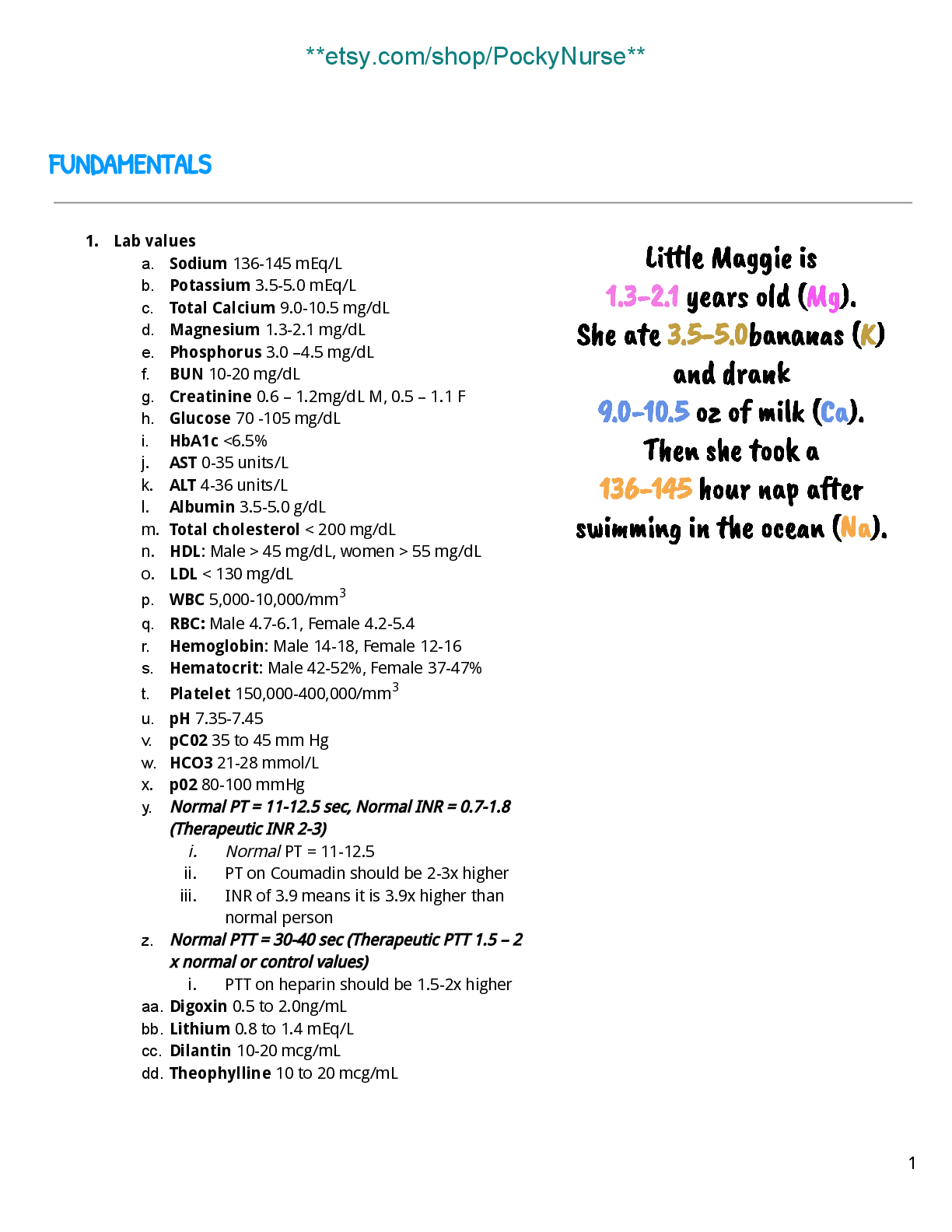
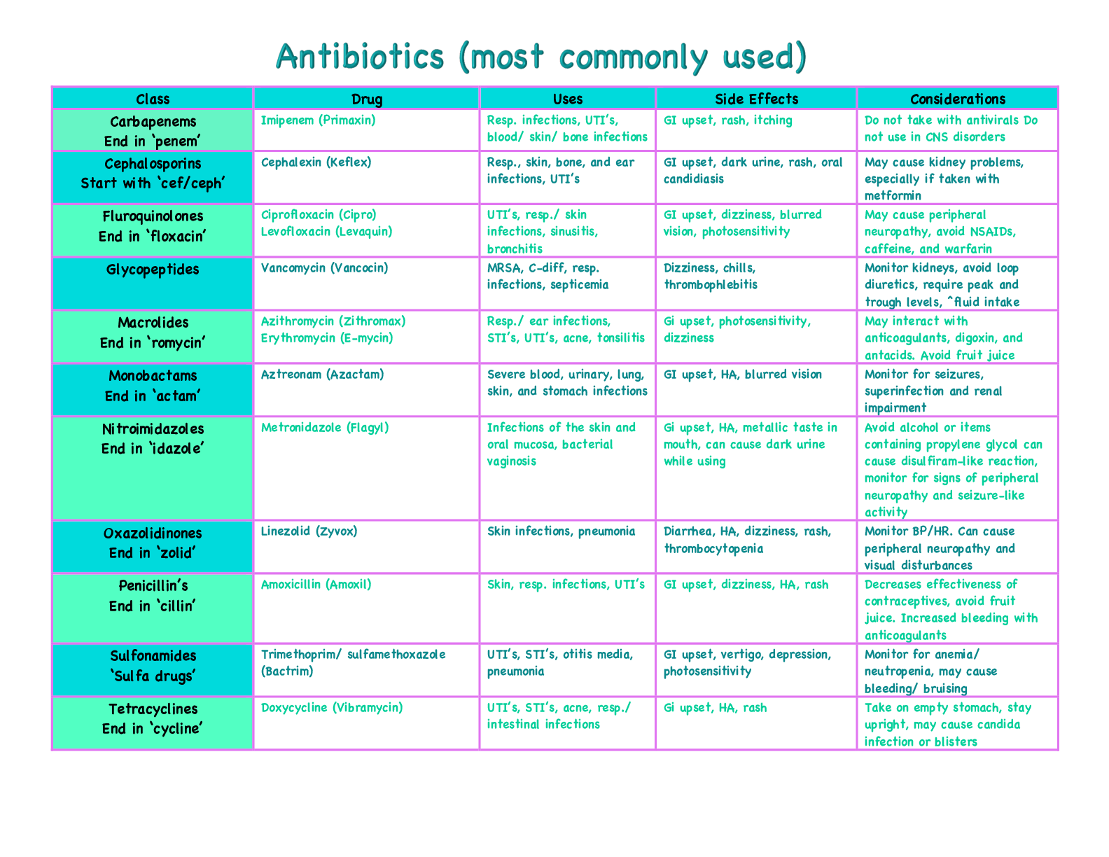

.png)