NCSBN – Lesson 8G: musculoskeletal System study Guide
Gouty arthritis is characterized by urate deposits and pain in the joints, especially those in the feet and legs. Urate deposits don't occur in septic or traumat
...
NCSBN – Lesson 8G: musculoskeletal System study Guide
Gouty arthritis is characterized by urate deposits and pain in the joints, especially those in the feet and legs. Urate deposits don't occur in septic or traumatic arthritis. Septic arthritis results from bacterial invasion of a joint and leads to inflammation of the synovial lining. Traumatic arthritis results from blunt trauma to a joint or ligament. Seasonal arthritis is not a condition.
Increasing pain that is not relieved by narcotic analgesics may be an indication of compartment syndrome. The nurse should immediately inform the charge nurse and emergency intervention will be required. Thromboembolic complications include deep vein thrombosis and pulmonary embolism, which are not characterized by increasing pain at the site of injury. Both pulmonary embolism and fat embolism present with sudden respiratory findings. Osteomyelitis is a bone infection that could occur some time after the initial injury, usually after at least 48-72 hours.
Developmental hip dysplasia will present with uneven gluteal folds and thigh creases. A nurse will be able to hear a clicking sound when when the infant is placed on their back, leg flexed and the affected hip is moved to abduction. This is typically seen in infants less than 4 weeks old and is refereed to as a positive Ortolani's sign. Additionally, the nurse will assess for a positive Barlow maneuver (abducting the hip while applying pressure on the knee will cause the hip to dislocate from the socket). Finally, a pelvic downward tip is not a finding a nurse will find with possible development dyspepsia.
The use of oral corticosteroids for a long period of time increases the risk for developing osteoporosis. Being postmenopausal and physically inactivity may also contribute, but are less significant. Other factors that increase the risk for osteoporosis and fracture include low bone mass and poor calcium absorption.
However, long-term steroid treatment is the most significant risk factor.
After having a total hip replacement, the client is positioned with an abduction wedge or pillow(s) between the legs. The abduction pillow helps prevent adduction and internal rotation of the affected leg, which could cause dislocation of the hip prosthesis. Some surgeons recommend clients use an abduction wedge for as long as 6-12 weeks postoperatively.
Osteoporosis is commonly thought of as a "woman's problem", but after age 65 men and women are losing bone mass at about the same rate and calcium absorption decreases. Treatment for osteoporosis includes regular weight-bearing exercises, such as walking, in which bones and muscles work against gravity; aqua aerobics will not make bones stronger. The client needs adequate intake of calcium and vitamin D; while supplements may be needed, calcium is best absorbed from natural food sources. There is a direct link between tobacco use and decreased bone density.
Trauma
Muscular and skeletal injuries are a common occurrence within the general population. This section reviews injuries such as contusions, sprains and fractures of the musculoskeletal system.
Contusions – Trauma
Contusions occur when a fall or blow breaks capillaries but not skin. Extravasation
(bleeding) occurs under the skin.
• Assessment Findings
o Ecchymosis (bruising)
o pain is found when the contusion is palpated.
• Management
o For the first 24-48 hours, apply ice for 15 minutes, three times a day.
▪ Wrap the area to compress.
▪ The color of a bruise changes from a blackish-blue to a greenish-yellow after 3-5 days and should completely heal within 7-10 days.
o Observe closely for extension or enlargement of the bruise if the client is at risk for bleeding due to thrombocytopenia or other coagulation disorder.
o Observe the client for changes in mental status if the bruise is related to a head injury. When the injury occurs near a structure (such as the eye), report any evidence that the integrity of the structure is not jeopardized.
Strains – Trauma
A strain is a mild injury to the muscle attachment to the bone. It is a lesser injury than a sprain.
• Etiology & Pathophysiology
o A strain is caused by overstretching, overexertion or misuse of muscles. Chronic
strains occur with long-term overstretching of the muscle or tendon or with the repeated use of the muscle beyond its physiologic limits. An acute strain is a recent injury to the muscle or tendon. Acute strains are classified by degree.
• Assessment Findings
o First Degree Strain
▪ A first degree strain is mild and has a gradual onset. It feels stiff and sore locally.
▪ The muscle will be tender to the touch and the client may experience muscle spasm.
▪ There will be no loss of range-of-motion and little to no edema or
ecchymosis will be seen.
• Management
o Comfort measures
o Apply ice
o Rest, possibly immobilize for short term and elevate
o Oral analgesics or NSAIDs
o Second Degree Strain
▪ Second degree strains occur with a moderate stretch of the muscle and have a sudden onset, with acute pain that eventually leaves the area tender.
▪ The client will have extreme muscle spasms and passive motion will increase the pain.
▪ Edema will develop early and later ecchymosis may appear.
• Management
o Keep limb elevated
o Apply ice for the first 24-48 hours, then apply moist heat
o Limit mobility using an Ace bandage wrap
o Muscle relaxants, analgesics and NSAIDs
o Physical therapy for strength and range-of-motion
o Third Degree Strain
▪ Third degree strains occur when there is severe stretching of the muscle
with a tear.
▪ The client feels a sudden, snapping or burning sensation.
▪ These strains cause muscle spasms, joint tenderness and edema (may be
extreme).
▪ The client cannot move the strained muscle voluntarily.
▪ Ecchymosis typically occurs but is delayed.
• Management
o Keep the limb elevated
o Apply ice for 24-48 hours, then apply moist heat
o Either immobilize or limit mobility of the limb
o Limit weight bearing on lower limbs
o Medication – muscle relaxants, analgesics and NSAIDs
o Physical therapy for strength and range-of-motion
Sprains – Trauma
Sprains are a more serious injury than a strain. Sprains involve injury to ligament structures
by stretching, exertion or trauma.
• Assessment Findings
o First Degree Sprain
▪ minimal tearing of ligament fibers and will present with localized edema
or hematoma.
▪ There will be some mild discomfort at the location of the injury, with
increased pain when the limb is palpated or bearing weight.
▪ There will be no loss of function or weakening of the joint structure.
• Management
o Compress the injured area with an Ace bandage to limit
swelling
o Keep the limb raised to decrease edema
o Apply ice 24-48 hours following injury
o Analgesics for discomfort
o Isometric exercises to increase circulation and resolve hematoma
o Second Degree Sprain
▪ Up to half of the ligamentous fibers are torn in a second degree sprain.
▪ The client will present with edema, possible hematoma, a decreased active range-of-motion, and mild weakening of the joint and pain.
• Management
o Protectively dress/splint the joint and immobilize it
o Elevate the limb to decrease edema
o For 24-48 hours, alternate:
▪ Ice to produce vasoconstriction in order to decrease swelling and pain
▪ Moist heat to decrease swelling and provide comfort
o Analgesics for discomfort
o Physical therapy to increase circulation and maintain nutrition to the cartilage
o Third Degree Sprain
▪ complete rupture of the ligamentous attachment.
▪ The client will present with severe edema with hematoma and with
severe pain.
▪ There will a dramatic decrease in the active range-of-motion
▪ loss of joint integrity and function.
• Management
o Casting/immobilization
o Surgery to restore integrity of joint
o See second degree treatment
Fractures – Trauma
A fracture is any alteration in the continuity of a bone. A fracture dislocation is a fracture in which the joint is dislocated in that position.
• Classification of Fractures
o Completeness
▪ A complete fracture is when the bone is broken in two or more pieces.
▪ An incomplete fracture is when the bone is broken but still in one piece.
o Wound
▪ A closed fracture is a simple fracture that does not break the skin.
▪ An open fracture or compound fracture is complex.
• Bone fragments break through the skin and injure the soft tissue, often causing infection.
o Fracture Line
▪ Longitudinal linear: the fracture runs parallel to the lengthwise direction of the bone
▪ Oblique: a fracture that is produced by a twisting force and requires traction to heal properly
▪ Spiral: a fracture that also results from twisting force and may accompany damage to soft tissue and requires traction or internal fixation
▪ Transverse: a fracture that is caused by angulation and is common in pathological fractures and generally stable after reduction
• Types
o Avulsion Fracture
▪ Avulsion fractures occur when bone fragments and soft tissue are pulled away from the bone.
• It is typically caused by a direct force on the bone.
o Comminuted Fractures
▪ Comminuted fractures are produced by a high energy force which results in two or more bone fragments.
▪ The fragments are splintered and cause severe injury to the soft tissue.
o Compression Fractures
▪ Compression fractures are often seen in the lumbar spine and at times
are pathological (a disease weakens the bone).
o Greenstick Fractures
▪ Greenstick fractures are caused by compression and/or angulation forces and result in an incomplete fracture. The cortex of the bone bends to one side and buckles on the other.
▪ Greenstick fractures are common in children who have relatively pliable bones. They require reduction or completion of the fracture line through the cortex. Pressure is applied with a cast to the apex of the deformity.
o Impacted Fractures
▪ Impacted fractures (telescoped) occur when direct force breaks bone and telescopes the fragments. Fracture fragments move in unison and rapid
o Stress Fractures
▪ Stress fractures are incomplete fractures and a result of repetitive trauma to the region.
▪ There are two types: fatigue and insufficiency.
• Fatigue stress fractures are caused from repeated trauma
• Insufficiency stress fractures are pathological fractures.
• Assessment Findings
o Swelling, pallor and ecchymosis of surrounding subq tissue
o Loss of sensation to body parts
o Deformity
o Pain and/or tenderness
o Muscle spasms
o Loss of function, abnormal mobility
o Crepitus (grating sound)
o Shortening of affected limb
o Decreased or absent pulses distal to injury
o Affected extremity colder than contralateral part
• Management
o Closed Reduction Procedure
▪ Closed reduction is a procedure to set (reduce) a broken bone without surgery. This allows the bone to grow back together. It works best when it is done as soon as possible after the bone breaks.
▪ Pre- and post-reduction X-rays are essential to determine whether the reduction has been successful.
o Immobilization
▪ Immobilization keeps the bone fragments from moving and relieves pain. Methods to keep the bones immobilized include casting, traction, splints, braces or external fixation.
o Traction
• Young woman lying in a hospital bed undergoing traction for a leg injury.
o Types of Traction:
▪ Manual: applied by pulling on the extremity – may be used during cast application
▪ Skin: applied by pulling force through the client's skin – used to relax the muscle spasm
▪ Skeletal: applied directly through pins inserted into the client's bone – used to align the fracture
o Skin Traction
▪ Uses 5-7 pound (2.27-3.18 kg) weights attached to the skin to indirectly apply force
▪ Weights are attached either through tape, using straps, boots or cuffs
▪ Examples:
▪ Buck's traction: used to immobilize, position, and align the lower extremity in the treatment of contractures and diseases of the hip
and knee
▪ Donlop's traction: used for children with fractures of the upper arm when the arm must remain flexed
▪ Pelvic traction
▪ Bryant's traction: used to immobilize both lower extremities in the treatment of a fractured femur or in the correction of a
congenital hip dislocation
o Skeletal Traction
▪ Skeletal traction uses pins inserted into bones:
▪ Used when more pulling force is needed than skin traction can provide
▪ Approximately 25-40 pounds (11.34-18.14 kg) of weight can be applied
▪ Requires the surgical placement of tongs, pins or screws into the bone
o Nursing Care
▪ Traction:
▪ Be sure to know if the traction is to be applied either intermittently or continuously
▪ Be sure to know the amount of weight to be applied
▪ Check ropes and knots, pulleys and weights at least once a
shift:
• Rope should move freely over pulleys
• Prevent friction, which will impair the efficiency of the traction
• Weights should hang freely
▪ Identify and maintain countertraction:
• Countertraction is the force opposing the pull of traction, which is generally provided by the client's body
• If countertraction is not maintained, the client is not in traction
• Sign of loss of countertraction is that the client slides down in bed
• Elevate the foot of the bed with shock blocks
▪ Maintain client in correct alignment and body position
▪ Skin care:
▪ Regular assessment for breakdown, especially coccyx and heels but also all bony prominences
▪ Assessment and care of pins, wires or tongs with skeletal traction and observe for signs of infection
▪ Assessment of skin by removing traction at least once per day for hygiene and reapplication (manual traction should be used while skin traction is off)
▪ Perform regular neurovascular checks approximately every four hours and document:
▪ Sensation
▪ Temperature
▪ Movement
▪ Distal perfusion
▪ Assist client to cough and deep breathe and to use incentive spirometer
▪ Assist with range of motion exercises
▪ Assist client with use of trapeze
▪ Assess for skin breakdown
• Stages of Bone Healing
o Stages of Bone Healing:
▪ Hematoma formation
▪ Fibrocartilage/granulation tissue formation
▪ Callus formation
▪ Ossification
▪ Consolidation/remodeling
Bone Healing Stages
The healing of a fracture occurs allows for the bone and surrounding tissues to be restored to its original physical and mechanical properties. There are three separate, but overlapping, stages of bone healing: 1) early inflammatory stage, 2) repair stage, and 3) late remodeling stage.
▪ Inflammatory stage: begins immediately after the bone is fractured and lasts several weeks after the injury; continues into the next stage
o Hematoma formation:
▪ One to three days following the injury
▪ Blood clot forms around the fracture site
▪ Bone necrosis occurs distal to the fracture site due to a loss of blood
o Inflammatory cells infiltrate the bone:
▪ Results in the formation of granulation tissue, vascular tissue, and immature tissue (which will specialize to form a bridge of tough connective tissue)
▪ Begins three days to two weeks after the injury
▪ Repair stage: bone production, in which inflammation is replaced with fibrous tissue and cartilage and the bone ends become joined and stabilized:
o Collagen matrix laid down
o Dead bone is resorbed
o Fibrocartilaginous callus formation:
▪ Develops over approximately two to six weeks
▪ Capillary growth in the hematoma
▪ Phagocytosis breaks down and removes the formed hematoma
▪ Fibroblasts and osteoblasts migrate into the site and begin reconstruction of the bone
▪ Size and shape of callus is in direct response to the amount of displacement of fracture fragments
▪ The fibrocartilaginous callus serves to splint the fracture
o Ossification:
▪ Continued migration and multiplying of osteoblasts and osteocytes results in the fibrocartilaginous callus turning into bony callus
▪ The gap in the bone is bridged and union occurs
▪ Begins from each end of the fracture and progresses toward the center of the fracture
o Delay at this stage results in delayed union or nonunion of the bone
o This stage can last one to two months after a fracture
▪ Remodeling stage: bone is restored to its original shape, structure and mechanical strength:
o Excess material of the bony callus is removed and compact bone is laid down in order to reconstruct the bone; callus becomes calcified and blends into the bone
o Occurs slowly over months to years
o Fracture line may still be evident on radiographs
• Complications
o Immediate
▪ Immediate complications of the injury are shock (higher risk with pelvic and femur factures), fat embolism (can occur 24 hours after the injury and is more common with pelvic/femur fractures), deep venous thrombosis (DVT) and pulmonary embolism (PE).
▪ Compartment syndrome is a complication that is considered a medical emergency.
▪ Compartment syndrome occurs when the nerve and blood vessels are compressed causing muscle and nerve damage. The client will experience a decrease in sensation, paleness and weakness. It is treated with a longitudinal incision to relieve pressure and/or the health care provider may loosen a tight cast or wrap.
▪ Orthopedic Complications
• Venous thromboembolic problems:
o Thrombophlebitis:
▪ Inflammation of a vein with the formation of a blood clot
▪ Incidence is greatest after trauma or surgery to legs or feet
o Deep venous thrombosis (DVT):
▪ Anterior tibial or femoral veins
▪ May be caused by immobility
▪ Findings include calf pain, positive Homan's sign
▪ Nursing care:
• Anticoagulant therapy
Paralysis and weak, diminished or absent Pulse (pulselessness)
• Anti-embolism stockings (usually thigh- high) – measure prior to surgery
• Sequential compression device(s)
o Pulmonary embolism (PE):
▪ Blood clot from systemic circulation enters pulmonary circulation
▪ Most commonly seen after hip fractures and total hip or knee arthroplasty
▪ May be caused by femoral vein manipulation during surgery and, therefore, may occur without signs of DVT
▪ Findings include chest pain (pleuritic), sudden shortness of breath, tachycardia, palpitations, or change in mental status
▪ Diagnosis confirmed via ventilation/perfusion scan or pulmonary angiography
▪ Nursing care:
• If PE suspected, do not leave client
• Notify health care provider immediately
• Continuous IV heparin therapy is usually prescribed
o Fat embolism:
▪ Fat cells enter pulmonary circulation
▪ Associated with:
• Multiple trauma accidents
• Multiple organ involvement
• Fractures of marrow producing bones, especially the femur
• Joint replacements
• Insertion of intermedullary rods
▪ Usually occurs 24-48 hours after the fracture
o Bleeding complications:
▪ Hemorrhage:
• Abnormal loss of a large amount of blood from the body
• Most common in fractures of long bones producing bone marrow
▪ Hematoma formation:
o Wound infection:
▪ May be superficial or deep wound
▪ Deep wound infection may lead to osteomyelitis
o Delayed
▪ Findings include erythema and swelling around suture line, increased drainage and elevated temperature
▪ Treatment:
• Antibiotics
• May require incision and drainage of wound or removal of prosthesis if severe infection
is present
o Special complications in hip joint replacement:
▪ Femoral fracture:
• Occurs near distal end of femoral-shaft part of prosthesis
• Occurs more frequently with elderly, clients with osteoporosis, or after revision to total hip replacement
• Primary finding is severe pain with ambulation
• Diagnosis is confirmed with X-ray
• Depending on severity, treatment will be immobilization or open reduction with
internal fixation
▪ Dislocation of hip prosthesis:
• Greatest risk during the first postoperative week but can occur at any time within the
first year
• Risk decreases as muscle tone of the hip increases
• Caused by flexion of the hip (beyond 90°) or poor prosthetic fit
• Findings include pain and external rotation of the leg
• Treated by closed reduction under conscious sedation (if it occurs within the first six weeks) or open surgical revision
o Special complication in knee joint replacement – flexion failure:
▪ Client cannot flex knee 90° two weeks postoperatively
▪ Treated with closed manipulation of the knee joint under general anesthesia
▪ Joint stiffness and post-traumatic arthritis (osteoarthritis, type II)
▪ Reflex sympathetic dystrophy is characterized by abnormal pain and swelling of the extremity
▪ Myositis ossificans is the formation of hypertrophic bone usually near the injured bone or muscles
▪ Malunion occurs when a fractured bone heals in an abnormal position associated with reduced functioning and pain, which may require an osteotomy to restore the appropriate alignment of the bones
▪ Delayed union is when the fractured bone is slow to heal and is usually due to inadequate reduction, inadequate immobilization, inadequate blood flow to the bone and/or infection
▪ Non-union occurs when the fracture does not heal Nursing Interventions
The nursing interventions appropriate to fractures correspond to the risk factors present.
▪ Risk for Peripheral Neurovascular Deficit
o When the client is at risk for peripheral neurovascular deficit, the nurse will need to check the client's neurovascular status often.
o Assessment includes palpating for pulses, sensation, skin temperature, ability to move appendages (fingers or toes) and proper skin color (no pallor or cyanosis).
o Elevate the limb above the level of the heart unless compartment syndrome is suspected. Apply cold to minimize edema.
▪ Risk for Moderate to Severe Pain
o Assess the level of pain and administer pain medications as ordered. Maintain appropriate traction if applied. Non-medical interventions include repositioning the client and using padding on bony prominences.
o Teach the client relaxation techniques, such as visual imagery and music therapy.
▪ Risk for Impaired Skin Integrity
o Impaired skin integrity is typically caused by open fractures, soft tissue injuries, pressure areas, inadequate nutrition and/or immobility. Additional factors that affect skin integrity include the age of the client, the general condition of the client and any other preexisting conditions the client may have.
o Nursing interventions that will decrease the risk for impaired skin integrity include immobilizing the client as soon as possible, turning the client every two hours while immobile and positioning the client with proper body alignment. You may also decrease your client's risk by utilizing special equipment (e.g. pressure air mattress) and evaluating risk status with a validated tool such as Braden Scale for Predicting Pressure Sore Risk© are also key actions.
▪ Risk for Infection
o The risk of infection is a concern if the client has open fractures, superficial or deep wounds or any kind of surgical intervention. To decrease the risk of
infection, the nurse will monitor for elevated temperature, redness or drainage from the surgery site. Providing proper wound care and administering antibiotic therapy (if indicated) can help decrease the risk of an infection.
▪ Impaired Gas Exchange
o Clients with a fracture are also at risk for developing a fat embolism or DVT. Mobilize the client as soon as possible. Have them use their incentive spirometer frequently and teach proper coughing and deep breathing techniques.
▪ Skeletal Traction Care
o Daily pin care; clear crusty drainage is normal
o Inspect the traction apparatus every eight hours for alignment and proper function
o Ensure weights are always hanging freely and are not obstructed by other objects
▪ Client teaching will include:
o How fractures heal
o Why the fracture is being immobilized
o How to bear weight and how much (if permitted)
o How to use assistive devices to walk – evaluate the client's ability to use the device and make adjustments according to your assessment
o Cast care – see Lesson 7 Reduction of Risk Potential (Procedures) for more information about cast care
▪ Keep the cast dry
▪ For itching, suggest blowing air into the cast with a hair dryer on a cool setting
▪ Report swelling, discoloration of digits, pain during motion and burning and tingling under the cast
o How to prevent complications – continuing to use an incentive spirometer, coughing and deep breathing exercises, movement/shifting of weight, neurovascular checks and reporting abnormal findings to their health care provider
Fractures & Healing – Trauma Factors that affect healing:
• Enhances Healing
o Fracture is near a good blood supply
o Minimal damage to soft tissue
o Anatomic reduction
o Fragments are in a good position to heal
o Immobilization
o Weight can be borne on long bones
• Delays or Inhibits Healing
o Poor blood supply to one or more bone fragments (mid-shaft fracture have less
blood supply)
o Severe damage to soft tissue
o Separation of fragments
o Traction pulls fragments apart
o Improper fixation allows bones to move or rotate
o Pre-existing factors, such as obesity, diabetes, steroid use or smoking
o Severely comminuted fracture
o Bone loss
o Infection
Fractures in Children – Trauma
The etiology of fractures in children is usually due to immature motor and cognitive skills, trauma, osteogenic diseases, birth injuries or child abuse. Children seldom experience complete breaks because their bones are flexible. If a greenstick or spiral fracture occurs, this may indicate child abuse.
The pediatric client is at risk for external hemorrhage with critical blood loss and a break at the epiphyseal plate that can affect the future growth of the limb.
Repetitive Use Injuries Trauma
Repetitive use injuries are a result of repeated twisting and turning of the affected joint. Examples include carpal tunnel syndrome, bursitis and epicondylitis.
• Carpal Tunnel Syndrome
o Carpal tunnel syndrome occurs when the carpal tunnel in the wrist narrows from repetitive irritation, then compresses and irritates the median nerve.
▪ The client experiences numbness and tingling of the thumb, index finger and middle finger of the affected hand.
• Bursitis
o Bursitis affects the bursae in the shoulder, hip, leg and/or elbow and is often associated with athletic endeavors such as pitching in baseball and playing tennis.
▪ The client will present with tenderness and pain in the joint that increases with movement.
• Epicondylitis
o Epicondylitis occurs when there is inflammation of a tendon where it inserts into the bone.
▪ Clients will present with point tenderness and pain radiating down the affected extremity.
• Nursing Interventions
o Encourage the client to rest and immobilize the involved joint. Ice therapy and
NSAIDs may be used to decrease pain and inflammation. In the case of surgery, nursing care focuses on the prevention and recognition of postoperative complications, as well as the support of rehabilitation.
Pediatric Osgood-Schlatter Disease – Trauma
Pediatric Osgood-Schlatter disease is a benign, self-limiting knee condition associated with pain and edema of the tibial tubercle. It is one of the most common causes of knee pain in adolescents.
• Etiology
o Osgood-Schlatter disease's etiology is idiopathic.
o It could possibly be due to repetitive stress from sports-related activity, plus
overuse of immature muscles and tendons. It is usually self-limiting.
• Assessment Findings
o Clients will present with knee pain or tenderness, edema of the tibial tubercle
and an exacerbation of symptoms if the client runs or jumps.
• Diagnostic Studies
o Diagnostic studies include a physical exam, history and radiographic studies.
• Management
o The client will avoid activities that aggravate the condition and use an elastic
bandage on the affected knee or joint for support. Rest, ice and NSAIDs are recommended.
Legg-Calvé-Perthes Disease – Trauma
Legg-Calvé-Perthes disease (osteochondritis) is an aseptic necrosis of the femoral head.
• Etiology
o Legg-Calvé-Perthes disease is caused by the disturbance of circulation (blood
supply) to the femoral epiphysis, creating ischemic aseptic necrosis of the femoral head. It is usually self-limiting.
• Assessment Findings
o This disease has an insidious onset and the client will complain of an
intermittent painful limp on one side.
o There will also be a decrease in range-of-motion in affected extremities.
• Diagnostic Studies
o Diagnostic studies may include a history, radiographic studies, bone scan
and/or MRI.
• Management
o Bed rest and non-weight bearing range-of-motion will be implemented.
Corrective devices such as braces, casts or traction will be used.
o Conservative therapy must be continued for 2-4 years. Surgery is a possibility, depending on the extent of the damage.
• Nursing Interventions
o The client will engage in mostly outpatient activities.
o Teaching points to consider include explaining the purpose, function, application and care of corrective devices. For a pediatric client, discuss appropriate play activities, emphasizing that the child must stay relatively inactive during treatment.
Rhabdomyolysis – Trauma
Rhabdomyolysis is a disease involving the breakdown of muscle tissue. The term was first used to describe crush injuries during World War II. This disorder often occurs after a major muscle trauma.
• Etiology & Pathophysiology
o Long distance running, certain severe infections, and electric shock can cause
muscle damage and release of myoglobin. There has also been a connection with the use of cerivastatin and gemfibrozil resulting in rhabdomyolysis.
o When trauma to the muscle compresses tissue, it causes ischemia and necrosis. Local edema continues to increase compartment pressure and tamponade causes the blood vessels to collapse. This leads to tissue hypoxia, muscle infraction, and neural damage. Myoglobin, potassium, creatine kinase and urate are released from the necrotic muscles fibers into the circulation.
• Assessment Findings
o Clients will present with both local and systemic symptoms.
o Local symptoms include:
▪ muscle pain, tenderness, swelling and weakness.
o Systemic symptoms are indicated by
▪ dark-red-brown urine, a result of myoglobin entering into the urine. Additional findings include, fever, malaise, nausea, vomiting, confusion, agitation, delirium and anuria.
• Diagnostic Studies
o Urine myoglobin level will be greater than 0.5 mg/dL and the creatine kinase
will be significantly elevated. Serum potassium, phosphate and creatinine levels will be elevated as well. CT scan and MRI will reveal muscle necrosis, venous pressure measurements will be elevated and a ECG will show cardiac changes from the elevated potassium levels.
• Management/Nursing Interventions
o Treating the underlying cause of rhabdomyolysis and preventing renal failure are
key priorities.
o IV hydration should be initiated as soon as possible to decrease the creatine kinase levels (normal saline).
o Best rest, anti-inflammatory agents and analgesics will help relieve pain.
o Corticosteroids will be administered in complicated cases if necessary.
o If compartment venous pressure is greater than 25 mm Hg, an immediate fasciotomy and debridement to relieve pressure and promote circulation will have to be performed
Degenerative Disorders
Degenerative bone disorder usually occur when there is a deterioration of bones. These disorders involve the gradual reduction of bone mass, which makes the bones more brittle. This section reviews some common degenerative bone disorders.
Osteoarthritis (OA) - Degenerative Disorders
Osteoarthritis (OA) is a degeneration of the articular cartilage and formation of new bone in the subchondral margins of the joint. It primarily involves the weight-bearing joints.
• Pathophysiology
• Stage One – Micro-fracture of the Articular Surface
• Articular cartilage is worn away
• Condyles of bones rub together – joint swells and is painful
• Cartilage loses cushioning effect – joint friction develops
• Prostaglandins may accelerate degenerative changes
• Stage Two – Bone Condensation
• Erosion of cartilage
• Cartilage may be digested by an enzyme in the synovial fluid
• Stage Three – Bone Remodeling
• Matrix synthesis and cellular proliferation fail
• Eventually the full thickness of articular cartilage is lost
• Bone beneath cartilage hypertrophy and osteophytes form at the joint margins
• Joints begin to degenerate
• Assessment Findings
o Joint stiffness after periods of rest
o Pain in a movable joint, typically worse with action and relieved by rest
o Paresthesia
o Joint enlargement – bones grow abnormally, spurs form and synovitis sets in
▪ Heberden's nodes: DISTAL interphalangeal (closest to the fingernail and furthest away from the body)
▪ Bouchard's nodes: PROXIMAL interphalangeal joint (middle joint of the finger…closest to the body)
o Joint deformities
o Tenderness on palpation
▪ May involve widely separated areas of the joint
▪ Mild synovitis may be felt – positive bulge sign may be found
o Limitations occur in the client's in active range-of-motion because:
▪ Joint surfaces no longer fit together
▪ Muscles spasm and contracture
▪ Joints are blocked by osteophyte, loose bodies
▪ Crepitation, crunching when joints are moved
▪ Eventual ankylosis or stiffening of the joint
o Changes to the client's gait occur:
▪ Abnormal antalgic gait: a modified gait (limp) that helps the client avoid pain on weight-bearing structures
▪ Shortened stance
▪ Widened base of support
▪ Shortened step length
• Diagnostic Studies
o Lab tests to rule out autoimmune disorders:
▪ Sedimentation rate – used to measure inflammation in the body:
• 0-15 milliliters per hour for males
• 0-20 milliliters per hour for females
• Higher for elderly clients
o Rheumatoid factor (RF):
▪ A lab test that is ordered when symptoms indicate that RA may be present
▪ A positive test may also be seen in lupus, endocarditis, tuberculosis (TB), cancer, sarcoidosis, syphilis, viral infection or disease of liver, lung or
kidneys
o C-reactive protein (CRP) – increased with inflammation, appears in higher amounts when there is swelling in the body
o Other lab tests:
▪ Complete blood count (CBC): analyze before NSAID therapy to determine if within normal limits
▪ Kidney and liver status (before using NSAIDs, especially in older adults)
▪ Test for TB with purified protein derivative (PPD)
▪ Antinuclear antigen (ANA) titer:
• A positive test may indicate an autoimmune disease
• May be lower in the elderly
• Does not necessarily prove a connective tissue disease
o Synovial fluid analysis distinguishes OA from rheumatoid arthritis (RA)
o X-rays, bone scans, magnetic resonance imaging (MRI) and/or computed tomography (CT) scan
• Management for Typically Conservative OA Treatment
o Provide education and counseling about the disease that includes the need to
implement proper exercise, relaxation techniques, appropriate nutrition and the maintenance of normal weight. Activity and rest management, preservation of joint motion through a balance of rest and activity, passive range-of-motion exercises and active stretching are also important management techniques.
o Pharmacologic Intervention:
▪ Aspirin – most often recommended:
▪ Advantages: relatively safe and inexpensive
▪ Disadvantage: tinnitus and gastrointestinal (GI) problems, including ulcers and bleeding
o Nonsteroidal anti-inflammatory medications (NSAIDs):
▪ Includes topical pain relieving creams, rubs and sprays, e.g., capsaicin cream, diclofenac 1% topical solution and diclofenac patch (also known as Flector patch)
▪ Reduce pain and inflammation
▪ May cause GI bleeding or gastric ulcers or cramping with diarrhea
o Corticosteroid injections
o Hyaluronic acid injections
o Mild narcotic pain relievers
• Non-pharmacologic Interventions
o Assistive devices, such as canes and walkers, are helpful supportive devices.
Transcutaneous electrical nerve stimulation (TENS) has been shown to be effective for pain relief.
• Complementary & Integrative Health
o Using practices such as acupuncture and massage therapy may help lessen pain
and improve function.
• Nursing Interventions
o Determine the client's level of functioning - mobility and ability to perform daily
tasks.
o Client teaching will include:
▪ Pain medications
▪ Assistive devices
▪ Prescribed heat therapies, posture and body mechanics
▪ Weight reduction (ensure client has adequate resources on the topic)
▪ Exercise
▪ Referral to support agencies
▪ Discuss with health care provider prior to taking any herbal or dietary supplements
▪ Home safety (rugs, lights, shoes and handrails) Inflammatory Disorders
The diseases in this section are often brought on by an autoimmune response or an environmental factor that results in an inflammatory response.
Rheumatoid Arthritis (RA) – Inflammatory Disorders
Rheumatoid arthritis (RA) is a chronic systemic inflammatory disease of the connective tissue.
• Etiology & Pathophysiology
o The cause of RA is not fully understood.
It is thought to be an autoimmune disorder and may involve a bacteria or virus (e.g., Epstein Barr virus). It does have a genetic tendency and is more common in women than men.
It strikes patients aged 20-50 years.
o Synovitis immune complexes initiate the inflammatory response and IgB antibodies are formed. The body does not recognize these antibodies as belonging to the "self," so it forms an antibody known as RF. As a result, pannus forms and there is a destruction of subchondral bone. The inflammatory response is activated and can go on indefinitely. It is irreversible and will lead to ankyloses of the joint.
• Assessment Findings
o General signs include
▪ Fatigue
▪ loss of appetite,
▪ weight loss
▪ enlarged lymph glands.
o Early in RA, clients will present with
o painful, stuff joints
o The capsules and soft tissues will be warm, red, swollen and incapable
of full range-of-motion.
▪ Plan of care in ACUTE PHASE
• Relieve pain pain is highest in this phase
• Preserving joint function
• Preventing joint deformity
o In late RA, joints will show
▪ bony ankyloses
▪ destruction of joint (reactive hyperplasia)
▪ adhesions
▪ inflammation and effusion
o Rheumatic nodules (firm, oval, non-tender masses under the skin) can be
observed. Clients may also present with Raynaud's syndrome.
o The client may present with ulnar deviations. These include deformed hand
and fingers, including swan-neck of the fingers and Boutonnière deformities.
• Diagnostic Studies
o History and physical, radiographic studies and/or aspiration of synovial fluid.
o Lab tests:
▪ Elevated erythrocyte sedimentation rate (ESR)
▪ Decreased red blood cells (RBC)
▪ Positive C-reactive protein – rise in response to inflammation
▪ Positive antinuclear antibody in 20% of cases
▪ Positive RF
• Management
o Pharmacologic Intervention:
▪ NSAIDs
▪ Hydroxychloroquine sulfate
▪ Immunosuppressive agents, e.g., azathioprine, cyclophosphamide, methotrexate
▪ Prednisone (steroids)
▪ Sulfasalazine, leflunomide and anakinra
▪ Biological response modifiers (BRMs), e.g., etanercept, infliximab and adalimumab
▪ Surgical interventions – joint replacement surgery
▪ Splinting – resting, correction or fixation
▪ Adequate rest
▪ Ice for joint inflammation; heat for joint stiffness
o Nutrition therapy:
▪ Weight reduction
▪ Calcium supplements
• Complementary & Integrative Health
o Herbal remedies or dietary supplements like gamma-linolenic acid (GLA) may
help reduce inflammation, fish oil (omega-3 fatty acids) may reduce the need for NSAIDs and other conventional RA medications.
o There is ongoing research on other supplements that could prove helpful, including ginger, boswellia, green tea and turmeric.
o Acupuncture, biofeedback and relaxation training may boost morale or reduce symptoms, although research results have been inconclusive. Mindfulness meditation may help RA clients with a history of depression.
• Nursing Interventions
o Psychological support
o Care of clients taking corticosteroids
o Promote self-care and independence
o Administer medications as prescribed
o Encourage balance of exercise, rest and energy conservation
o Teach client how to use assistive devices
o Teach client to speak with a health care provider before using over-the-counter (OTC) medications or home remedies
Juvenile Idiopathic Arthritis – Inflammatory Disorders
Juvenile idiopathic arthritis (formerly known as juvenile rheumatoid arthritis-JAR) is an
inflammation of the joints.
• Etiology & Pathophysiology
o Juvenile idiopathic arthritis is an autoimmune disorder with a probable genetic predisposition.
• Pathophysiology Process
o Trigger inflames synovium – chronic inflammation
o Effusion of the joint and increased fluid
o Erosion and fibrosis of the articular cartilage
o Further deterioration occurs with bone erosion
o Decrease in the joint's range-of-motion and function
• Assessment Findings
o The client may develop
▪ a sudden inability to walk on one leg, with intermittent joint pain,
▪ stiffness and swelling.
▪ There will be a decrease in range-of-motion, with morning stiffness.
• This can result in a significant disability.
• Diagnostic Studies
o Lab tests – there are no definitive serologic tests:
▪ Increased ESR and CRP – has to be C-reactive protein (sign of rheumatic fever)
▪ Anemia
▪ Leukocytosis in early stages
▪ Human leucocyte antigen (HLA) testing
o X-rays – widening of joint spaces, followed by gradual fusion and articular
destruction, with soft tissue swelling
• Management
o The treatment objectives include
▪ preserving the joint function
▪ preventing physical deformities and pain relief.
o Physical and occupational therapy referrals will help the client maintain
mobility.
o Teaching points will include balancing rest with activity, positioning and
splinting.
o Ultrasound, electrical stimulation, heat and whirlpool therapies will also help with mobility and pain relief.
o Surgery may be necessary for joint replacement.
o Pharmacologic Intervention:
▪ NSAIDS e.g., naproxen
▪ Slow acting antirheumatic drugs (SAARDS)
▪ Corticosteroids
▪ Cytotoxic agents, e.g., methotrexate
• Nursing Interventions
o Monitor for compliance with physical and occupational therapy
o Prevent contractures
o Monitor pain and discomfort – possible application of moist heat
o Provide family and child support services
o Support the client around body image and decreased mobility issues
o Assess the client for altered patterns of growth and development due to decreased activity and a decreased ability to perform activities of daily living (ADL)
Systemic Lupus Erythematosus (SLE) – Inflammatory Disorders
Systemic lupus erythematosus (SLE), also known as lupus, is a chronic, systemic, inflammatory disease of the collagen tissues.
• Etiology & Pathophysiology
• SLE is an autoimmune disorder.
• African Americans, Hispanics, Asians, and Native Americans are 2-3 times more likely than Caucasians to have lupus. Most cases of SLE are found in women.
• Antigens stimulate antibodies, which form soluble immune complexes. Those complexes
are deposited in tissues. The number of T-suppressor cells dwindles, and the immune complex inflames the tissue. The intensity and location of the inflammation affects what the nurse will find on assessment. Clients with central nervous system (CNS) or renal involvement have a poorer prognosis.
• Assessment Findings
• SLE is most likely present if the client has four or more of these findings:
• Arthritis: characterized by swelling, tenderness and effusion involving two or more peripheral joints
• Malar rash: characteristic butterfly rash over cheeks and nose
• Discoid lupus skin lesions
• Photosensitivity
• Oral ulcers
• Serositis: pleuritis (inflammation of the membranes in surrounding lungs)
• Renal disorder: persistent proteinuria
• Neurologic disorder: seizures or psychosis in the absence of drugs or pathology
• Hematologic disorder: hemolytic anemia with reticulocytosis (increased immature RBC) or leukopenia
• Immunologic disorder: positive lupus erythematosus (LE) cell preparation or anti-DNA or false positive serologic test for syphilis
• Antinuclear antibody: abnormal titer of antinuclear antibody by immunofluorescence or equivalent assay
• Positive lupus erythematosus (LE) cell reaction
• Management
• The goals of management are to control system involvement, monitor findings, and induce remission. The health care team will work to prevent negative effects of therapy and recognize flare-ups promptly.
• Pharmacologic Intervention:
• Salicylates
• Nonsteroidal anti-inflammatory agents (NSAIDs)
• Corticosteroids, anti-infectives and anti-cancer drugs
• Antimalarial drug
• Nursing Interventions
• Nursing actions include effective pain management strategies, implementing actions for the care of clients taking corticosteroids and maintaining skin integrity. Teaching points include weight management, energy conservation techniques and advising the client about sun protection.
Gout – Inflammatory Disorders
Gout is a metabolic disease marked by a painful inflammation of the joints, deposits of urates in and around the joints and usually an excessive amount of uric acid in the blood.
• Etiology & Pathophysiology
• Gout primarily affects men and its peak incidence is between 40-60 years old. Gout does have a familial tendency.
• Abnormal purine metabolism or excessive purine intake results in formation of uric acid crystals which are deposited in the joints and connective tissue. Deposits are most often found in the metatarsophalangeal joint of the great toe or in the ankle.
• Assessment Findings
• Tight, reddened skin over the inflamed joint
• Elevated temperature
• Edema of the involved area
• Severe pain in the affected joint
• Hyperuricemia
• Acute attacks commonly begin at night and last 3-5 days
• Gout attacks may follow trauma, diuretics, increased alcohol consumption, consuming a high purine diet, stress (both psychological and physical) or suddenly stopping maintenance medications
• Warning signs of flare-up include the exacerbation of previous findings or the development of a new one
• Systemic manifestations may include fever, renal disease and tophus
• Diagnostic Studies
• Synovial fluid analysis will reveal uric acid crystals and blood work will show hyperuricemia.
• Management
• Expected outcomes include symptom control and the prevention of attacks.
• Pharmacologic Intervention:
• NSAIDS
• Colchicine – used when NSAIDs are contraindicated, enhances the excretion of uric acid
• To prevent flare-ups: antihyperuricemic agents such as allopurinol or probenecid to minimize the production of uric acid
• Heat or cold therapy
• Dietary changes
• Avoid purine-rich foods such as meats, organ meats, shellfish, sardines, anchovies, yeast and legumes
• Control weight
• Drink less alcohol – all types
• Increase daily fluid intake
• Nursing Interventions
• Pain management strategies
• Elevate the affected limb; provide bed rest and immobilize joint
• Avoid pressure or placing bedding on affected joint
• Reinforce dietary management and weight control
• Administer anti-gout medications as ordered
• Increase fluid intake to prevent renal calculi (kidney stones)
• Cautious use of aspirin and diuretic products (avoid if possible) Fibromyalgia – Inflammatory Disorders
Fibromyalgia is a common rheumatic syndrome of musculoskeletal pain, stiffness and
tenderness.
• Etiology
• Fibromyalgia has a possible autoimmune or environmental cause. Pain and tenderness are thought to be from overactive nerves.
• Assessment Findings
• Clients will present with:
• chronic achy muscle pain local or systemic
• tenderness at trigger points.
• They often complain of fatigue, sleep deprivation, headaches and irritable bowels. Pain and fatigue seem to be aggravated by exertion.
• Management
• Heat, massage and stretching can help relieve the symptoms.
• Promote sleep improvement (amitriptyline has been found to promote sleep and relieve symptoms).
• Complementary & Integrative Health
• There is some evidence that transcranial magnetic stimulation (TMS) can help. Vitamin D supplements may reduce pain in people with fibromyalgia who are deficient in this vitamin.
• Nursing Interventions
• Reinforce the client's understanding of this disorder. Assure the client that fibromyalgia is a real disorder and not psychosomatic. Support prescribed therapy as ordered.
Metabolic Bone Disorders
Metabolic bone disorders affect bone strength and can be caused by abnormalities of minerals, hormones, pathogens or unknown etiologies.
Osteomalacia – Metabolic Bone Disorders
Osteomalacia is a metabolic bone disorder that occurs when mineralization is delayed, resulting in a soft and weak bone.
• Etiology & Pathophysiology
• Rickets is caused by a vitamin D deficiency, which results in the bones not having enough calcium and phosphorus. There will then be less serum calcium than normal, which triggers an increase in parathyroid hormone and renal phosphorus clearance.
• Assessment Findings
• The client will complain of:
• generalized muscle
• skeletal pain in the hips
• similar pain in the lower back.
The client will have a waddling gait with a wide stance – and may not want to walk at all.
The bones will present
with deformities in the weight-bearing bones and scoliotic or kyphotic deformities of the spine. There is generalized muscle weakness and the bones tend to break easily.
• Diagnostic Studies
• Radiographic findings will detect generalized demineralization, pseudo fractures and bending deformities.
• Lab tests will show a decrease in serum calcium and phosphorus.
• The alkaline phosphatase level will be moderately elevated.
• Management
• Pharmacologic Intervention:
• Calcium gluconate
• Vitamin D daily, until signs of healing take place
• Implement a high-protein diet
• Ultraviolet radiation therapy
• Nursing Interventions
• Reinforce teaching about dietary vitamin D intake and remind the client of the importance of safe sun exposure to gain vitamin D.
Osteoporosis – Metabolic Bone Disorders
Osteoporosis is a multifactorial disease that results in a reduction of bone mass, loss of bone strength and an increased risk of fracture.
• Etiology & Epidemiology
• Osteoporosis has an insidious onset and its exact cause is unknown.
• There are two types of osteoporosis:
• estrogen-related
• age-disease related.
• One in two women and one in four men older than 50 are affected. Contributing factors include aging, heredity, nutrition, lifestyle, medications and other illnesses.
• Risk factors:
• Genetic risk factors:
• Race and ethnicity:
• Caucasian
• Asian
• Latino
• Small body frame and thin-boned
• Short stature and low body fat
• Family history of hip fracture
• Reproductive and endocrine factors:
• Hypo-estrogenism associated with increased bone remodeling, faster bone loss
• Early or surgically induced menopause
• Amenorrhea in athletes and/or due to anorexia nervosa:
• Hypogonadism
• Weakens the bones
• Decreases bone mass
• Dysmenorrhea
• Nulliparity (no pregnancies)
• Low testosterone levels in men
• Premature menopause
• Hyperthyroidism increases bone turnover and remodeling
• Hyperparathyroidism:
• Increases bone turnover and remodeling
• Increased parathyroid hormone (PTH):
• Stimulates osteoclast activity
• Depresses osteoblast activity
• Result is an increase in serum concentration of calcium
• Hyperadrenocorticalism
• Type I diabetes mellitus
• Medications that may cause bone loss:
• Glucocorticoids, e.g., cortisone and prednisone
• Aluminum-containing antacids
• (Some) anti-seizure medications, e.g., Dilantin or phenobarbital
• Cancer chemotherapeutic drugs
• Cyclosporine A and FK506, an immunosuppressive drug
• Gonadotropin releasing hormones (GnRH), e.g., leuprolide acetate, used to treat prostate cancer; goserelin, used to treat endometriosis and breast cancer
• Heparin
• Lithium carbonate
• Methotrexate
• Proton pump inhibitors (PPIs), e.g., omeprazole; lansoprazole
• Selective serotonin reuptake inhibitors (SSRIs), e.g., escitalopram oxalate; fluoxetine HCl; sertraline HCl
• Diseases and conditions that cause bone loss:
• Anorexia nervosa and other eating disorders (see above)
• Celiac disease – trouble digesting foods with gluten; problems absorbing nutrients such as calcium and vitamin D
• Depression
• Hyperparathyroidism (see above)
• Hyperthyroidism
• Inflammatory bowel disease (IBD) – trouble absorbing calcium and vitamin D
• Multiple myeloma (cancer of the bone marrow)
• Organ transplants
• Rheumatoid arthritis
• Assessment Findings
• The client's history will reveal a history of falls, acute fractures and a prior history of a traumatic fracture.
• Pain will be greater when the client is active and typically occurs in the
mid-to-low thoracic spine early in the disease.
• Assess the client's anxiety. Many clients are anxious about further falls and
fractures, as well as their ability to perform ADL.
• Clients later in the illness will present with kyphosis, also known as a
"dowager's hump." Clients may lose two or more inches in height
• Diagnostic Studies
• Blood tests include complete blood counts (serum levels of calcium, phosphate and alkaline phosphatase)
• X-rays to identify fractures, kyphosis and pre-fracture osteoporosis
• Bone densitometry is the best means of measuring risk for fracture
• Quantitative CT scan measures pure vertebral trabecular bone
• Dual energy X-ray absorptiometry (DXA, previously DEXA):
• Assesses cortical and trabecular bones in the spine and hip
• T-score of -2.5 or lower indicates osteoporosis
• Single photon absorptiometry measures the cortical bone in long bone
• Management
• Restorative exercise to increase bone density and decrease risk for fracture
• Nutritional changes and/or supplements to increase calcium and vitamin D intake
• Pharmacologic Intervention:
• Anti-resorptive agents – do not increase bone mass but may prevent further bone loss
• Estrogen therapy, calcitonin and peptide hormone
• Bisphosphonates – inhibit bone resorption:
• Sustained use for the prevention and treatment of osteopenia, osteoporosis and Paget's disease
• Client may need supplemental calcium and vitamin D while taking these drugs
• Alendronate – can be taken daily or once weekly (frequency of administration determined by dosage)
• Ibandronate – can be taken daily or once monthly (frequency of administration determined by dosage)
• Risedronate – can be taken daily, once weekly, or once a month (frequency of administration determined by dosage)
• Zoledronic acid – intravenous (IV) medication, infused once a year to reduce risk of fractures; may impact kidney function
• Androgens – taken long-term to increase bone mass in osteoporotic women
• Surgical intervention – treats vertebral compression by injecting a cement-like mixture into the vertebrae (vertebroplasty, kypoplasty)
• Nursing Interventions
• Client teaching points include prevention and safety.
• Prevention education includes increasing awareness of the disease, discouraging risk-related behaviors and reinforcing positive behaviors and lifestyles.
• Reduce the risk of falling by teaching proper lifting and movement techniques, proper footwear, recommending a home safety evaluation and installing safety equipment in the home.
Paget's Disease – Metabolic Bone Disorders
Paget's disease (osteitis deformans) causes a slowly progressing resorption and irregular remodeling of bone.
• Etiology & Pathophysiology
• The cause is unknown. However, viral implications and family tendencies are suspected. Bone is resorbed and new bone develops poorly. The bone is weak
and fractures easily. The disease affects the skull, femur, tibia, pelvis and
vertebrae.
• Assessment Findings
• Clients are initially asymptomatic.
• As the disease progresses
• there is pain and point tenderness of the affected limbs.
• Pathologic fractures can occur (typically in the femur or tibia), as well as deformity of the long bones.
• Diagnostic Studies
• Radiographic studies show a bowing of long bones, thickened areas of bone, pathologic fractures and sclerotic changes.
• Laboratory analysis will show an increase in alkaline phosphatase (osteoblasts are more active and an increase in urinary hydroxyproline (osteoblasts are more active). The serum calcium level will be normal.
• Management
• Pharmacologic Intervention:
• NSAIDs
• Bisphosphonates – alendronate – slows bone reabsorption
• Calcitonin-slows bone resorption and allows normal lamellar bone development
• Plicamycin is a chemotherapeutic agent used when nerves are damaged and the client is unresponsive to other treatments
• Surgical interventions may be used to reduce pathological fractures, relieve pain, correct secondary deformity or relieve neurologic impairment.
• Nursing Interventions
• Administer medications as prescribed
• Provide pain management
• Increase mobility
• Heat therapy
• Collaborate with physical therapy for exercise regimen
• Collaborate with nutrition therapy for balanced diet plan
Osteomyelitis – Metabolic Bone Disorders
Osteomyelitis is a bacterial infection of the bone.
• Etiology & Pathophysiology
• Osteomyelitis is caused by endogenous sources or direct entry via an open fracture or external fixation devices. Staphylococcus aureus is the most common pathogen. The microbe affects the metaphysis of the long bone. Inflammation and pus forms and spreads along the shaft of the bone. New bone starts to form, but the existing cortex loses blood vessels and the necrotic area will detach (sequestrum).
• Assessment Findings
• Pain, localized tenderness, erythema over involved bone
• Decreased range-of-motion at the affected bone
• Irritability, restlessness
• Fever
• Diagnostic Studies
• Lab tests:
• Erythrocyte sedimentation rate (ESR) will increase
• Blood cultures and bone aspirate cultures
• Complete blood count – increased white blood cells
• Radiographic studies (often negative for 10-14 days)
• Bone scan
• Management
• IV antibiotics will be administered and may require long-term IV access (4-6 weeks). Surgery and/or immobilization may be required. Encourage bed rest.
• Nursing Interventions
• Monitor antibiotic levels
• Monitor level of comfort
• Encourage the client to perform range-of-motion exercises if possible; no weight bearing exercises
• Monitor nutrition – recommend high calorie liquids
• Client teaching will include:
• IV therapy and physical therapy at home
• For children, perform school work at home if appropriate and design play activities within limits of physical ability
• Immobilization of the affected leg helps to decrease pain and reduce the risk for pathologic fractures.
• Weight-bearing exercise increases the risk for pathologic fractures.
• NSAIDs are frequently prescribed to treat pain.
• Avoid flexing the affected limb to prevent contractures. Pediatric Structural Musculoskeletal Disorders
The following section covers conditions that affect the bone structure of the pediatric population. Common disorders include club foot, bowleg, hip dysplasia and scoliosis.
Children's Musculoskeletal Differences Pediatric Structural Musculoskeletal Disorders
• A child's musculoskeletal system differs from the adult musculoskeletal system until maturation is complete.
• Children's bones are more pliable and porous – they bend, buckle and absorb shock
• Children's tendons and ligaments are more flexible and dislocation and sprains are less common
• Children under one year of age generally do not experience fractures
• Bones produce callus that speeds healing
• Children have a thicker periosteum, so there is stronger and more active osteogenesis
• The skull is pliable during infancy:
• Anterior fontanel fuses at 18 months
• Posterior fontanel fuses at two months
• Skeletal maturation completes when the epiphysis fuses with the diaphysis (usually at 18- 21 years of age)
Clubfoot – Pediatric Structural Musculoskeletal Disorders
Clubfoot is a congenital malformation of one or both feet.
• Etiology
• The true etiology of congenital clubfoot is unknown. Most infants who have clubfoot have no identifiable genetic, syndromal or extrinsic cause.
• Assessment Findings
• The client will present with:
• plantar-flexed foot or feet
• with an inverted heel and adducted forefoot.
• Management
• Serial manipulation and casting after birth and possible surgery.
Genu Varum (bowleg) & Genus Valgum (knock knees)
Pediatric Structural Musculoskeletal Disorders
Bowlegs is a condition in which a person's legs appear bowed out, meaning their knees stay wide apart even when their ankles are together.
Knock knees is a condition in which the knees angle in and touch each other when the legs are straightened.
• Etiology
• Both genu varum and genus valgum are congenital conditions.
• Assessment Findings
• Bowlegs (genu varum) is common in infants and toddlers.
• Knock knees (genus dalgum) are common in preschool age and older.
• Management
• Bowleg and knock knees usually resolve spontaneously.
• Pathologic forms may require night splints, manual manipulation and/or
casting or surgery.
Hip Dysplasia – Pediatric Structural Musculoskeletal Disorders
Hip dysplasia is the developmental dysplasia (DDH) or dislocation of the hips.
• Etiology & Pathophysiology
• Hip dysplasia could be genetic. It may be related to maternal hormone secretion and intrauterine positioning of the fetus.
• A mechanical cause could be related to a breech presentation, oligohydramnios (amniotic fluid that is less than expected for gestational age) or a large infant.
• Hip dysplasia occurs when the head of the femur is improperly seated in acetabulum in the hip socket of the pelvis with varying degrees of dislocation.
• Assessment Findings
• The client will present with
• limited abduction
• a short femur on the affected side (Galeazzi's sign)
• asymmetry of the gluteal skin folds.
• They will have a waddling gait (bilateral dislocations).
• Diagnostic Studies
• Physical exam and screening at birth
• Monitor for hip dysplasia throughout first year of life
• Check for dysplasia through Ortolani's test and the Barlow maneuver from
birth to 2-3 months of age
• Radiographic studies
• Management
• Surgery and immobilization of the joint via Pavlik harness, spica cast or
traction.
• Concerns include compliance, skin integrity and avascular necrosis from
improper positioning of the harness.
Scoliosis – Pediatric Structural Musculoskeletal Disorders
Scoliosis is the lateral curvature of the spine and rotation of the vertebral bones.
• Etiology & Pathophysiology
o Scoliosis has an idiopathic etiology and is associated with neuromuscular
disorders or trauma. It may also be congenital.
o This condition is most commonly diagnosed during the adolescent growth spurt through routine health screenings.
o The pathology is dependent on type (idiopathic, congenital or paralytic). The curved spine deforms the rib and the body develops a compensatory curve to maintain posture and balance.
• Assessment Findings
o Visible curve (either C- or S-shaped curves)
o "Rib hump" or asymmetric rib cage
o Legs are different lengths
o Waist angles uneven
• Diagnostic Studies
o Health screening and radiographic studies are diagnostic measures.
• Management
o Initially, exercise and bracing to hold curve:
▪ Milwaukee brace (rarely used)
▪ Wilmington and Boston orthosis (plastic shell)
▪ Thoracolumbosacral orthosis (TLSO) – a custom-molded jacket
o If the curve progresses: surgery is required (arthrodesis – surgery is quite
complex).
o Concerns include body image and self-esteem, long term pain and discomfort, compliance with exercises and bracing, skin integrity and airway clearance.
Health Promotion & Health Screening
The following programs and health screenings are currently recommended and/or mandated as part of a healthy person's regular health assessment in the U.S.
Everyone
• Dental exam:
o Regular visits will help to identify any tooth or gum problems before they
progress
o Should begin within six months of a child's first tooth and no later than the first birthday
o Regular check-ups and cleanings should be performed every six months
• Hearing test:
o Ear problems can be signs of health, development or communication issues:
▪ Electrophysiologic test: used to measure newborn's hearing ability based on electrical information from the auditory nervous system
▪ Pure tone audiometry: used for children aged 4 years and older
o Specific candidates for screening includes: perinatal infection (rubella, herpes, cytomegalovirus), chronic ear infections, Down syndrome, low birth weight infants, family history of hearing impairment
o Mandated by school districts or a state's education or health department
o Recommended every 10 years; every three years after age 50
• Vision test:
o Regular examinations can prevent many leading causes of blindness and can help
correct poor vision
o The American Optometric Association suggests that:
▪ Children under the age of 3-years-old should be screened during regular pediatric appointments
▪ School-age children have their vision check every two years
▪ Adults up to age 40 should be checked every 2-3 years
▪ Adults after age 40 should have their vision checked every other year or more frequently if they have diabetes or hypertension
o Basic vision testing typically includes:
▪ Visual acuity: tested using the Snellen eye chart, using either letter of the alphabet or the letter "E" for younger children
▪ Glaucoma screening
o Mandated by school districts or a state's education or health department
• Blood pressure test:
o According to the American Heart Association, men and women aged 18 and older
should be screened for high blood pressure at least once every two years (unless there is a family history of cardiovascular disease)
o Screening for children and adolescents is also recommended but an optimal interval has yet to be determined
o Recommended screening method
▪ Auscultatory method with a properly calibrated sphygmomanometer and correctly fitting cuff
▪ Person should be seated quietly in a chair for at least five minutes with feet on the floor and arms supported at heart level
▪ At least two measurements are taken, two minutes apart
▪ Be aware of "white coat hypertension"
o Prehypertensive individuals (systolic pressure 120-139 mm Hg and diastolic
pressure 80-89 mm Hg) should be counseled on lifestyle modifications such as weight reduction, exercise, diet and smoking cessation
o Systolic pressure greater than 140 mm Hg and/or diastolic greater than 90 mm Hg should be referred to a health care provider for possible antihypertensive drug therapy
• Cholesterol test: baseline at age 20; every five years if normal
• Well-child care:
o Well-child care (birth to age 6 years) includes routine care, comprehensive health promotion and disease prevention exams; vision and hearing screenings; height, weight, and head circumference; routine immunizations; and developmental and behavioral appraisal in accordance with the American Academy of
Pediatrics (AAP) and the Centers for Disease Control and Prevention (CDC) guideline
o Scoliosis screening:
▪ Early detection and intervention is important because untreated scoliosis can lead to disfigurement, impaired mobility, and cardiopulmonary complications
▪ Recommendations vary but generally performed at onset of adolescence
▪ Screenings (typically in 6th grade) are mandated by school districts or a state's education or health department
• Physical exam:
o Every 1-5 years depending on risk factors and health concerns
o Rectal exam: annually over age 40
o Stool check for blood (Stool Occult Blood): annually
• Skin cancer screening and self-exam
o The American Cancer Society encourages periodic self-examinations by visually
inspecting any new, misshapen or discolored moles or lesions
o Regular screenings are included in a routine physical exam
• Colonoscopy:
o Screening used to check for cancer or precancerous growths in the colon or
rectum
o The average person should have a colonoscopy once every 10 years after turning 50 (unless there is a family history of colon cancer)
• Immunizations (non-childhood):
o Tetanus immunization booster: every 10 years
o Influenza vaccine: annually
o Pneumococcal vaccine: at age 65 (or all persons aged 19-64 years with chronic or immunosuppressive medical conditions, e.g., asthma)
Men
• Testicular self-exam:
o Testicular cancer is the most common type of cancer in men between the ages of
15-24 and is highly curable when caught early
o Men should visually inspect and palpate the skin on the scrotum and testicles in front of a mirror, following a warm bath or shower
• Digital rectal exam:
o The most direct way for a health care provider to screen for prostate and
colorectal cancer
o Men age 50 and older (or earlier for those at high risk for cancer) may benefit from an annual digital rectal exam as part of the routine physical exam
• Prostate-specific antigen (PSA) test:
o This blood test measures the amount of PSA in a man's blood:
▪ As men age, PSA levels naturally rise
▪ Elevated PSA levels means there is an enlarged prostate, which may be an indicator of prostate cancer
o Typically combined with the digital rectal exam
o Formerly an annual screening for all men over 50 was recommended; routine screening is no longer recommended unless a risk exists
Women
• Pap smear:
o Detects the earliest signs of cervical cancer by checking for any changes in the
cells of the cervix
o The American College of Obstetricians and Gynecologists (ACOG) recommends that women should have their first Pap test three years after first having sex, but no later than age 21
▪ The test should be performed yearly until the age of 30
▪ Women ages 30-65 should have the test every 2-3 years after three consecutive normal Pap smears
▪ Women 70 years and older can stop having Pap smears after three consecutive normal Pap smears without any abnormal Pap smears in the last 10 years
• Clinical breast exam:
o Helps health care providers discover breast cancer in its early stages
o Women in their 20s and 30s should have a clinical breast exam as part of the regular, routine physical, at least every three years
o Women ages 40 and older should have yearly clinical breast exams
• Mammogram:
o Used to detect and diagnose breast cancer
o The American Cancer Society recommends that "women age 40 and older should have a screening mammogram every year and should continue to do so for as long as they are in good health"
• Self breast exam:
o Monthly breast exams should be performed to detect any changes in their breasts
and underarm areas
o Should be performed throughout one's life, beginning in the 20s
o Should be done at the same time each month (preferably seven days after onset of the menstrual cycle, when the breasts are less tender)
o It should be emphasized that self-exams are not a substitute for mammography or regular exams conducted by a health care professional
• Bone density test:
o Used for screening for osteoporosis, the test uses bones that are more likely to
break due to osteoporosis, e.g. hip and lower spine
o Most popular bone density test is dual energy X-ray absorptiometry (DEXA)
o A baseline bone density test should be done at age 50 or at a time coinciding with menopause
Orthopedic Surgery
Common orthopedic surgical procedures are discussed in this section. It is important for nurse to have the knowledge and skills to care for postoperative clients.
Total Hip Replacement – Orthopedic Surgery
• Indications for Surgery
o OA
o RA
o Femoral neck fractures
o Avascular necrosis of femoral head caused by steroids
o Failure of previous prosthesis
• Surgical Modalities
o Total hip replacement (hip arthroplasty): replacement of both articular surfaces of
the hip joint, the acetabular socket and the femoral head and neck
o Acetabular socket is screwed into pelvis
o Femoral shaft may be either cemented into the femur or may have a special coating that promotes bone growth around the prosthesis
o Hemiarthroplasty of the hip is the replacement of one of the articular surfaces, usually the femoral head and neck
• Surgical & Immediate Postoperative Care
o Postoperative Drainage
▪ In the first 24 hours, expect the wound to drain blood and fluid up to 500 mL. At around the 48-hour point, the wound drainage should be minimal. Clients may require transfusion due to blood loss during surgery.
▪ The best pain management approach is using a patient-controlled analgesia (PCA) for the first 48 hours and advancing to non-narcotic oral analgesics by the fourth or fifth postoperative day.
▪ Monitor for signs of DVT and PE or fat embolism. Also monitor the neurovascular status of the affected limb, including color, temperature, presence of pulses and paresthesia.
o Nursing Interventions to Manage Postoperative Complications
▪ Prevent abduction of the hip – an abduction device is used during the
first postoperative week while the client is in bed or sitting in a chair
▪ Turn the client by logrolling – keep the abduction device in place
▪ Use a fracture bedpan to prevent flexion of the hip
▪ Monitor for findings of hip dislocation
▪ Administer low molecular weight heparin (LMWH) as prescribed or
other therapies to prevent DVT
▪ Prevent complications of immobility – turning every two hours (logroll), encourage the client to cough, breathe deeply and perform leg exercises as tolerated
▪ Collaborate with physical therapy to help the client begin early ambulation and exercise
▪ Monitor the surgical site for excessive bleeding
▪ Administer blood products as prescribed
▪ Provide pain management
o Client teaching should include:
▪ How to use assistive devices
▪ Methods to prevent dislocation
▪ Avoid sitting for more than one hour at a time
▪ Wear a support stocking on the unaffected leg
▪ An ACE bandage is usually prescribed for the AFFECTED LEG
▪ When the client can resume sexual activity
▪ Avoid flexing the hip for 3-6 months
▪ Do not drive for 6 weeks unless HCP told so
Total Knee Replacement – Orthopedic Surgery
Indications for surgery include osteoarthritis (OA) and rheumatoid arthritis (RA) if the
condition warrants a replacement. Trauma can also be an indicator for surgery.
• Surgical Modalities
o The choice of prosthesis depends on the strength of surrounding ligaments
that provide joint stability.
o The appropriate type of prosthesis (metal or acrylic and hinged or semi- constrained) will also be determined.
• Nursing Interventions
o For the first 24-48 hours postoperative, apply ice to the knee to minimize
bleeding and edema
o Postoperative drainage in the first eight hours can be up to 200 mL
▪ by 48 hours, expect minimal wound drainage
o Transfusions are rarely required
o Within 24 hours, start aggressive physical therapy to promote knee flexion
o The health care provider may prescribe a continuous passive motion (CPM) device
o The health care provider prescribes the amount of flexion and extension, measured in degrees; amount of flexion is increased as tolerated
o When the CPM machine is not in use, a knee immobilizer is used
o Remember to turn off the CPM when the client is eating or using the bedpan
o Keep the leg elevated when the client is out of bed
o On the first postoperative day, collaborate with physical therapy to teach the client to use crutches or a walker
o Best pain management – PCA for the first 48-72 hours, advancing to non- narcotic oral analgesia by the fourth or fifth postoperative day (pain management can vary depending on client factors)
o Monitor the limb's neurovascular status, color, temperature and pulses and look for signs of DVT or PE
Amputation – Orthopedic Surgery
Amputation is the removal of an entire extremity or part of an extremity. The purpose is to relieve symptoms, improve overall functioning, save the client's life or improve the client's quality of life.
The objective of surgery is to eradicate the disease process while conserving as much of the extremity as possible.
• Toes and portion of the foot:
o Usually as a result of trauma or infection
o Causes minor changes in gait or balance
• Symes amputation:
o Disarticulation of ankle; level of amputation is generally at the distal tibia and fibula
o Stump can bear full weight, with prosthesis
• Below knee (BKA):
o Amputation is approximately at the junction of the proximal/middle thirds of the calf to the mid-calf
o Preserves knee joint which facilitates use of prosthesis when the stump is about 12.5-17.5 cm in length
• Knee disarticulation:
o At level of knee joint
o Most often used in children and young adults; usually avoided in the elderly
o Increased stability of joint for use with prosthesis
• Above knee amputation (AKA):
o The level of amputation is chosen based on blood supply
o Measures undertaken to provide as much length to limb as possible so as to facilitate use of a prosthesis
• Hip disarticulation:
o Amputation through the hip joint capsule
o Indications:
▪ Failed vascular procedures following lower-level amputations
▪ Massive trauma with crush injuries to the lower extremity
▪ In the past, hip disarticulation was performed when there was a malignancy in the lower extremity, i.e., soft-tissue sarcomas,
osteosarcomas, but newer treatments for tumors are preferred
o Client cannot walk with prosthesis; will use crutches and/or a wheelchair
• Below elbow amputation (BEA):
o Transradial
o Preserves elbow joint, thus eases use of prosthesis
o Forearm rotation and strength are proportional to retained length
• Above elbow amputation (AEA):
o Transhumeral
o Measures undertaken to provide as much length to limb as possible
• Staged amputation:
o Used for infection
o Guillotine amputation to remove infectious and necrotic tissue is performed
o After intensive antibiotic therapy, a second operation is performed for skin closure
Surgical Indications
• Lower extremity indications for amputation include:
o progressive peripheral vascular disease (often secondary to diabetes mellitus)
o gangrene
o trauma
o congenital deformities
o malignant tumors
• Upper extremity indications include:
o Trauma
o malignant tumor
o infection
o congenital deformities. Levels of Amputation
• Often termed now as the "retained limb," the extremity will be amputated to the most distal point that will heal successfully.
o This will also be determined by the limb's circulation and functional status. Types of Prosthesis
• Hydraulic
• Pneumatic
• Biofeedback – controlled
• Myoelectrically controlled
• Synchronized
Potential Postoperative Complications
• Complications can include hemorrhage, infection, skin breakdown and contractures. Nursing Interventions
• Pain management – usually relieved with narcotic analgesics
• May require evacuation of accumulated fluid or hematoma
• Muscle spasms may be relieved by heat or changing position
• Beta-adrenergic blocking agents for burning, dull pain
• Anticonvulsants for sharp and cramping pain
• Ultrasound therapy, massage and biofeedback
Management of Phantom Limb Pain
• Phantom limb pain may occur any time up to three months post-amputation and is
most common with above-knee amputations (AKA).
o This type of pain is relieved with stump desensitizing by kneading or massage, transcutaneous electrical nerve stimulation (TENS) and distraction.
• Beta-adrenergic blocking agents for burning, dull pain
• Anticonvulsants for sharp and cramping pain
• Ultrasound therapy, massage and biofeedback
Other Client Concerns
• Wound Healing
o Nursing actions that promote healing include using aseptic dressing change technique, compression dressing wrapped in a figure-eight fashion or casting to control edema.
• Altered Body Image
o The client's emotional response and altered body image may take months or even years to resolve.
o The nurse conveys acceptance and respect for the individual, and then fosters independence by encouraging the client to look at, feel and eventually care for the limb.
• Grief
o Many clients go through a mourning process which includes shock, anger and
depression. Health care providers should support and actively listen to clients.
• Restoring Physical Mobility
o Early rehabilitation with muscle strengthening exercises and prostatic preparations is key to restoring the client's mobility.
• Prosthesis Preparation
o Once the sutures or staples have been removed and the surgical wound has healed, the health care provider, working in conjunction with the client, can begin to prepare the residual limb for a prothesis.
o The residual limb must be shrunk and shaped into a conical form to secure a proper fit within the prosthesis
▪ Bandaging of the stump in a figure eight manner
▪ A compression stocking (shrinker sock) should be worn 24 hours a day
and removed only for bathing
▪ An air splint
o Residual limb positioning
▪ When seated, the residual limb should be supported and not allowed to dangle
▪ When lying down, the client should keep the residual limb flat
and not supported on pillows or blankets
▪ Clients should spend about 15 to 20 minutes every day lying in a prone position to stretch the muscles at the front of the hip
o The client should perform exercises to stretch and strengthen the residual limb
o The client should massage, rub and/or tap the end of the residual limb to
desensitize the area to touch, slowly increasing pressure
o The client should perform daily limb care and hygiene
▪ Wash the residual limb at least once a day, using warm water and a
mild antibacterial soap
▪ Use a washcloth to gently scrub over all surfaces of the residual limb
▪ Dry limb thoroughly
▪ Wear a clean shrinker sock
o Problems that delay prosthetic use may include:
▪ Non-shrinkage of the residual limb
▪ Flexion deformities
▪ Abduction deformities of the hip
▪ Skin separation along the surgical scar
Preventing Contractures
• With a below-knee amputation (BKA), the client should lie supine with the affected leg fully extended for 20-30 minutes, 3-4 times per day.
• With above-knee amputation (AKA) the client should lie prone with the affected leg fully extended for 20-30 minutes, 3-4 times per day.
Arthroscopy – Orthopedic Surgery
Arthroscopy is an endoscopic procedure that allows direct visualization of the joint. It is most
often performed on knees and shoulders.
• Surgical Indications
o Torn medial and lateral meniscus include S/S of pain on the inner aspect of
knee, especially when sleeping in side-lying position
o Chondromalacia patellae
o Synovitis
o Torn cruciate ligament
o Subluxation patella
o Intra-articular soft tissue mass
o Pyarthrosis: bacterial infection of the joint may require drainage of the joint
• Postoperative Care
o Compression dressing wrapped in a figure-eight fashion to control edema
o Ice may be applied
o Oral analgesics for pain management
o Weight bearing depends on procedure
o Knee immobilizer
• Postoperative Complications
o Complications are rare but can include infection, stiffness or thrombophlebitis.
External Fixation – Orthopedic Surgery
External fixation is a device used for holding complex, unstable fractures in place. Pins or screws are placed into the bone on both sides of the fracture, and these are secured externally by clamps and rods.
• Surgical Indication
o The device will stabilize a fracture with soft
tissue injury, e.g., crush fractures.
• Procedure
o The fracture is aligned and immobilized by
pins of Kirschner wires inserted in the bone and
attached to a rigid frame outside the body.
• Nursing Interventions
o Monitor the client's neurovascular status every
two hours
o Elevate the extremity to reduce edema
o Assess pin insertion sites for infection: erythema, drainage and increased warmth
o Provide pin care and cleaning daily as prescribed
o Teach the client isometric and active exercises as prescribed
o The amount of allowed non-weight bearing ambulation depends on the level of soft tissue injury
o Discharge teaching includes use of assistive devices, care of pin site and
repositioning by lifting the frame instead of the extremity
[Show More]


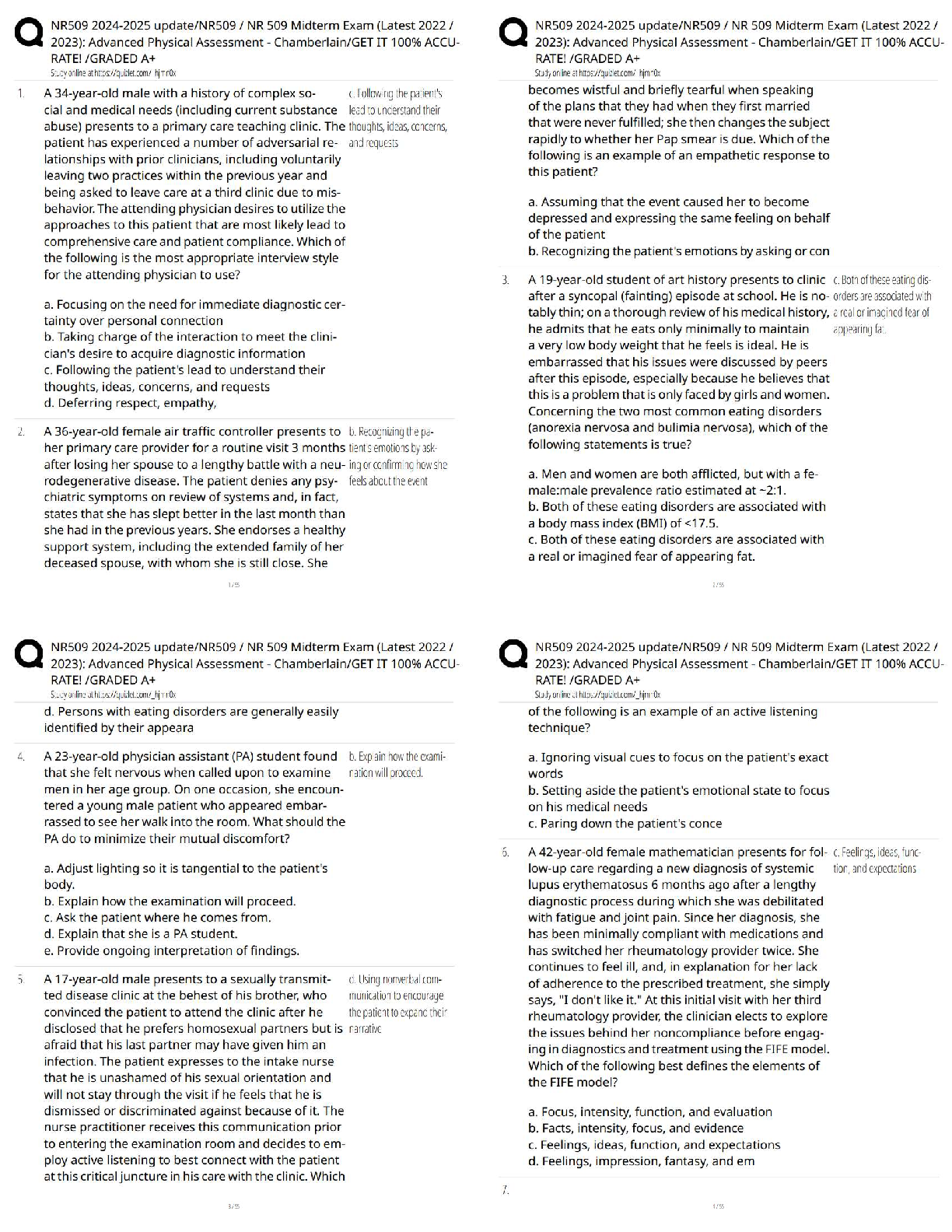

.png)