*NURSING > Final Exam Review > NURSING 112023 basic adult health Final Exam Review- Keiser University (All)
NURSING 112023 basic adult health Final Exam Review- Keiser University
Document Content and Description Below
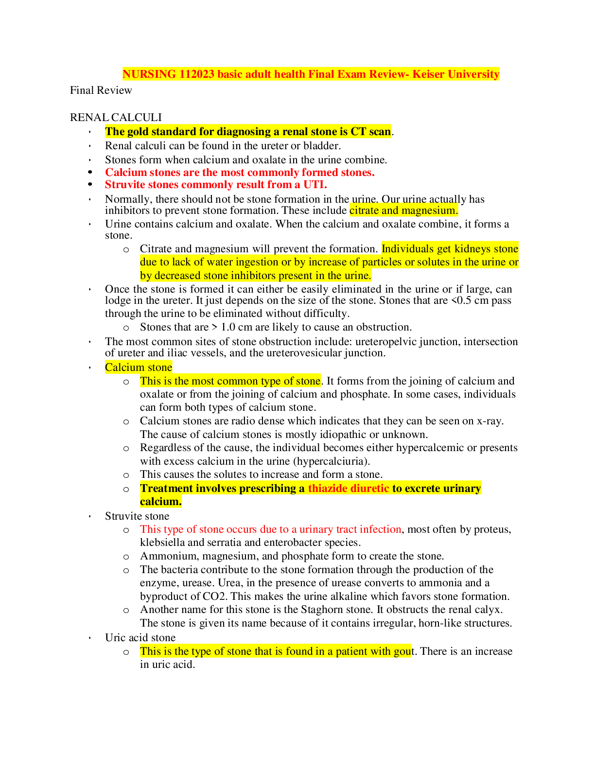
NURSING 112023 basic adult health Final Exam Review- Keiser University-Final Review RENAL CALCULI • The gold standard for diagnosing a renal stone is CT scan. • Renal calculi can be found in ... the ureter or bladder. • Stones form when calcium and oxalate in the urine combine. • Calcium stones are the most commonly formed stones. • Struvite stones commonly result from a UTI. • Normally, there should not be stone formation in the urine. Our urine actually has inhibitors to prevent stone formation. These include citrate and magnesium. • Urine contains calcium and oxalate. When the calcium and oxalate combine, it forms a stone. o Citrate and magnesium will prevent the formation. Individuals get kidneys stone due to lack of water ingestion or by increase of particles or solutes in the urine or by decreased stone inhibitors present in the urine. • Once the stone is formed it can either be easily eliminated in the urine or if large, can lodge in the ureter. It just depends on the size of the stone. Stones that are <0.5 cm pass through the urine to be eliminated without difficulty. o Stones that are > 1.0 cm are likely to cause an obstruction. • The most common sites of stone obstruction include: ureteropelvic junction, intersection of ureter and iliac vessels, and the ureterovesicular junction. • Calcium stone o This is the most common type of stone. It forms from the joining of calcium and oxalate or from the joining of calcium and phosphate. In some cases, individuals can form both types of calcium stone. o Calcium stones are radio dense which indicates that they can be seen on x-ray. The cause of calcium stones is mostly idiopathic or unknown. o Regardless of the cause, the individual becomes either hypercalcemic or presents with excess calcium in the urine (hypercalciuria). o This causes the solutes to increase and form a stone. o Treatment involves prescribing a thiazide diuretic to excrete urinary calcium. • Struvite stone o This type of stone occurs due to a urinary tract infection, most often by proteus, klebsiella and serratia and enterobacter species. o Ammonium, magnesium, and phosphate form to create the stone. o The bacteria contribute to the stone formation through the production of the enzyme, urease. Urea, in the presence of urease converts to ammonia and a byproduct of CO2. This makes the urine alkaline which favors stone formation. o Another name for this stone is the Staghorn stone. It obstructs the renal calyx. The stone is given its name because of it contains irregular, horn-like structures. • Uric acid stone o This is the type of stone that is found in a patient with gout. There is an increase in uric acid. o Individuals who are at risk for getting gout include those with leukemia and myeloproliferative disorder; those undergoing chemotherapy. Chemotherapy destroys the cancer cells. o DNA cells contain purine. When broken down, purine will increase uric acid levels that can lead to uric acid stone formation. o Uric acid increases the acidity of the urine with resultant decrease in urine pH. o Uric acid stones are radiolucent, meaning that the stones cannot be seen on x- ray. o Treatment includes hydration and increasing the alkaline of the urine by giving potassium bicarbonate. Individuals will also be prescribed allopurinol, an anti-gout medication. • Cystine stone o This is a rare type of kidney stone that is found mostly in children. It is caused by a genetic renal tubule defect that prevents the amino acid, cystine, from being reabsorbed that leads to the formation of a cystine stone. o This stone can also form Staghorn shaped stones. Symptoms o Renal colic: this is flank or costovertebral angle (CVA) pain. It is caused by the passing of the stone through the ureter with obstruction and spasm. o The characteristic of this pain begins mild and then greatly increases causing great discomfort to the patient. The pain begins in the flank and radiates to the groin. As the stone moves, the pain will be in the location of where the stone is located. o Hematuria: Hematuria will be found in 90% of individuals who have a kidney stone. While passing through the urinary tract, the stone will injure the urinary structures. It can also be associated with nausea and vomiting. • Diagnosis of the renal stone is confirmed through urinalysis. The patient can have either microscopic or gross hematuria. • The pH of the urine will be identified to help determine the type of stone. If the patient passes the stone, it should be taken for analysis to determine the type of stone. • X-ray of the kidney, ureter, and bladder (KUB) will also be performed to help determine the type of stone. Uric acid and cysteine stones will not be visible on x-ray since they are radiolucent. • The gold standard for diagnosing a renal stone is CT scan because all types of stones can be seen. An ultrasound can be performed for those individuals that cannot tolerate radiation (pregnancy). It is not the best test for diagnosing a kidney stone because it poorly isolates the stone. • Overall management of the patient includes providing an analgesic for pain and increased fluid intake to increase hydration. o Lithotripsy, a non-invasive procedure, will be performed if the stone lodges on the way out. It breaks the stone down into smaller fragments so that they can be eliminated. [Show More]
Last updated: 2 years ago
Preview 1 out of 71 pages
Buy this document to get the full access instantly
Instant Download Access after purchase
Buy NowInstant download
We Accept:

Also available in bundle (1)

BUNDLE: NURSING 11 basic adult health Final Exam, Questions & Answers
BUNDLE: NURSING 11 basic adult health Final Exam, Questions & Answers
By PROF 2 years ago
$25.5
3
Reviews( 0 )
$10.50
Can't find what you want? Try our AI powered Search
Document information
Connected school, study & course
About the document
Uploaded On
Jul 26, 2023
Number of pages
71
Written in

Additional information
This document has been written for:
Uploaded
Jul 26, 2023
Downloads
0
Views
142

.png)