Module 11: Actin Filaments
1. Draw an actin monomer and an actin filament and label all the parts, molecular weights,
etc..
2. Explain the geography of actin filaments. Explain the geography of stress cable, filapodia
...
Module 11: Actin Filaments
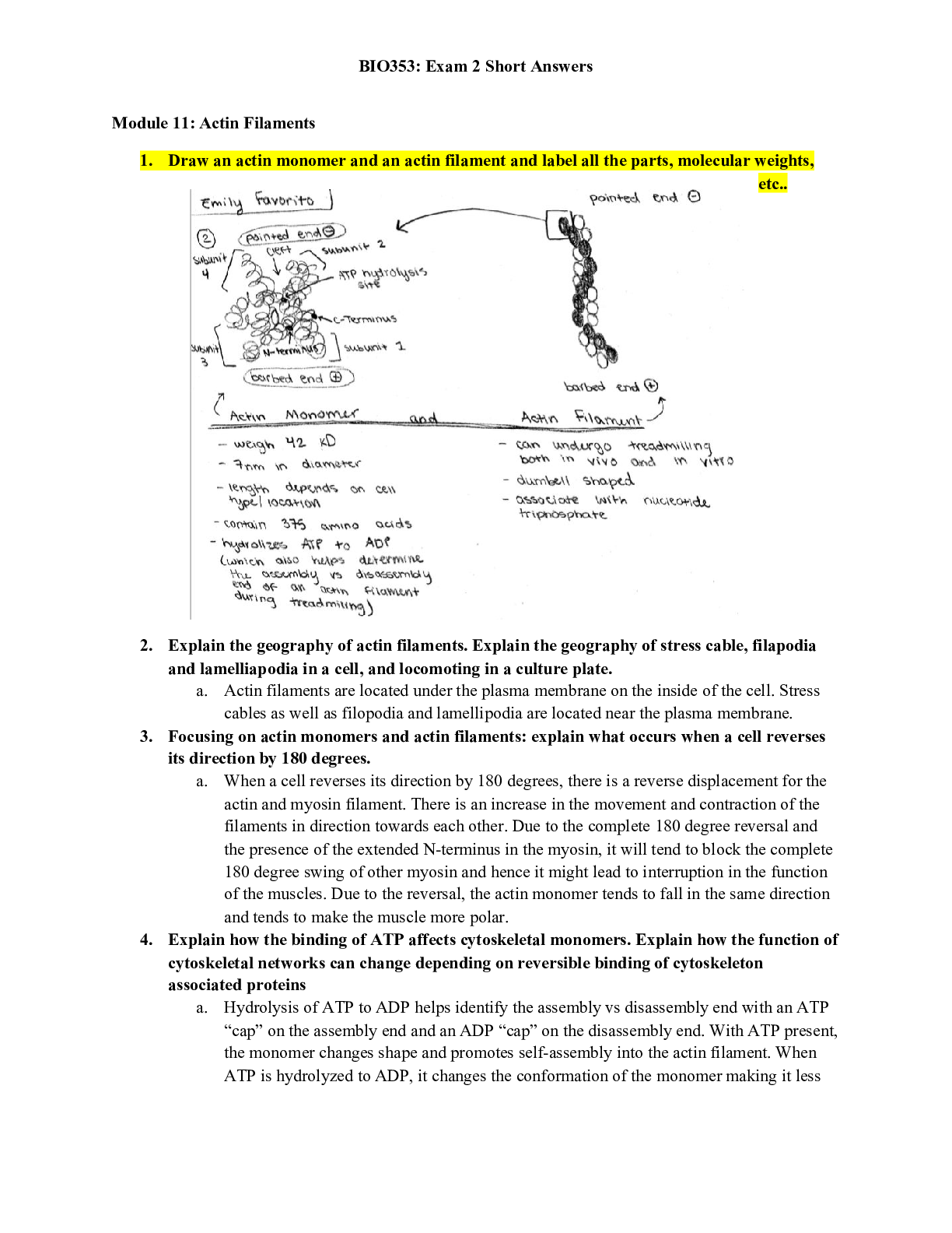
1. Draw an actin monomer and an actin filament and label all the parts, molecular weights,
etc..
2. Explain the geography of actin filaments. Explain the geography of stress cable, filapodia
and lamelliapodia in a cell, and locomoting in a culture plate.
a. Actin filaments are located under the plasma membrane on the inside of the cell. Stress
cables as well as filopodia and lamellipodia are located near the plasma membrane.
3. Focusing on actin monomers and actin filaments: explain what occurs when a cell reverses
its direction by 180 degrees.
a. When a cell reverses its direction by 180 degrees, there is a reverse displacement for the
actin and myosin filament. There is an increase in the movement and contraction of the
filaments in direction towards each other. Due to the complete 180 degree reversal and
the presence of the extended N-terminus in the myosin, it will tend to block the complete
180 degree swing of other myosin and hence it might lead to interruption in the function
of the muscles. Due to the reversal, the actin monomer tends to fall in the same direction
and tends to make the muscle more polar.
4. Explain how the binding of ATP affects cytoskeletal monomers. Explain how the function of
cytoskeletal networks can change depending on reversible binding of cytoskeleton
associated proteins
a. Hydrolysis of ATP to ADP helps identify the assembly vs disassembly end with an ATP
“cap” on the assembly end and an ADP “cap” on the disassembly end. With ATP present,
the monomer changes shape and promotes self-assembly into the actin filament. When
ATP is hydrolyzed to ADP, it changes the conformation of the monomer making it less
suited to fit into an actin filament. Position of ATP gives the monomer a polarity that
steps up the polarity of the actin filament.
5. Describe how myosin II can be made into a tool to analyze the different polarities of actin
filaments in the cell.
a. Myosin II can be made into a tool by scientists using biotechnology to determine the
orientation of an actin filament in stress cables by the winding of the heavy and light
chains.
6. Draw the structure of a myosin II molecule when its light chains are not phosphorylated.
7. Explain the geography of actin filaments as it relates to a living cell.
8. Explain how myosin II can be made into a tool by scientists using biotechnology. And
explain how this can be used to determine the orientation of an actin filament in:
a. stress cables - anti-parallel bundles of actin filaments, hence the myosin barbs will point
in opposite directions along the two filaments.
b. Filopodia - the barbs will all be directed at the tip as that is the positive end of the actin
(polymerizing/growing end)
9. Explain what cell permeabilization is.
a. Making holes in the cell to let aqueous solutions of cleaved myosin heads in. Done with a
non-ionic detergent and isotonic buffer
[Show More]


 Correct Study Guide, Download to Score A.png)
 Correct Study Guide, Download to Score A.png)