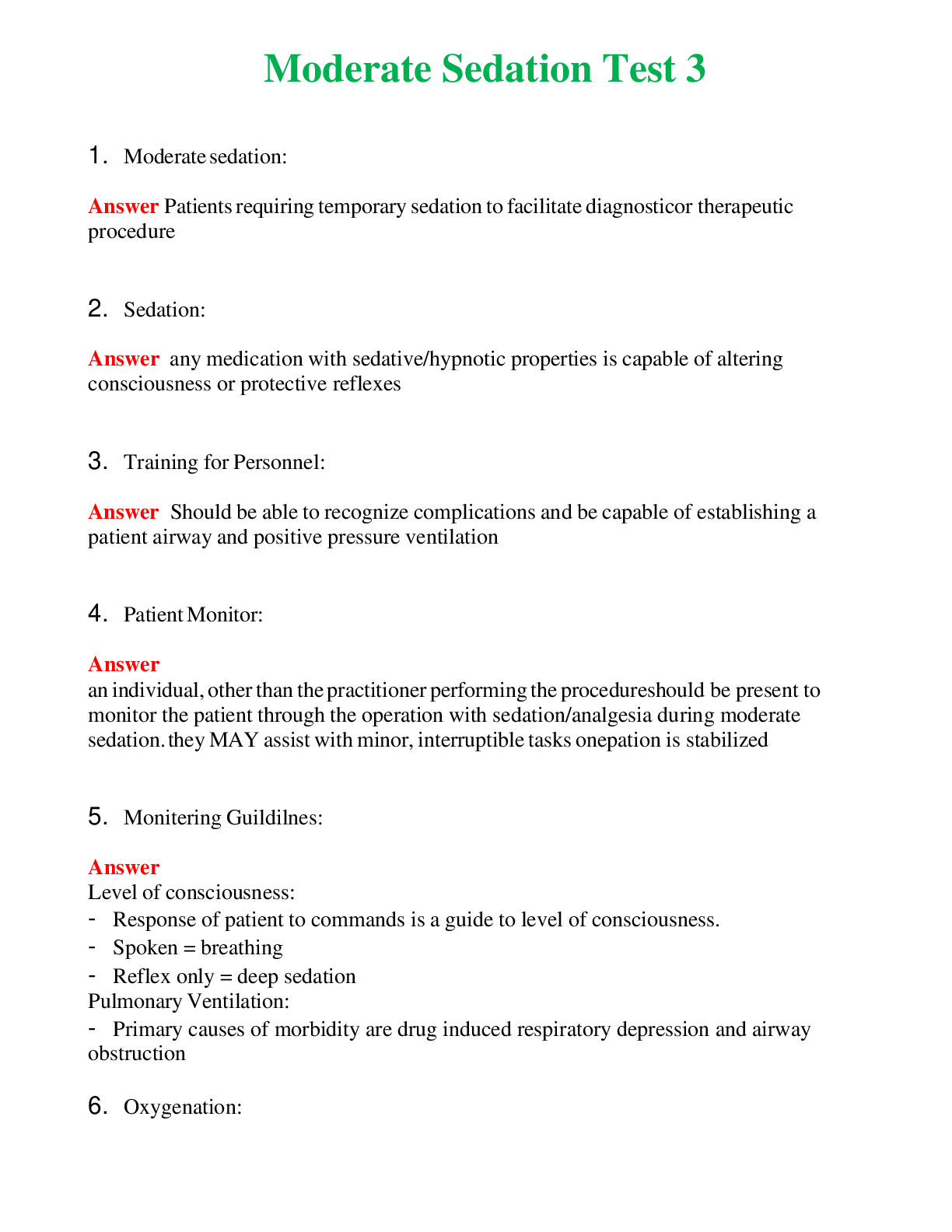
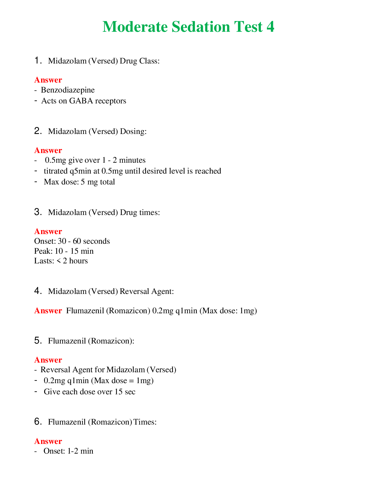
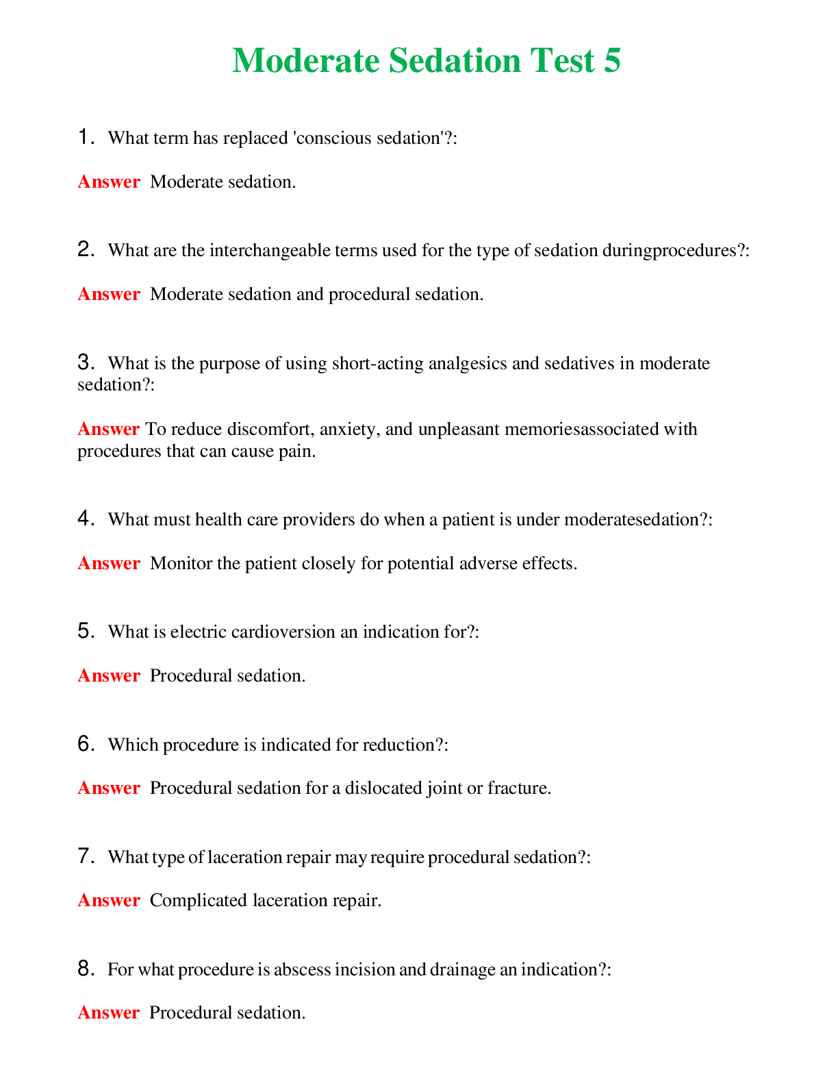
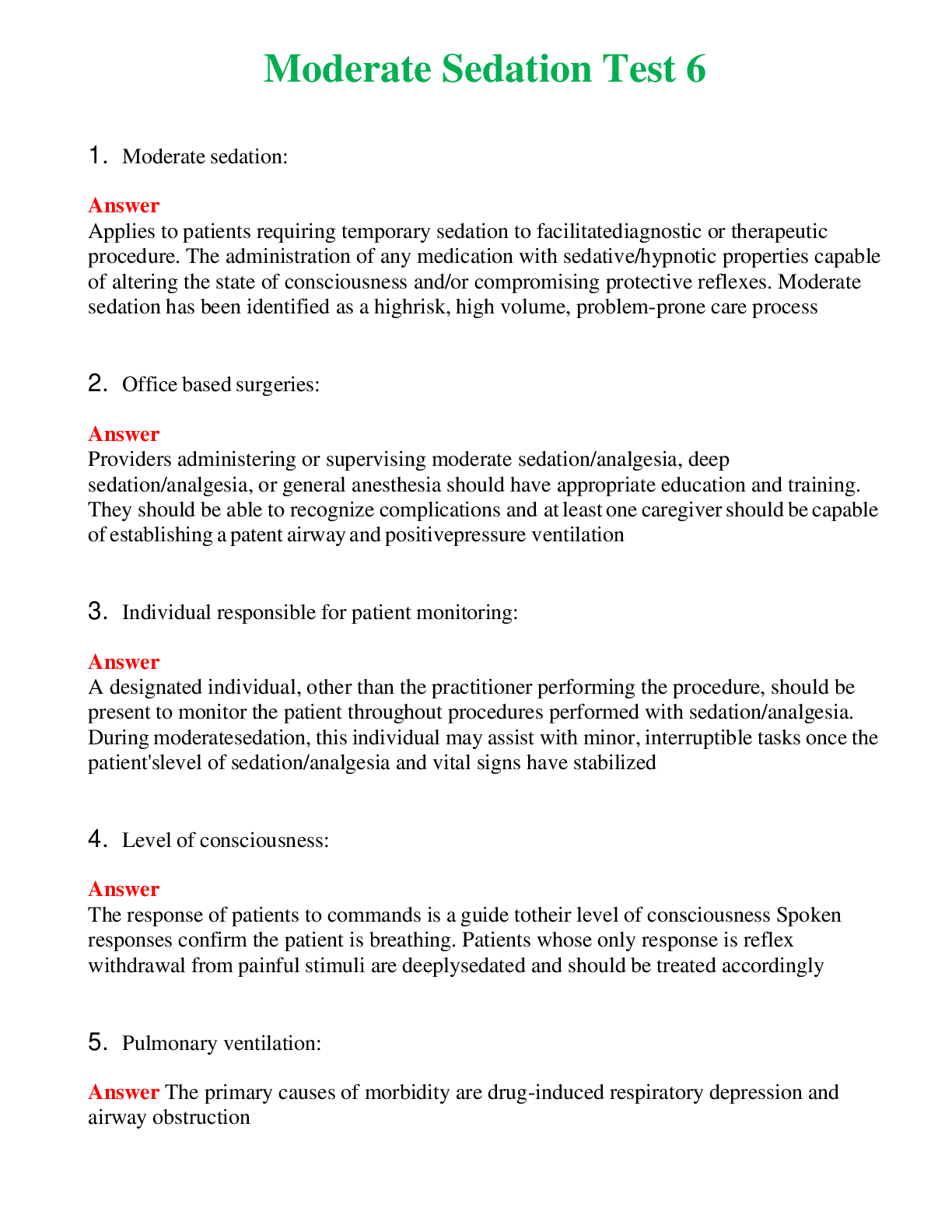
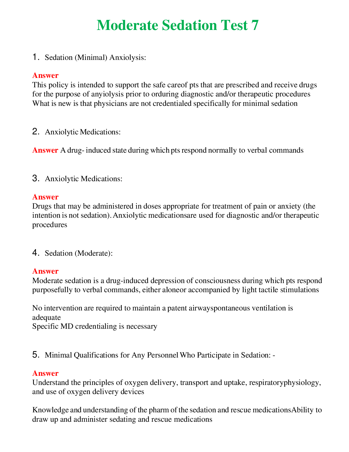
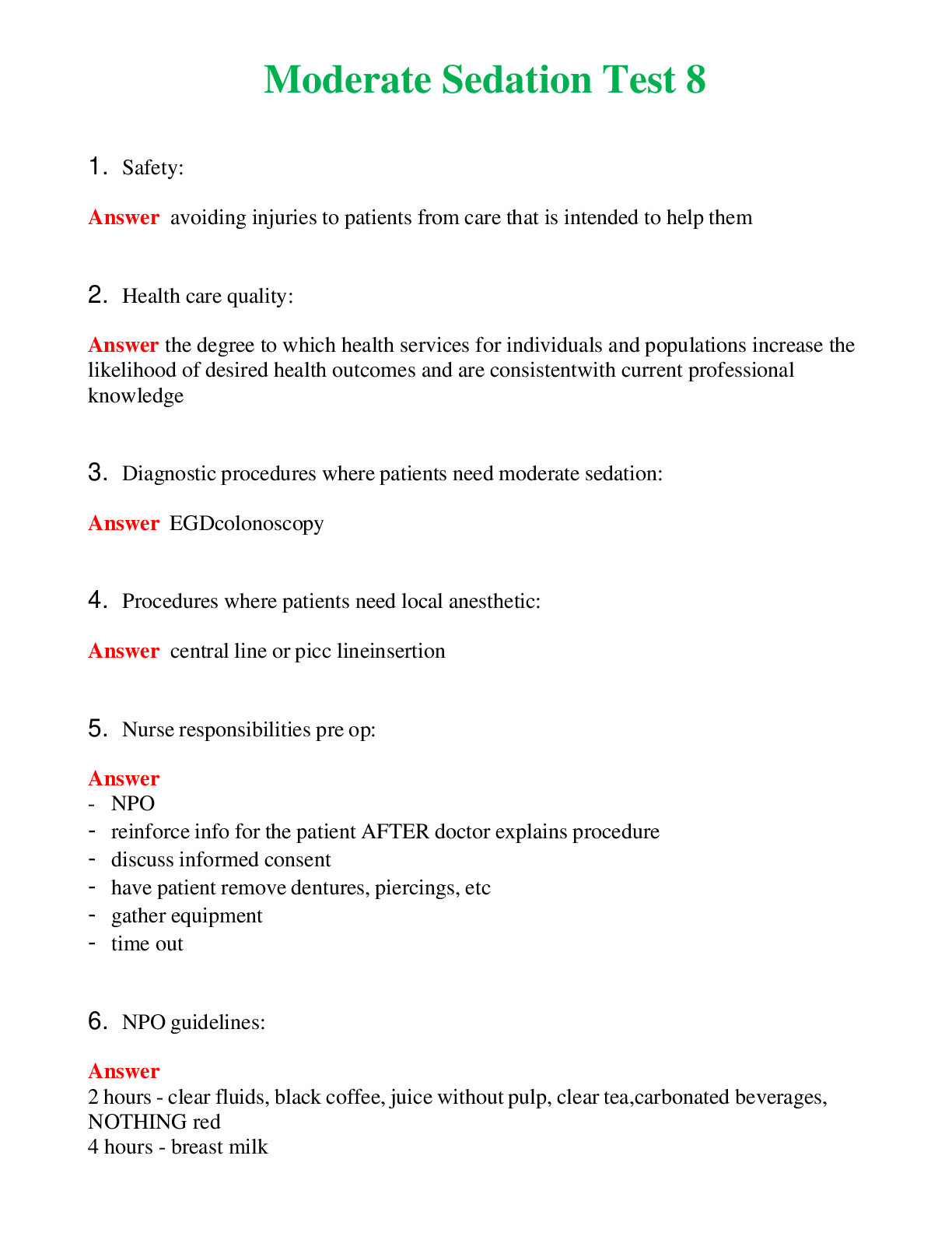
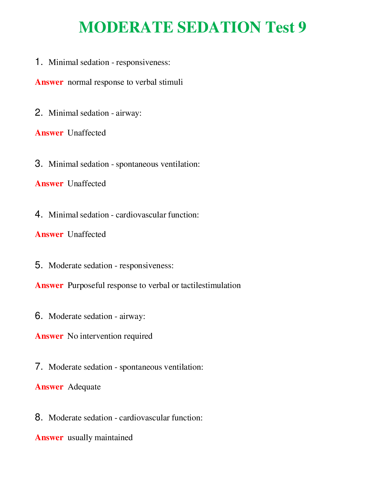
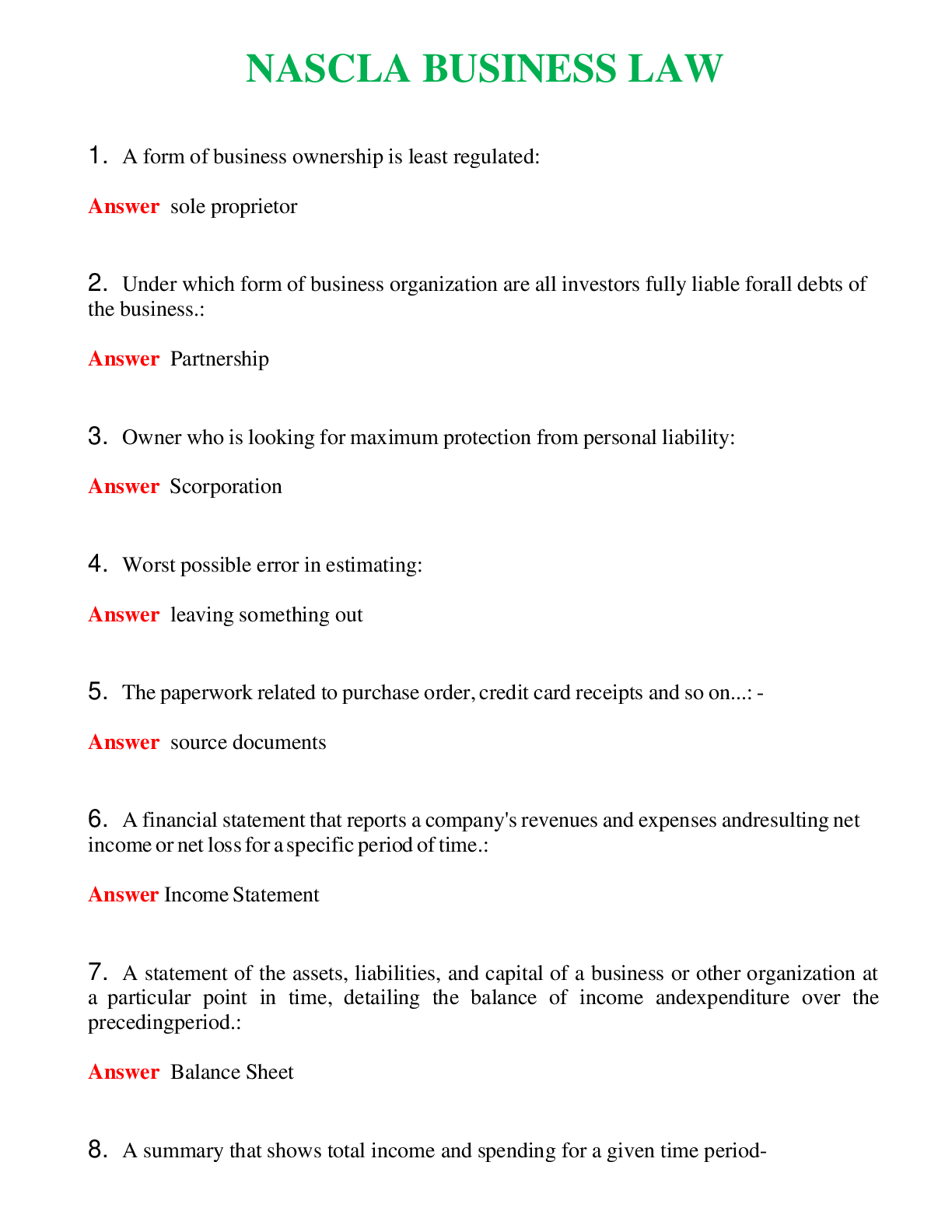
Pediatric Primary Care PNCB 1, Exam Questions & Answers
Document Content and Description Below
Pediatric Primary Care PNCB 1, Exam Questions & Answers-The child at highest risk for having an elevated blood lead level is a: 3 month old exclusively breastfed infant 6 month old who lives in a ... home built after 1970 2 year old with iron deficiency anemia 2 year old who is a picky eater D. - 2 year old with iron deficiency anemia The amount of lead absorbed from the gut is increased in children with nutritional deficiencies such as iron deficiency anemia (IDA). Iron deficiency anemia is often a comorbidity of lead poisoning. The hand-to-mouth behavior of infants and young children increases their lead exposure. However, living in a home built after 1970 reduces the risk since residential paint used in that era should not have been lead based. Infants more than 4 months of age exclusively breast fed without supplemental iron are at increased risk of IDA. A child who is a picky eater may or may not be at high risk for IDA, depending on foods actually eaten.Which laboratory assessment is the BEST indicator of vitamin D deficiency? Which laboratory assessment is the BEST indicator of vitamin D deficiency? 25(OH)-D (cholecalciferol) 1,25(OH)2-D (calcitriol) PTH (parathyroid hormone) 25(OH)-D (cholecalciferol) - 25(OH)-D (cholecalciferol) The best diagnostic study of vitamin D deficiency is the level of 25(OH)-D (cholecalciferol). 1,25(OH)2-D (calcitriol) is the active metabolite of 25(OH)-D, but due to its short half-life it is not a good indicator of vitamin D sufficiency. The parathyroid hormone releases calcium from bone. Rachitic changes can be seen at growth plates and decreased calcification leads to thickening of the growth plate. Serum calcium and phosphorous are initial screening tests but not the best indicator of vitamin D deficiency. In a 2 month old with visible rib fractures on radiograph, the NEXT most critical evaluation to obtain is a: CT scan of the head long bone series coagulation profile retinal ophthalmologic exam - CT scan of the head Posterior rib fractures associated with accidental trauma are rare. Posterior fractures can be seen in infants who have been shaken as the perpetrator hands are typically wrapped around the infant's thorax during the shaking, with the vertebrae acting as a fulcrum. These findings should alert the provider to consider shaken baby syndrome (SBS). Subdural and subarachnoid hemorrhages are the most common acute intracranial injuries seen in SBS and are associated with high rates of morbidity and mortality. Thus, the most important study to do next is a CT scan. Studies have shown that nearly one third of confirmed abusive head trauma cases were missed on initial presentation, and many infants then sustain additional brain injury along with poorer neurologic outcomes because of the delay in diagnosis. Long bone studies will be needed as part of a thorough work-up of non-accidental trauma, but the skull would be the most critical area to image first. Coagulation studies are done to rule out any coagulation problem associated with injury to the brain and are important for medico-legal reasons, but again, brain studies take precedence. A thorough ophthalmologic exam is needed in suspected cases of SBS—preferably done by a pediatric ophthalmologist. The MOST common barrier related to transitioning health care for an adolescent with special needs or chronic illness is finding an adult health care provider for transition. resistance of the family and adolescent to transition of care. lack of health care provider time to plan for transition of care. difficulty in talking with patients about transitioning care. - finding an adult health care provider for transition. Finding an adult health care provider, one who is qualified to care for young adults with special health care needs, is the most commonly perceived barrier to the successful transition of health care as identified by family and young adults, pediatric health care providers, and adult internists. Transitioning of care requires time and communication with the parents and adolescents involved. Many families may be hesitant to leave the nurturing environment of pediatric care, and may perceive differences in adult practices as a difficult adjustment. Internists may lack the training and qualifications to address many of the complicated health care needs of adolescents with chronic illnesses. Because of the delicate nature of such conversations, some pediatric providers may not be comfortable in dealing with the complexities of transitioning care. A toddler is unable to use the right arm normally after the caregiver pulled her arm to prevent the child from falling. Which finding would confirm the diagnosis of subluxation of the radial head? severe swelling and bruising of the elbow elbow flexed with pronated forearm point tenderness at ulnar aspect of elbow obvious deformity of the forearm - elbow flexed with pronated forearm Subluxation of the radial head, also called nursemaid's elbow, must be differentiated from a fracture prior to reducing the annular ligament of the elbow. Radiographic examination is not necessary if the child's physical findings and history are consistent with subluxation. The typical presentation of this injury includes the following: age 2-5 years; history of a longitudinal traction injury, possible "pop" and immediate pain, inability to use the arm normally, and arm splinted against the side. On examination the elbow appears normal, is flexed with a pronated forearm against the body, is tender laterally over the radial head, and has limited flexion with no supination. If the child fell on his/her elbow or there is no history of a traction injury, suspect a fracture and order the appropriate radiograp Education for caregivers whose child has sickle cell disease should include that the majority of pain crises are triggered by which of the following? no identifying cause temperature changes cigarette smoke exposure stressful situations - no identifying cause Sickle cell disease is a common genetic hematologic disorder. Pain is the most common and disabling symptom of sickle cell disease. Environmental temperature and second-hand smoke exposure have been studied as possible precipitating factors, but have not been supported by the research. Negative emotions can facilitate the pain cycle. In general, pain episodes are erratic and unpredictable and occur for various, unknown reasons. A 5 year old complains of a painful left eye after being accidentally scratched by a sibling two hours ago. Fluorescein exam shows a small central corneal abrasion. The MOST appropriate management during the first 24 hours is frequent application of topical antibiotic. observation of the injured eye. frequent application of topical nonsteroidal anti-inflammatory drops. occlusive patching of the injured eye. - frequent application of topical antibiotic. Accidental abrasion of the corneal epithelium causes pain, tearing, and photophobia and is a common eye injury in children. An abrasion can be detected by examining the eye with a Wood's lamp after instillation of fluorescein dye. The one time use of a topical ophthalmic anesthetic may be useful in gaining cooperation for an adequate eye exam. The goal of treatment is rapid healing of the abrasion. Until such healing occurs, the eye should be protected from infection by the use of a topical ophthalmic antibiotic every 4-6 hours for a few days. The repeated use of a topical anesthetic is not recommended, as these medications can cause corneal toxicity and inhibit the blinking reflex. Topical steroids are not recommended as they lower the eye's resistance to infection. Oral acetaminophen or ibuprofen and intermittent cool compresses may manage discomfort. Narcotics are not recommended because of frequent side effects. The use of topical nonsteroidal anti-inflammatory drops is being studied in the treatment of some sterile corneal abrasions, such as those acquired during laser treatment of refractive errors in adults, but are not recommended in management of traumatic corneal abrasions in children. Patching is no longer recommended for most corneal abrasions, as it does not reduce discomfort or speed healing and makes instillation of antibiotic medication more difficult. Most corneal abrasions heal steadily over the first 24-48 hours. Persistent or increasing pain or discomfort after the first 24 hours indicates the need for further ophthalmologic evaluation. The parents of a 2 year old are concerned that their child is having temper tantrums in public settings. Which is the BEST response? The temper tantrum is an indication that the child is tired and needs to go home. Make sure to ensure safety while ignoring the child's display of behavior. You can pick the child up and take him to a quiet place for a time out. In a public place, it is okay to give into the child's desires to maintain peace. - Make sure to ensure safety while ignoring the child's display of behavior. Temper tantrums are common in children between the ages of 12 months and four years of age and may occur as often as once a week in this age group. A temper tantrum is often the sign that a child is frustrated and trying to achieve autonomy, yet is still immature in the reactions to the outcome of the situation. Appropriate responses and interventions by the parents can assist the child in attaining developmental mastery. Some suggestions include trying to prevent a tantrum by offering the child achievable choices, maintaining an environment that provides positive reinforcement for desired behavior, and fighting only those battles which need to be won. If a child does exhibit a tantrum, provide a safe environment and do not over-react to the behavior. Stay nearby the child during the temper tantrum and provide positive reinforcement when the behavior improves. A two year old is too young to understand the ramifications of a time out, so removing the child from the scene may not be helpful. The child may need to be restrained if in danger from the environment, so holding the child may be appropriate. Giving in to the child reinforces the behavior, despite where the tantrum occurs. Five weeks after joining the cross-country team, a 15 year old complains of pain in the front of his left shin that intensifies 15 minutes into each practice run. Plain radiographs of the left leg are normal. Which subsequent test would be MOST appropriate for diagnosis? bone scan MRI of left leg repeat plain films in 1 week repeat plain films in 2 weeks - MRI of left leg Tibial stress fractures are common in runners. An adolescent who complains of pain in the front of the shin which intensifies 15 minutes into running has symptoms consistent with an anterior tibial stress fracture. It is important to diagnosis this quickly and to make the appropriate orthopedic referral. A bone scan would be positive in this type of stress fracture, but it remains positive for the subsequent 1-2 years. Therefore, it is not useful for assessing healing or ability to return to play. MRI has replaced bone scans as the most sensitive tool for diagnosing stress fractures in long bones. Repeating plain films in 2 weeks can demonstrate periosteal reaction if a stress fracture is present. There is no reason to wait 1-2 weeks for a diagnosis and delay treatment. [Show More]
Last updated: 1 year ago
Preview 5 out of 38 pages

Loading document previews ...
Buy this document to get the full access instantly
Instant Download Access after purchase
Buy NowInstant download
We Accept:

Also available in bundle (1)
Click Below to Access Bundle(s)
BUNDLE: Pediatric Primary Care PNCB 1, Pediatric Primary Care PNCB 2, Pediatric Primary Care PNCB Final Exam, PedsCE PNP-PC Exam, Exam Questions & Answers, All Rated 100%
BUNDLE: Pediatric Primary Care PNCB 1, Pediatric Primary Care PNCB 2, Pediatric Primary Care PNCB Final Exam, PedsCE PNP-PC Exam, Exam Questions & Answers, All Rated 100%
By PROF 1 year ago
$29.5
4
Reviews( 0 )
$14.50
Can't find what you want? Try our AI powered Search
Document information
Connected school, study & course
About the document
Uploaded On
Dec 30, 2024
Number of pages
38
Written in
All

Additional information
This document has been written for:
Uploaded
Dec 30, 2024
Downloads
0
Views
22