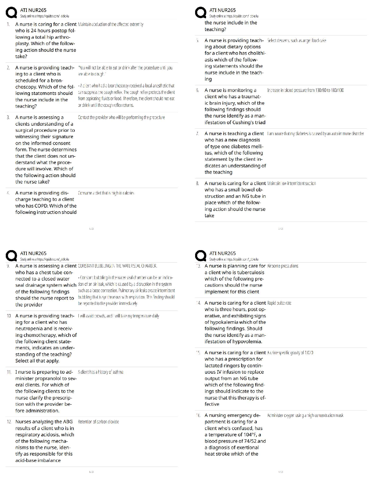
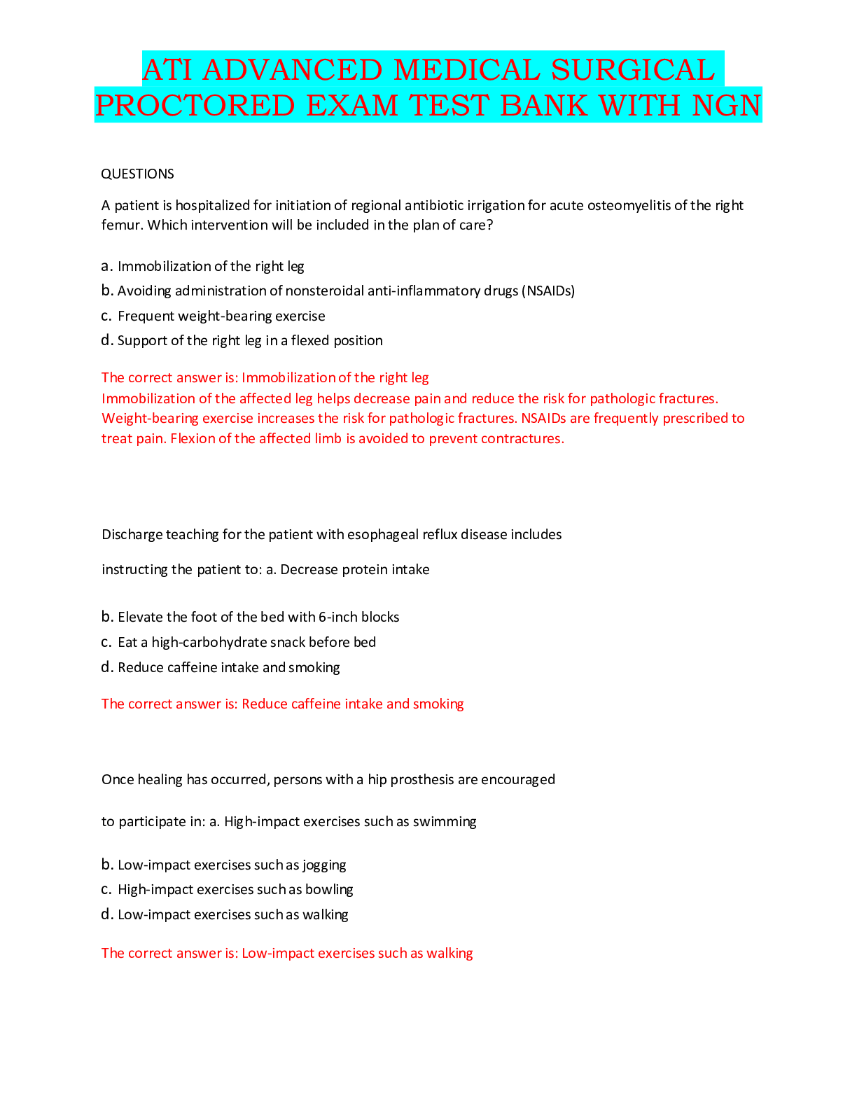
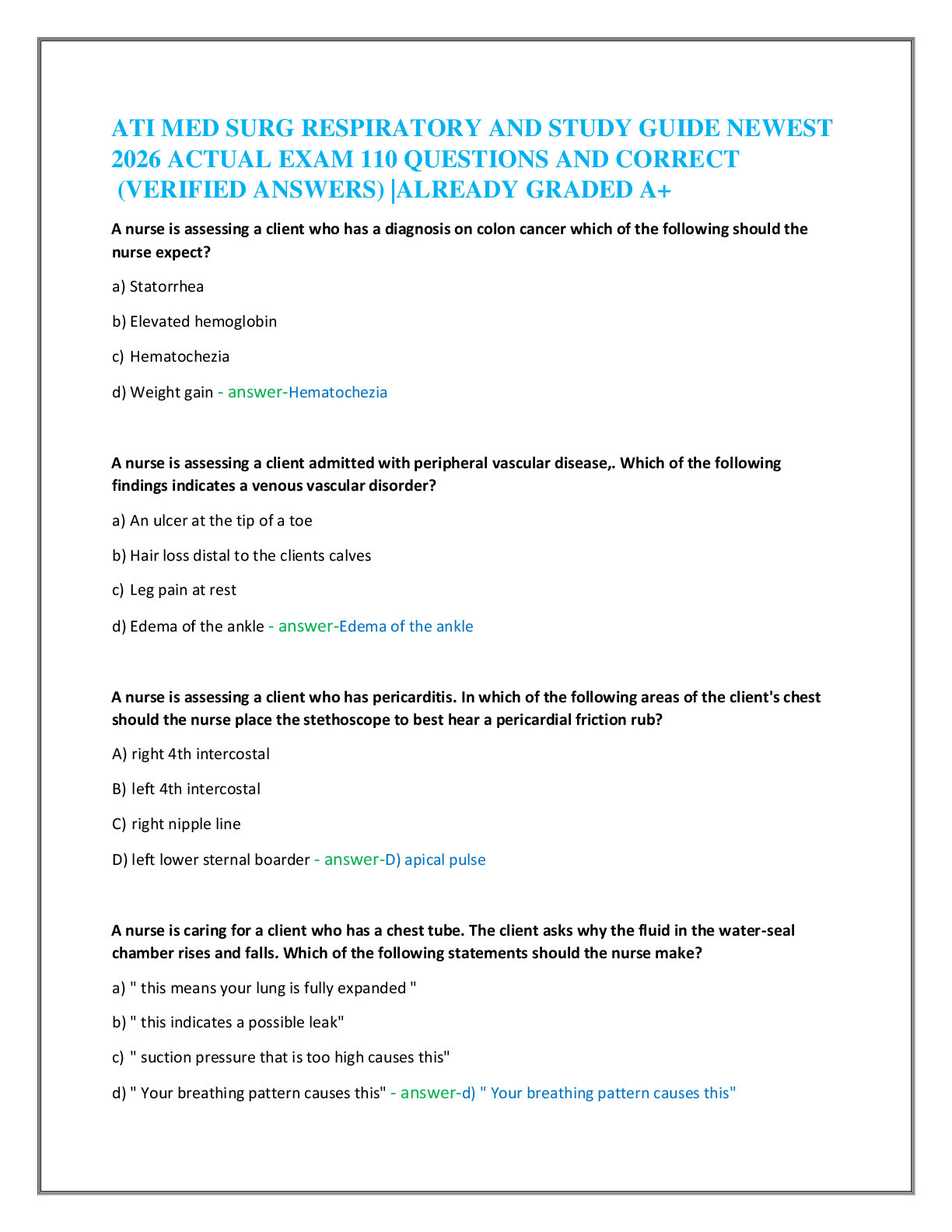
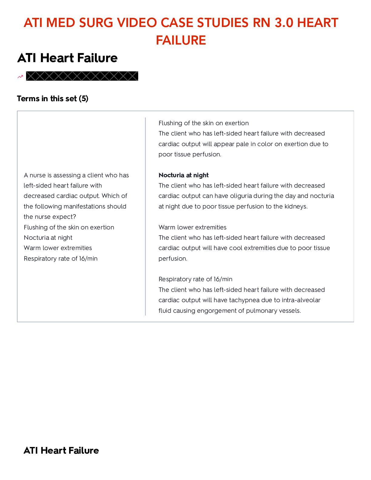
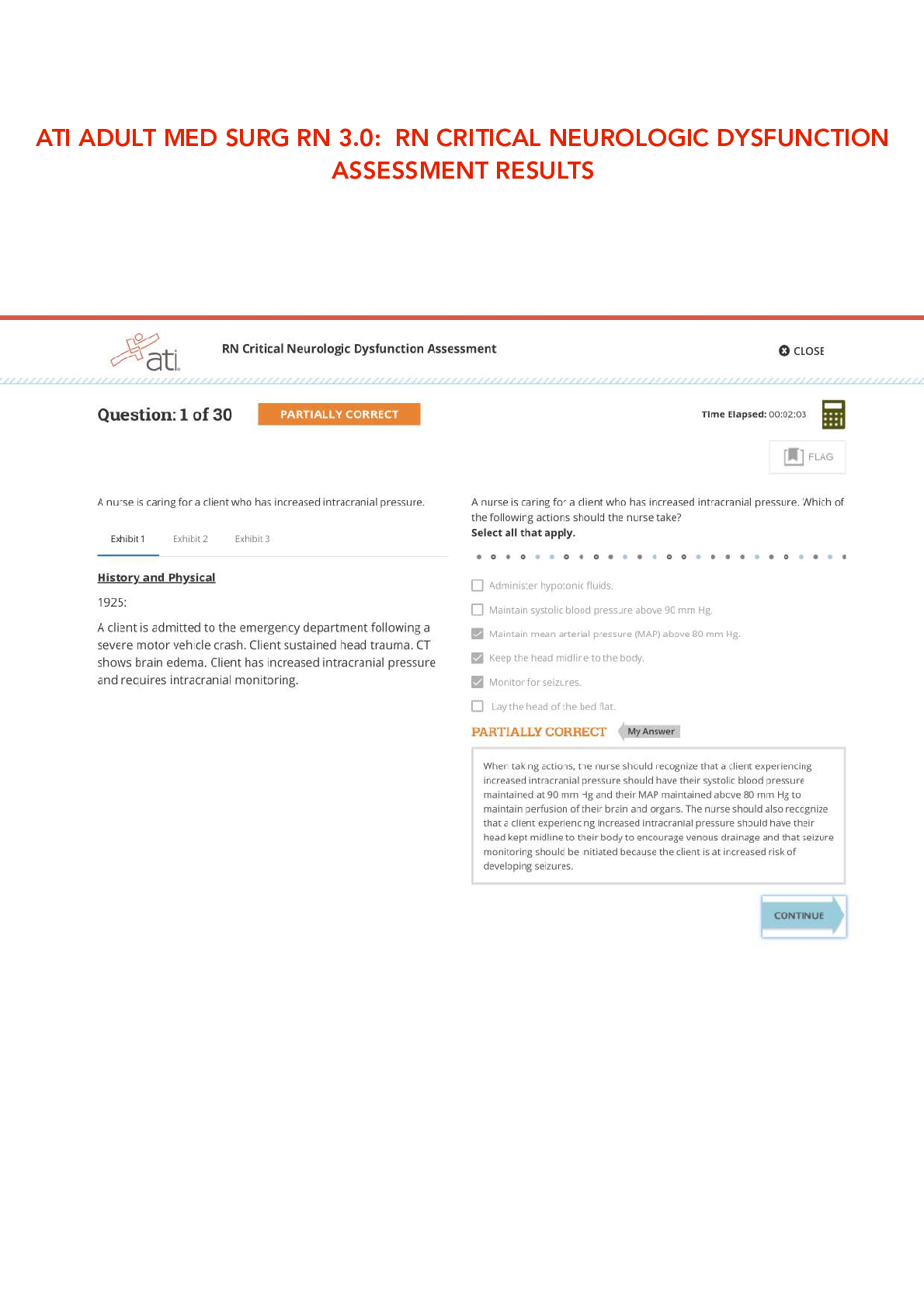
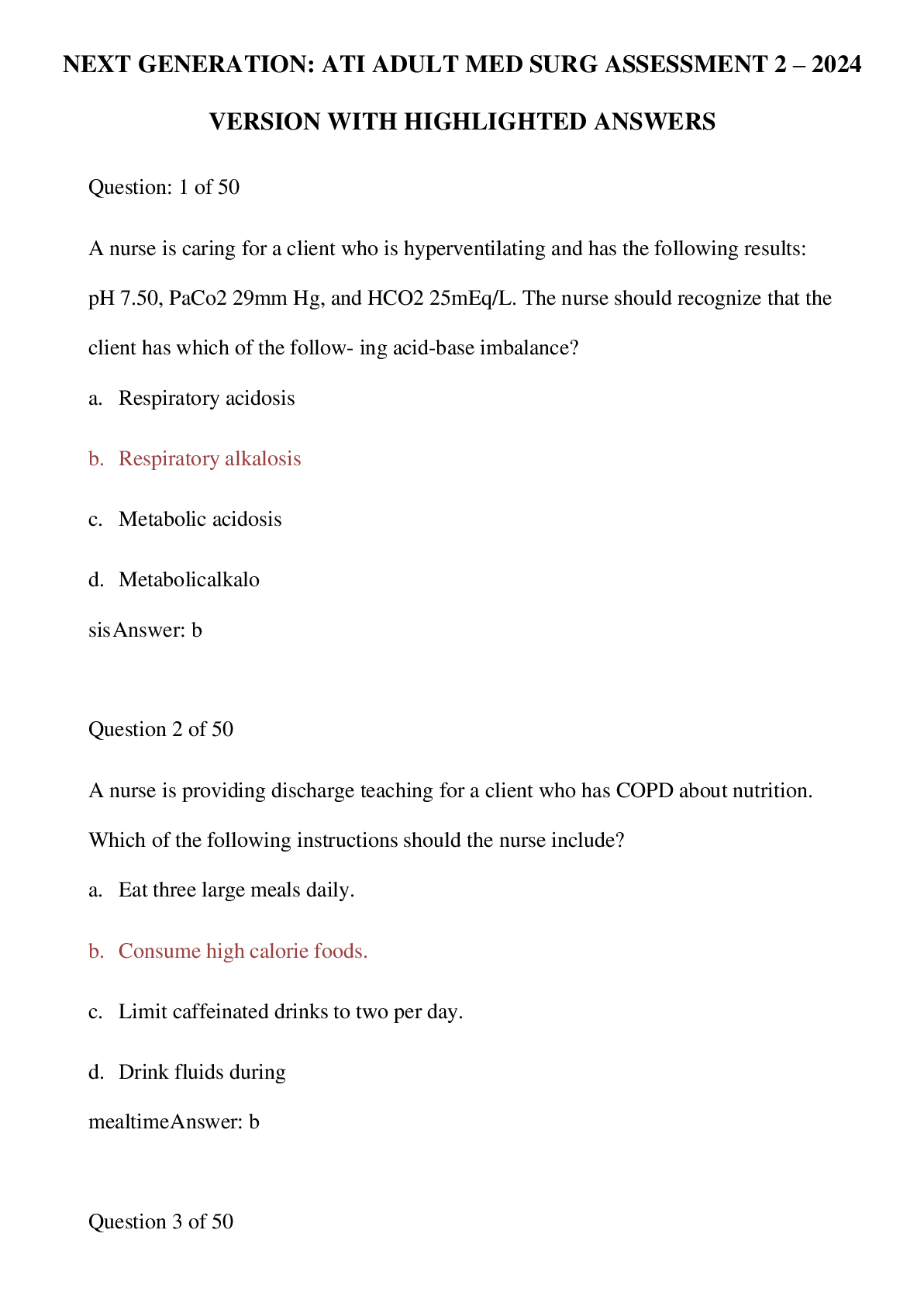
MED SURG 2 EXAM 1 STUDY NOTES
UNIT
TOPIC
TB CHAPTER
Unit 9: Musculoskeletal
Function
Assessment of
Musculoskeletal
Function
39
ATI CHAPTER
67-72
Pharm: 34,45
Musculoskeletal Care
Modalities
...
MED SURG 2 EXAM 1 STUDY NOTES
UNIT
TOPIC
TB CHAPTER
Unit 9: Musculoskeletal
Function
Assessment of
Musculoskeletal
Function
39
ATI CHAPTER
67-72
Pharm: 34,45
Musculoskeletal Care
Modalities
40
Management of
Patients w/
Musculoskeletal
Disorders
41
Management of
Patients w/
Musculoskeletal
Trauma
42
Unit 3: Concepts &
Challenges in Patient
Management
Shock & Multiple Organ
Dysfunction Syndrome
14
37
CHAPTER 39: ASSESSMENT OF MUSCULOSKELETAL FUNCTION
Anatomic & Physiologic Overview
Downloaded by Morris Muthii (
[email protected])
Structure & Function of the Skeletal System
● 206 bones divided into 4 categories: long, short, flat, irregular -
Long bones: found in upper & lower extremities (e.g. femur). Rod shaped or shafts w/ rounded
ends - - -
Short bones: irregularly shaped located in ankle & hand (e.g. metacarpals, phalanges)
Flat bones: extensive protection of underlying structures (e.g. sternum, skull)
Irregular bones: cannot be categorized (e.g. vertebrae & jaw bones)
● Cortical bone (compact bone) exists where support is needed & cancellous bone (lattice like
bone structure; trabecular bone) is found where hematopoiesis & bone formation occur.
● Bones composed of cells, protein matrix, and mineral deposits. 3 cells include: -
Osteoblasts: bone-forming cell; secretes bone matrix - -
Osteocytes: mature bone cell for bone maintenance; located in lacunae
Osteoclasts: located in shallow Howship’s lacunae. Multinuclear cells that destroy, resorb, &
remodel bone.
● Joint: where bone ends meet; provides for motion & flexibility
Bone Formation & Maintenance
● Osteogenesis: process of bone formation
● Ossification: process of formation of the bone matrix & deposition of minerals.
● Bone is a dynamic tissue in a constant state of turnover. -
During childhood & adolescent years, new bone is added faster than old bone is removed. - - - -
Continues until peak bone mass is reached (20 y/o)
Complete skeletal turnover occurs every 10 years
Balance between bone resorption & formation influenced by: exercise, diet (calcium), hormones
(calcitriol, parathyroid hormone, cortisol, growth hormone, sex hormones)
Weight-bearing activity supports bone maintenance; any activity done while person is on their
feet that works bones & muscles against gravity (e.g. walking, tennis)
● Daily intake of 1,200 mg calcium essential to maintain adult bone mass -
Calcium sources: low-fat milk, yogurt, cheese, OJ, cereals, bread
● Young adults need vitamin D intake of 600 IU & adults 50 y/o+ need daily intake of 800-1000 IU -
Vitamin D sources: fortified milk, cereals, egg yolks, saltwater fish, liver
● Calcitriol increases blood calcium by promoting calcium absorption from GI tract & facilitates
mineralization of osteoid tissue.
● PTH & calcitonin regulates calcium concentration in blood. -
Increased PTH prompts calcium mobilization - -
Calcitonin inhibits bone resorption & increases calcium deposit into bone
SQ/ IM/ IN administration. Watch for bloody nose. Alternate nostrils
● Excessive thyroid hormone & cortisol production can result in increased bone resorption &
decreased bone formation
● Long-term cortisol or corticosteroid therapy increases risk for osteopenia & fractures
Bone Healing
● Stage 1: Hematoma formation: 1-2 days after fracture. Bleeding & local vasoconstriction.
Hematoma forms at fracture site. Cytokines released, initiating fracture healing process (fibroblast
Downloaded by Morris Muthii (
[email protected])
proliferation causing angiogenesis to occur)
● Stage 2: Fibrocartilaginous callus formation: granulation tissue formation. Fibroblasts &
osteoblasts migrate to fracture site & begin bone reconstruction
● Stage 3: Bony callus formation (Ossification): 3-4th week of fracture. Continues until a firm
bony union is formed. Mature bone gradually replaces fibrocartilaginous callus & excess callus
reabsorbed by osteoclasts. Fracture site feels immovable. Safe to remove cast.
● Stage 4: Remodeling: osteoclasts remove necrotic tissue. May take months to years.
Functions of the Musculoskeletal System
● Protection of vital organs
● Framework to support body structures, mobility
● Movement; produce heat & maintain body temperature
● Facilitate return of blood to the heart
● Reservoir for immature blood cells
● Reservoir for vital minerals
Structure & Function of the Articular System
● Synarthrosis: immovable joints
● Amphiarthrosis: allow limited movement (e.g. vertebrae or symphysis pubis)
● Diarthrosis: freely movable -
Ball & socket: permit full freedom of movement (e.g. hip, shoulder) - - - -
Hinge joints: permit bending in one direction via flexion/ extension (e.g. elbow, knee)
Saddle joints: allow movement in 2 planes at right angles. Joint at base of thumb is saddle
(biaxial joint)
Pivot joints: allow one bone to move around central axis (e.g. articulation between radius & ulna)
Gliding joints: allows limited movement in all directions (e.g. wrist carpal bones)
● Joint capsule: tough fibrous sheath surrounds articulating bones
● Ligaments: ropelike bundles of collagen fibrils bind articulating bones together
● Tendons: cords of fibrous tissue that connect muscle to bone
● Bursa sac: filed w/ synovial fluid that cushions movement of tendons, ligaments, & bones
Muscles
● Composed of parallel groups of cells (fasciculi) encased in fascia. More fasciculi in muscle, more
precise movements are
● Sarcomere: contractile unit of skeletal muscle that contains actin & myosin
● Contraction of muscle fibers result in isotonic or isometric contraction: -
Isometric contraction: length of muscles remains constant but force generated by muscles is
increased (e.g. pushing against wall) - - -
Isotonic contraction: shortening of muscle w/o tension increase (e.g. forearm flexion)
During sedentary activity, ATP is synthesized from oxidation of glucose to water & Co2
During strenuous activity, glucose is metabolised to lactic acid. Stored muscle glycogen used to
supply glucose via anaerobic pathways
● Tendons connect muscle to bone & ligaments connect bone to bone
Muscle Maintenance
● Muscle tone -
Flaccid: limp & w/o tone -
Spastic: muscle w/ greater-than-normal tone
Downloaded by Morris Muthii (
[email protected])
Atonic: soft & flabby - - -
Upper motor neuron lesions produces increased tone (e.g. cerebral palsy)
Lower motor neuron lesions produces decreased tone (e.g. muscular dystrophy)
● Hypertrophy: muscle repeatedly develops maximum tension over long time, causing cross-
sectional area & muscle fiber size to increase
● Atrophy: decrease in muscle size
Assessment of the Musculoskeletal System
● Health Hx -
Rest relieves most musculoskeletal pain - - - - - - - - -
Pain that increases w/ activity may indicate joint sprain, muscle strain, or compartment syndrome
Steadily increasing pain may indicate progression of infectious process (e.g. osteomyelitis,
malignant tumor, neurovascular complications)
Rheumatic dx & tendonitis pain usually occurs in morning whereas osteoarthritis worsens as day
progresses
Rheumatoid arthritis: ulnar deviation of fingers & swan neck deformity: hyperextension of
proximal interphalangeal joints w/ flexion of distal interphalangeal joints. Bilateral. Pain at rest.
Increased pain in morning.
Osteoarthritis: pain w/ activity & improves w/ rest. Heberden nodules are classic sign. Crackles.
Osteoarthritis Risk Factors: female, smoker, ages, genetics, med use
Treatment: NSAIDs, glucosamine, tylenol, injections into knees
Rhabdomyolysis: the death of muscle fibers and release of their contents into the bloodstream,
which causes renal (kidney) failure & concentrated urine.
Paresthesias: sensations of burning, tingling, or numbness
● Physical Assessment -
Normal curvature of spine is convex through thoracic portion & concave through cervical &
lumbar portions - - - - - - - -
Kyphosis: Humpback. Increased forward curvature of thoracic spine that causes bowing or
rounding of back, leading to hunchback or slouching posture
Lordosis: lumbar curvature. Swayback, exaggerated curvature of lumbar spine (common
causes: pregnancy, excessive visceral fat)
Scoliosis: lateral curvature deviation of spine
Examiner inspects spinal curves & trunk symmetry by standing behind pt & instructs pt to bend
backward supporting pt by placing hands on posterior iliac spine
Effusion: excessive fluid within capsule; swelling & increased temperature that may reflect
inflammation
Balloon sign: milk downward, apply medial pressure, tap & watch for fluid wave. Feel for fluid
entering space directly below patella.
Ballottement sign: medial & lateral aspects of extended knee milked firmly in downward motion.
Push patella toward femur & observes for fluid return to region superior to patella.
Fasciculation: involuntary twitching of muscle fiber groups
● Neurovascular Status -
Compartment syndrome (caused by pressure within muscle compartment that increases until
microcirculation diminishes, leading to nerve & muscle anoxia & necrosis). Function cab be lost if
anoxic situation continues > 6 hrs
Diagnostic Evaluation
● X- Ray studies: determine bone density, texture, erosion, & changes in bone relationships.
● CT: shows detailed cross-sectional image of body to visualize & assess tumors; injury to soft
tissue, ligaments, or tendons, & severe trauma. Identifies location & extent of fractures
● MRI: uses magnetic fields & radio waves to create high-resolution pics of bones & soft tissues to
Downloaded by Morris Muthii (
[email protected])
visualize torn muscles, ligaments, cartilages, herniated discs. -
Noisy, may take 30-90 min to finish. Pts w/ metal implants & pacemakers cannot take this test
● Arthrography: identifies cause of unexplained joint pain & progression of joint dx. Radiopaque
contrast agent injected into cavity to visualize joint structures. Joint is put through ROM to
distribute contrast while in x-rays. If tear is present, contrast leaks out of joint -
Compression elastic bandage & joint rested for 12 hrs post-procedure -
Expect clicking or crackling in joint for 1-2 days after procedure until contrast agent/ air absorbed
● Bone densitometry: evaluates bone mineral density (e.g. DEXA). BMD of heel can be used to
dx & monitor osteoporosis, bone fracture risk
● Bone scan: detects metastatic tumors, osteomyelitis, fractures, aseptic necrosis, & progression
of degenerative bone dx. Requires injection of radioisotope via IV -
Assess for ax to radioisotopes. Encourage fluids to distribute isotope. -
Flushing & warmth to be expected. Drink fluids post-procedure & empty bladder pre-procedure
● Arthroscopy: direct visualization of joint via fiberoptic endoscope. Biopsy & treatment of tears,
defects, & dx processes performed through arthroscope. - -
Post procedure: Wrap joint w/ compression dressing to control swelling & report signs of
complications (e.g. fever, excessive bleeding, swelling, numbness, cool skin)
Monitor neuro status
● Arthrocentesis: obtain synovial fluid for purposes of examination or to relieve pain due to
effusion. Dx septic arthritis & inflammatory arthropathies -
Ice given 1-2 days post-procedure
● Electromyography: assesses electrical potential of muscles & nerves leading to them to
evaluate muscle weakness, pain, & disability -
C/I in pts receiving anticoagulants, extensive skin infections. Electrodes may cause bleeding. -
Avoid using lotions/ creams on day of test
● Biopsy: determines structure & composition of bone marrow, muscle, or synovium to help dx
specific dx -
Monitor site for edema, bleeding, pain, hematoma formation, & infection
● Laboratory studies -
Phosphorus decreased in osteomalacia - - - - - - - -
Acid phosphatase increased in Paget’s dx & metastatic cancer
Alkaline phosphatase increased during early fracture healing & dx w/ increased osteoblastic
activity (e.g. metastatic bone tumors)
Bone metabolism increased via calcitonin, PTH, vitamin D levels
Creatine kinase, aspartate aminotransferase increased w/ muscle damage
Serum osteocalcin indicates rate of bone turnover
Urine calcium levels increased w/ bone destruction
Urinary N-telopeptide of type I collagen & deoxypyridinoline reflect increased osteoclast activity &
increased bone resorption
Increased serum levels of bone specific alkaline phosphatase, osteocalcin, & intact N- terminal
propeptide of type 1 collagen reflect increased osteoblast activity & enhanced bone remodeling
activity
CHAPTER 40: MUSCULOSKELETAL CARE MODALITIES
Casts
● Rigid external immobilizing device molded to contours of body.
● Used to: immobilize reduced fracture, correct deformity, apply uniform pressure to soft tissues,
and support to stabilize joint.
● Most common casting materials consist of fiberglass or plaster of Paris