NURS 5315 UTA Exam 1 | Questions and Answers Atrophy E. Cells decrease in size P. Still functional Physiologic: thymus gland in early childhood Pathological: disuse Hypertrophy E. Increase in cell size P. Increased wo
...
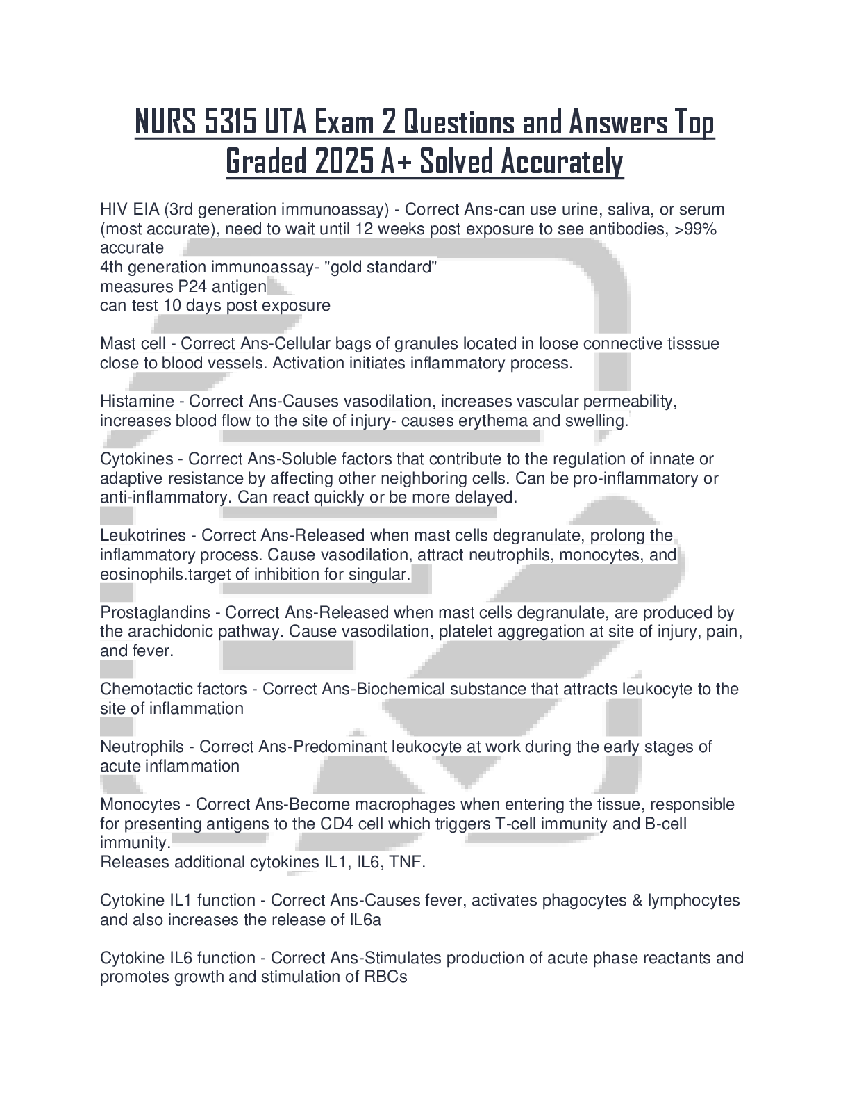
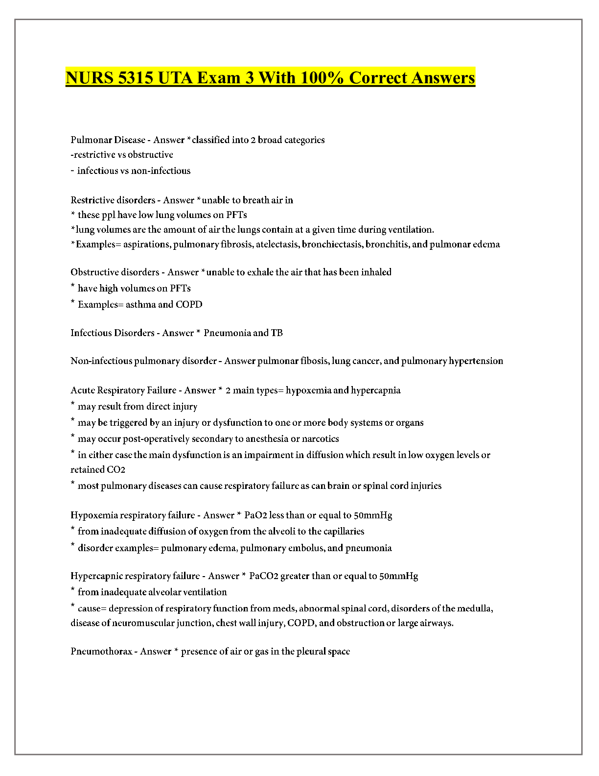
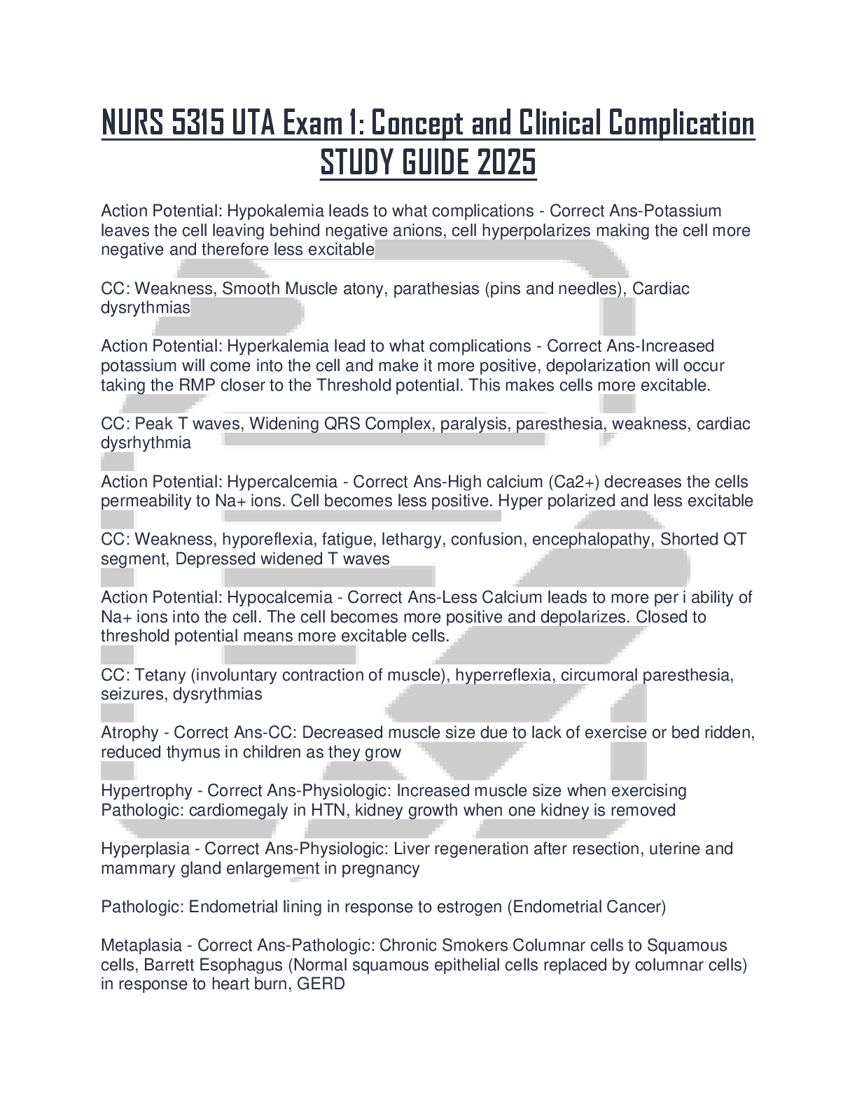
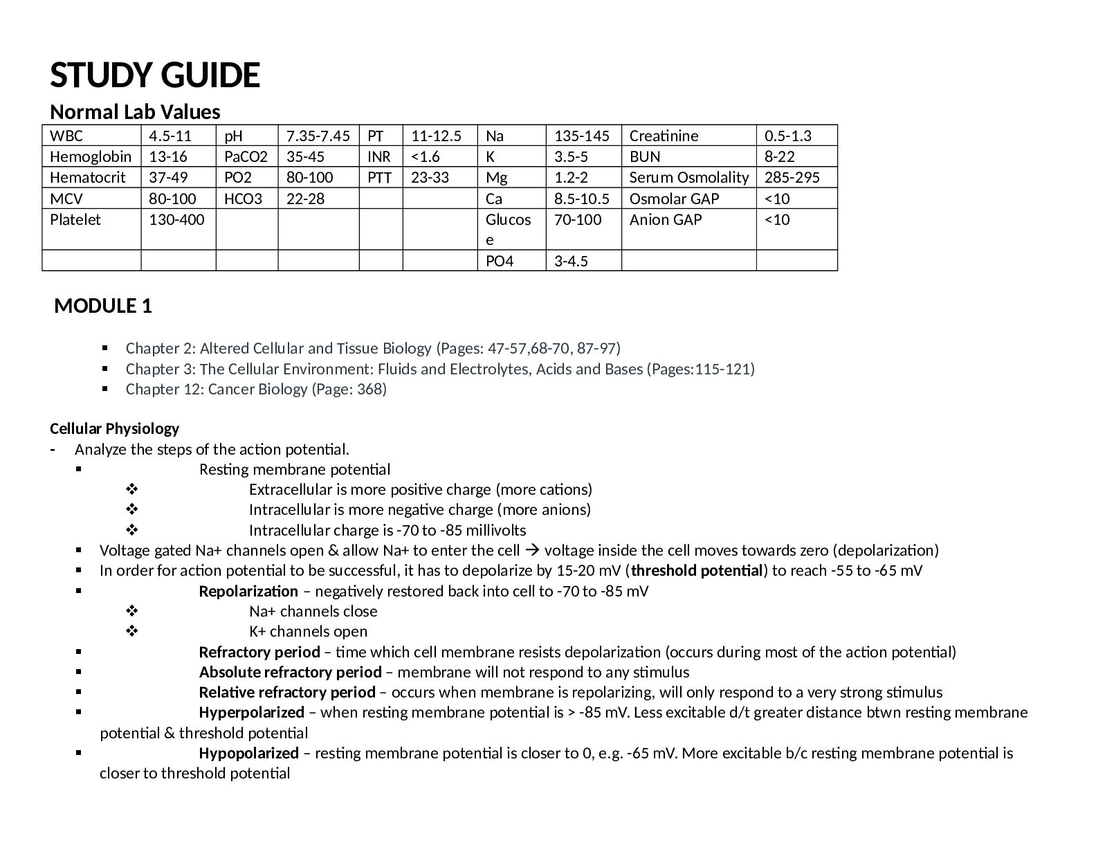
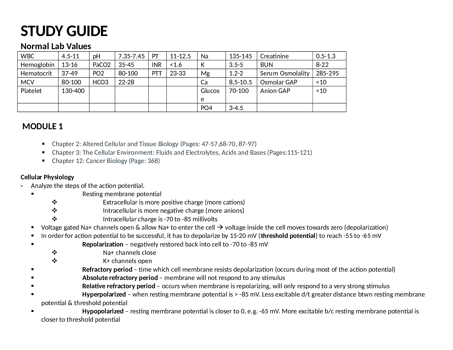
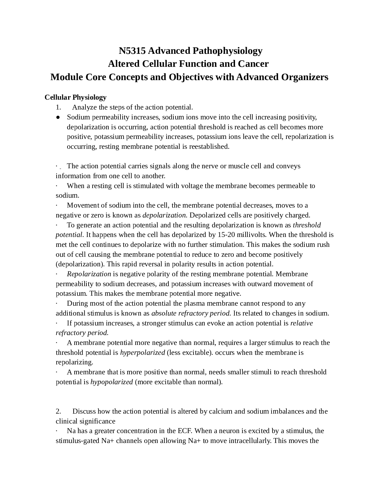
NURS 5315 UTA Exam 1 | Questions and Answers Atrophy E. Cells decrease in size P. Still functional Physiologic: thymus gland in early childhood Pathological: disuse Hypertrophy E. Increase in cell size P. Increased workload Physiologic: weightlifting Pathologic: cardiomegaly from HTN Hyperplasia E. Increase in cell number P. Increased cellular division Physiologic: liver regeneration Pathologic: endometrial- usually r/t hormones Dysplasia E. Cells change in size, shape, organization P. AKA atypical hyperplasia, a disorderly proliferation Physiologic: N/A Pathologic: squamous dysplasia of cervix from HPV Metaplasia E. one cell type replaced with another P. reprogramming of stem cells, reversible Physiologic: N/A Pathologic: stratified squamous cells in bronchial lining r/t cigarette smoke Hypoxia injury E. inadequate oxygenation of tissues P. decrease in mitochondrial function, decreased production of ATP increases anaerobic metabolism. eventual cell death. C.M. hypoxia, cyanosis, cognitive impairment, lethargy Free radical and ROS E. normal byproduct of ATP production, will overwhelm the mitochondria- exhaust intracellular antioxidants P. lipid peroxidation, damage proteins, fragment DNA C.M. development in Alzheimer's, heart disease, Parkinson's disease, Amyotrophic Lateral Sclerosis Ethanol E. mood altering drug, long term effects on liver and nutritional status P. metabolized by liver, generates free radicals C.M. CNS depression, nutrient deficiencies-Mag, Vit B6, thiamine, PO4, inflammation and fatty infiltration of liver, hepatomegaly, leads to liver failure irreversible Oncosis Na and H2O enter cell and cause swelling. Organ increases in weight, becomes distended and pale. Associated with high fever, hypocalcemia, certain infections Fatty Infiltration intracellular accumulation of lipids in the liver liver fails to metabolize lipids. usually from ETOH or high fat diet. can lead to cirrhosis dystrophic calcification accumulation of Ca in dead or dying tissues calcium salt clump and harden- interfere with cellular structure and function r/t pulmonary TB, atherosclerosis, injured heart valves, chronic pancreatitis metastatic calcification accumulation of Ca in normal tissue result of hypercalcemia r/t hyperparathyroidism, hyperthyroidism, toxic levels of Vit D. Can also r/t hyperphosphatemia in renal failure urate accumulation sodium urate crystals are deposited in tissues- group of disorders collectively called gout- acute arthritis, chronic gouty arthritis, tophus, nephritis Coagulative Necrosis kidneys, heart, adrenals- secondary to hypoxia Liquefactive Necrosis nerve cells- brain- accumulation of pus Caseous Necrosis lung disease- usually TB- tissue looks like clumped cheese Fat Necrosis breast, pancreas, abdominal structures- creates soaps Gangrenous Necrosis Dry- dark shriveled skin Wet- internal organs- can lead to death Gas- from clostridium- antitoxins and hyperbaric therapy Gout E. disturbances in serum urate levels. uncommon for < 30 years old. P. uric acid is deposited in the tissues of kidney, heart, earlobes, and joints. C.M. inflammation, painful joints. result of diuretic use or diet high in cream sauces, red wine, or red meat Rhabdomyolysis E. cell hypoxia caused by severe muscle trauma, hyperthermia, crush injuries, or severe dehydration P. hypoxia leads to cell death, cellular contents are released in bloodstream. C.M. CK is 5x upper normal limit, muscle pain, weakness, dark, reddish-brown urine, hypercalcemia, renal failure Alpha Fetoprotein Origin Liver and germ cell tumors Carcinoembryonic Antigen GI, pancreas, lung, breast tumors Beta Human Chorionic gonadotropin origin germ cell tumors Prostate Specific Antigen prostate tumors Carcino- from epithelial tissue- renal cell carcinoma Sarco- from connective tissue- chondrosarcoma -oma benign tumor- lipoma Carcinoma in situ preinvasive epithelial malignant tumors of glandular or squamous cells- cervix -blastoma from blasts or immature cells- neuroblastoma Lung ca metastasis Multiple organs including brain Colorectal ca metastasis Liver, lungs Testicular ca metastasis Liver, lungs, brain Prostate ca metastasis Bones (especially lumbar spine), liver Breast ca metastasis Liver, lungs, brain, bones Head and neck ca metastasis Liver, bones, lymphatics Ovarian ca metastasis Peritoneal surfaces, diaphragm, omentum, liver Sarcoma metastasis Lungs Melanoma metastasis In transit lymphatics, lung, liver, brain, GI tract Mechanisms of ca metastasis Local invasion, followed by invasion of surrounding tissues. Cells then may invade blood and lymphatic vessels. They must survive in circulation, then enter and survive in a new location. Then the cells can multiply and form a new tumor. TNM staging system T= tumor size >/= correlates with metastatic ability N= whether lymph nodes are involved M= extra nodal involvement (liver, lungs) Intravascular fluid compartment In venous system- 20% Osmolality The measure of solute concentration in a fluid. 280-295 mOsm Interstitial fluid compartment Surrounds the cells and bathes them in nutrients- 20% Intracellular fluid compartment Within the cells- 40% uk Osmosis Passive- the movement of water from an area of low concentration of solute to one of higher concentration Osmotic pressure Pulling- the amount of pressure or force that is exerted by solute molecules of a given compartment Hydrostatic pressure Blood pressure- pushes fluid outside of the vessels, the force of fluid against the walls of a compartment- venous obstruction, Na and water retention Oncotic pressure Colloid pressure keeps water inside the compartment, attracts water from interstitial space back into the capillary- losses or diminished albumin Effective arterial blood volume The amount of blood within the arterial space- ECF changes will cause changes in the EABV in the same direction Antidiuretic hormone Secreted by pituitary gland in response to water deficit, Na excess, or hypotension. Causes kidneys to reabsorb water, increasing plasma volume Renin Angiotensin Aldosterone System Activated by low blood volume, triggers release of renin which converts angiotensinogen to angiotensin 1. ACE converts angiotensin 1 to angiotensin which causes arterial vasoconstriction and stimulates release of aldosterone. Aldosterone stimulates renal Na reabsorption and K+ excretion. Water is retained, less urine is produced, blood volume increases. Natriuretic hormones ANP and BNP- released by heart- works opposite RAAS to decrease blood volume, promotes urinary excretion of Na and water Fluid volume deficit Dehydration- intake is not enough for body's needs C.M. Poor skin turgor, dry mucous membranes, sunken eyes, sunken fontanelles, decreased urine output, fatigue Fluid volume excess Fluid intake exceeds body's needs C.M. Edema, rales, HTN, weight gain, bounding pulses, intake> output, JVD, restlessness or anxiety
[Show More]