Pathophysiology > EXAM > NURS 5315 Advanced Pathophysiology Altered Cellular Function and Cancer | Module Objectives with Ad (All)
NURS 5315 Advanced Pathophysiology Altered Cellular Function and Cancer | Module Objectives with Advanced Organizers
Document Content and Description Below
Last updated: 3 years ago
Preview 1 out of 11 pages
Instant download

Buy this Document to get the Full Access Instantly
Provided by Students Who Aced it
We Verify Document Content to Gurantee Accuracy
Reviews( 0 )
Document information
Connected school, study & course
About the document
Uploaded On
Jan 17, 2021
Number of pages
11
Written in
All

Additional information
This document has been written for:
Uploaded
Jan 17, 2021
Downloads
0
Views
123
Document Keyword Tags
Recommended For You
Get more on EXAM »
NURS 5315 Advanced Pathophysiology Cancer | Module Objectives...

NURS 5315 Advanced Pathophysiology Disorders of the Immune Sys...

NURS 5315 Advanced Pathophysiology Gastrointestinal | Core Kno...

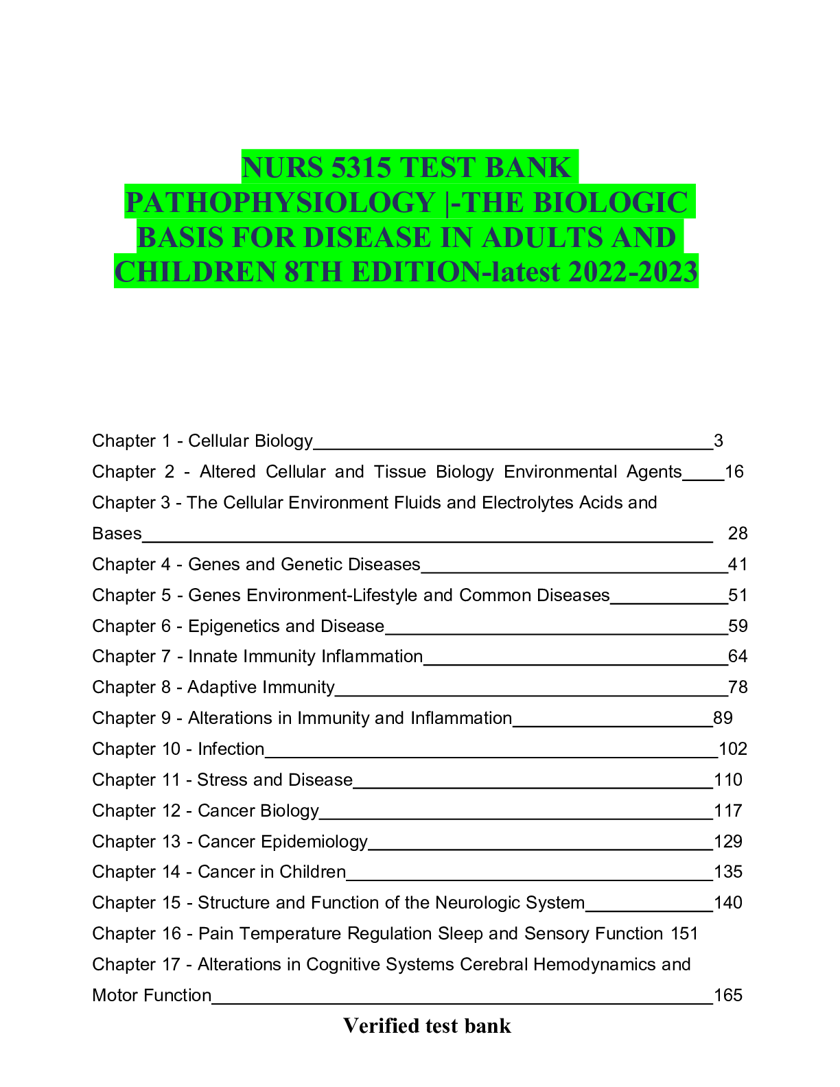
NURS 5315 Pathophysiology | The Biologic Basis for Disease in...

NURS 5315 PATHO Module 7 N5315 Advanced Pathophysiology Neurol...

NURS 5315 Advanced Pathophysiology (Latest 2025 / 2026 Up...


NURS 5315 Pathophysiology | The Biologic Basis for Disease in...
NURS 5315 Pathophysiology | The Biologic Basis for Disease in...

NURS 5315 Pathophysiology The Biologic Basis for Disease in Ad...
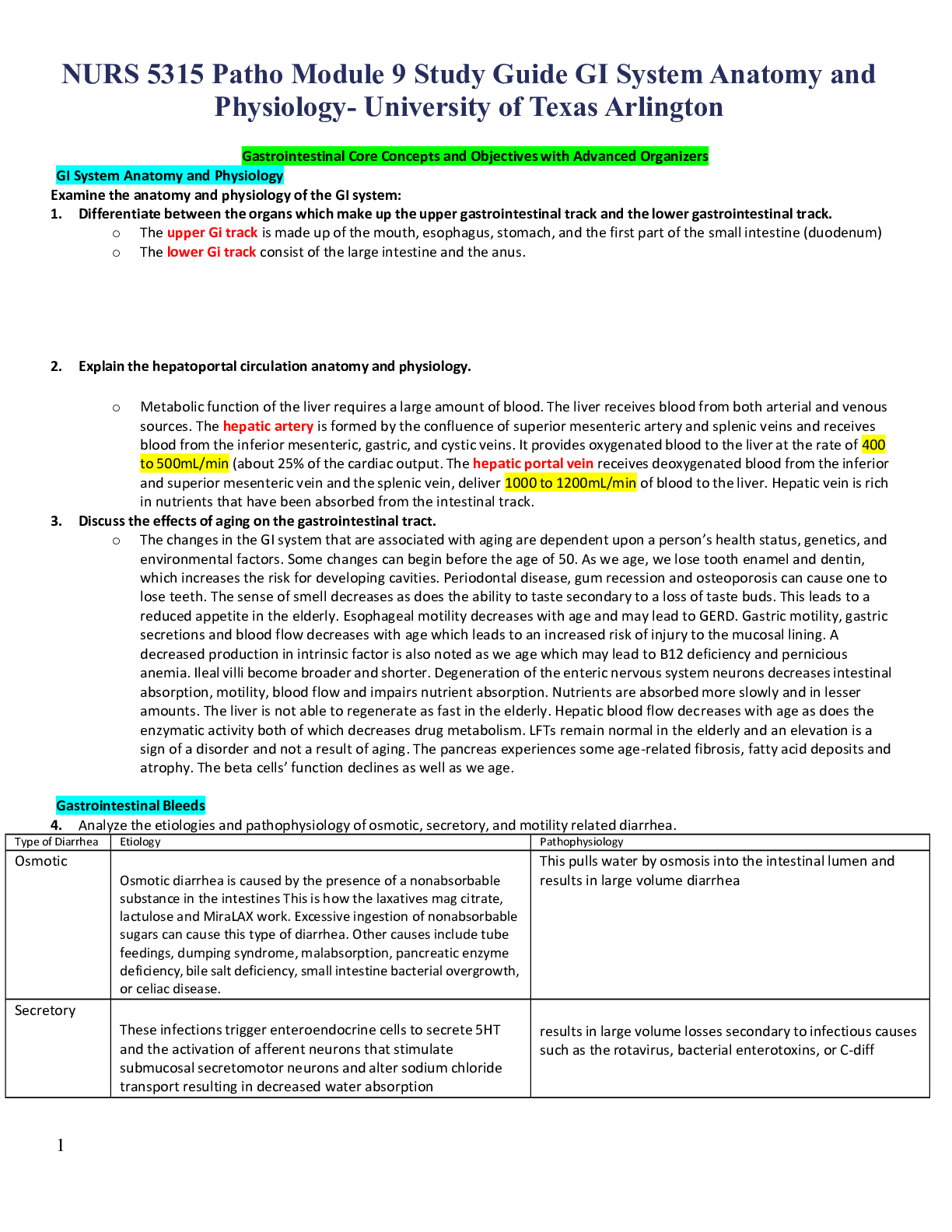
NURS 5315 Patho Module 9 Study Guide GI System Anatomy and Phy...
NURS 5315 Patho Module 9 Study Guide GI System Anatomy and Phy...

 – Chamberlain College of Nursing.png)
 – Chamberlain College of Nursing.png)