Biology > Presentation > Cardiovascular System Review_Chapter 9 | NURS101 Cardiovascular System Review_Updated (All)
Cardiovascular System Review_Chapter 9 | NURS101 Cardiovascular System Review_Updated
Document Content and Description Below
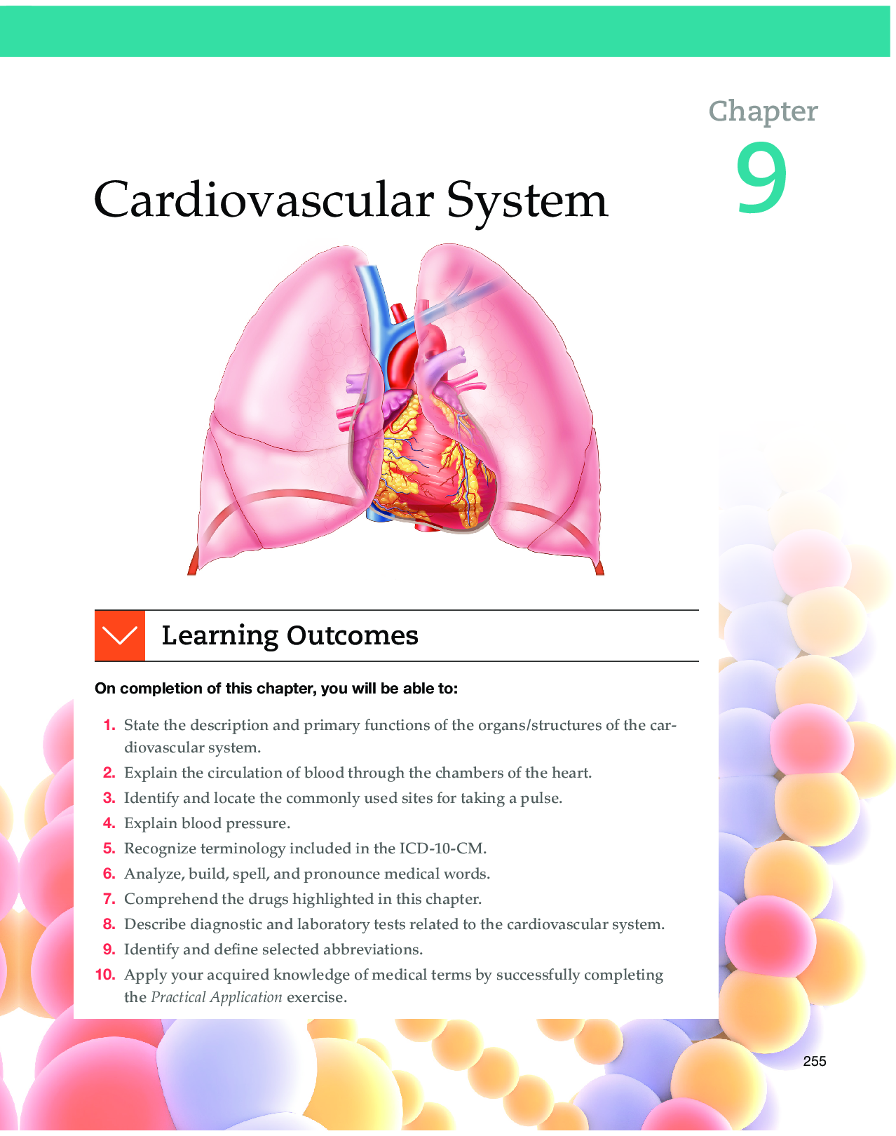
Cardiovascular System 9256 Anatomy and Physiology The cardiovascular (CV) system, also called the circulatory system, circulates blood to all parts of the body by the action of the heart. This proc ... ess provides the body’s cells with oxygen and nutritive elements and removes waste materials and carbon dioxide. The heart, a muscular pump, is the central organ of the system. It beats approximately 100,000 times each day, pumping roughly 8,000 liters of blood, enough to fill about 8,500 quart-sized milk cartons. Arteries, veins, and capillaries comprise the network of vessels that transport blood (fluid consisting of blood cells and plasma) throughout the body. Blood flows through the heart, to the lungs, back to the heart, and on to the various body parts. Table 9.1 provides an at-a-glance look at the cardiovascular system. Figure 9.1 shows a schematic overview of the cardiovascular system. Heart The heart is the center of the cardiovascular system from which the various blood vessels originate and later return. It is slightly larger than a person’s fist and weighs approximately 300 g in the average adult. It lies slightly to the left of the midline of the body, behind the sternum (see Figure 9.2). The heart has three layers or linings. • Endocardium. The inner lining of the heart. • Myocardium. The muscular middle layer of the heart. • Pericardium. The outer membranous sac surrounding the heart. Circulation of Blood through the Chambers of the Heart The heart is a pump and is divided into the right and left heart by a partition called the septum. Each side contains an upper and lower chamber. See Figure 9.3. The atria, or upper chambers, are separated by the interatrial septum. The ventricles, or lower chambers, are separated by the interventricular septum. The atria receive blood from the various parts of the body. The ventricles pump blood to body parts. Valves control the intake and outflow of blood in the heart chambers. Figure 9.4 shows the functioning of the heart valves and flow of blood through the heart. Table 9.1 Cardiovascular System at-a-Glance Organ/Structure Primary Functions/Description Heart The muscular pump that circulates blood through the heart, the lungs (pulmonary circulation), and the rest of the body (systemic circulation) Arteries Branching system of vessels that transports blood from the right and left ventricles of the heart to all body parts; transports blood away from the heart Veins Vessels that transport blood from peripheral tissues back to the heart Capillaries Microscopic blood vessels that connect arterioles with venules; facilitate passage of life-sustaining fluids containing oxygen and nutrients to cell bodies and the removal of accumulated waste and carbon dioxide Blood Fluid consisting of formed elements (erythrocytes, thrombocytes, leukocytes) and plasma. It is a specialized bodily fluid that delivers necessary substances to the body’s cells (oxygen, foods, salts, hormones) and transports waste products (carbon dioxide, urea, lactic acid) away from those same cells. Blood is circulated around the body through blood vessels by the pumping action of the heart. See Chapter 10, Blood and Lymphatic System, for a further discussion of blood.Cardiovascular System • 257 Figure 9.1 Schematic overview of the cardiovascular system. AIR (OXYGEN) Lung capillaries Trachea Left pulmonary artery Lung capillaries Left pulmonary vein Heart (blood) Veins Right pulmonary vein Arteries Venules Arterioles BODY CAPILLARIES Right lung Left lung Right pulmonary artery Blood high in oxygen and low in carbon dioxide (oxygenated) = = Blood low in oxygen and high in carbon dioxide (deoxygenated) Figure 9.2 Location of the heart in the chest cavity. Midsternal line Second rib Sternum Diaphragm Mediastinum (contains the organs between the pleural cavities) Superior vena cava Pericardium Diaphragm Apex of heart Aorta Pulmonary trunk Left lung258 • Chapter Nine Figure 9.3 Interior view of the heart chambers with tissues of the heart (endocardium, myocardium, and pericardium). Left ventricle Endocardium Myocardium Pericardium Superior vena cava Aorta Pulmonary trunk Right atrium Right ventricle Inferior vena cava Left atrium Pulmonary valve Tricuspid valve Aortic valve Mitral (Bicuspid) valve Figure 9.4 The functioning of the heart valves and blood flow. To lung Left pulmonary artery (branches) To lung Right pulmonary artery (branches) From body From body To body Right atrium Tricuspid valve Right ventricle Inferior vena cava From lung Right pulmonary vein (branches) Aorta Left ventricle Interventricular septum Myocardium (heart muscle) Apex Left atrium Aortic valve Mitral (bicuspid) valve Descending aorta From lung Left pulmonary vein (branches) 1 2 3 4 5 6 Superior vena cava Pulmonary valveCardiovascular System • 259 right atrium The right upper portion of the heart is called the right atrium (RA). It is a thin-walled space that receives blood from the upper and lower parts of the body (except the lungs). Two large veins, the superior vena cava and inferior vena cava, bring deoxygenated blood into the right atrium. Deoxygenated blood fills the right atrium before passing through the tricuspid (atrioventricular) valve and into the right ventricle. right ventricle The right lower portion of the heart is called the right ventricle (RV). It receives blood from the right atrium through the tricuspid valve. When filled, the RV contracts. This creates pressure, closing the right atrium and forcing open the pulmonary (semilunar) valve, sending blood into the left and right pulmonary arteries, which carry it to the lungs. The pulmonary artery is the only artery in the body that carries blood deficient in oxygen. In the lungs, the blood gives up wastes and takes on oxygen as it passes through capillary beds into veins. Oxygenated blood leaves the lungs through the left and right pulmonary veins, which carry it to the heart’s left atrium. The pulmonary veins are the only veins in the body that carry oxygen-rich (oxygenated) blood. The circulation of blood through the vessels from the heart to the lungs and then back to the heart again is the pulmonary circulation. left atrium The left upper portion of the heart is called the left atrium (LA). It receives blood rich in oxygen as it returns from the lungs via the left and right pulmonary veins. As oxygenated blood fills the LA, it creates pressure that forces open the mitral (bicuspid) valve and allows the blood to fill the left ventricle. left ventricle The left lower portion of the heart is called the left ventricle (LV). It receives blood from the left atrium through the mitral valve. When filled, the LV contracts. This creates pressure closing the mitral valve and forcing open the aortic valve. The oxygenated blood from the LV flows through the aortic valve and into a large artery known as the aorta and from there to all parts of the body (except the lungs) via a branching system of arteries and capillaries. fyi Pediatric cardiologists have recognized more than 50 congenital heart defects. If the left side of the heart is not completely separated from the right side, various septal defects develop. If the four chambers of the heart do not develop normally, complex anomalies form, such as tetralogy of Fallot (TOF), a congenital heart condition involving four defects: pulmonary artery stenosis, ventricular septal defect (VSD), displacement of the aorta to the right, and hypertrophy of the right ventricle. Heart Valves The valves of the heart are located at the entrance and exit of each ventricle and, as you learned in the preceding section, control the flow of blood within the heart. See Figure 9.5.260 • Chapter Nine tricuspid valve The tricuspid or right atrioventricular valve guards the opening between the right atrium and the right ventricle. In a normal state, the tricuspid valve opens to allow the flow of blood into the ventricle and then closes to prevent any backflow of blood. pulmonary (semilunar) valve The exit point for blood leaving the right ventricle is called the pulmonary (semilunar) valve. Located between the right ventricle and the pulmonary artery, it allows blood to flow from the right ventricle through the pulmonary artery to the lungs. mitral (bicuspid) valve The left atrioventricular valve between the left atrium and the left ventricle is called the mitral valve (MV) or bicuspid valve. It allows blood to flow to the left ventricle and closes to prevent its return to the left atrium. aortic (semilunar) valve Blood exits from the left ventricle through the aortic (semilunar) valve. Located between the left ventricle and the aorta, it allows blood to flow into the aorta and prevents its backflow to the ventricle. Vascular System of the Heart Due to the membranous lining of the heart (endocardium) and the thickness of the myocardium, the heart has its own vascular system to meet its high oxygen demand. The coronary arteries supply the heart with oxygen-rich blood, and the cardiac veins, draining into the coronary sinus, collect the blood (oxygen poor) and return it to the right atrium. See Figure 9.6. Figure 9.5 Valves of the heart. Anterior Posterior Aortic valve (left semilunar valve) Tricuspid valve (right atrioventricular valve) Mitral valve (left atrioventricular valve) Pulmonary valve (right semilunar valve)Cardiovascular System • 261 Conduction System of the Heart The autonomic nervous system controls the rate and rhythm of the heartbeat. It is normally generated by specialized neuromuscular tissue of the heart that is capable of causing cardiac muscle to contract rhythmically. This tissue of the heart comprises the sinoatrial node, the atrioventricular node, and the atrioventricular bundle. See Figure 9.7. Figure 9.6 Coronary circulation. (A) Coronary vessels portraying the complexity and extent of the coronary circulation. (B) Coronary vessels that supply the posterior surface of the heart. Anterior interventricular artery (descending branch) Right coronary artery Small cardiac vein Anterior cardiac veins Marginal branch A Great cardiac vein Circumflex branch Coronary sinus Posterior cardiac vein Right coronary artery Small cardiac vein Marginal branch Posterior interventricular vein Middle cardiac vein (descending branch) B Pulmonary trunk Left coronary artery Great cardiac vein Aortic arch Sinoatrial node (pacemaker) Internodal pathway Atrioventricular node Atrioventricular bundle (Bundle of His) Bundle branches Purkinje fibers Aorta Purkinje fibers Interventricular septum Superior vena cava Left atrium 1. 2. 3. 4. 5. Figure 9.7 Conduction system of the heart.262 • Chapter Nine sinoatrial node (sa node) Called the pacemaker of the heart, the SA node is located in the upper wall of the right atrium, just below the opening of the superior vena cava. It consists of a dense network of Purkinje fibers (atypical muscle fibers) considered to be the source of impulses initiating the heartbeat. Electrical impulses discharged by the SA node are distributed to the right and left atria and cause them to contract. atrioventricular node (av node) Located beneath the endocardium of the right atrium, the AV node transmits electrical impulses to the bundle of His (atrioventricular bundle). atrioventricular bundle (bundle of his) The atrioventricular bundle or bundle of His forms a part of the conduction system of the heart. It is a collection of heart muscle cells specialized for electrical conduction that transmits the electrical impulses from the AV node to the point of the apex of the fascicular branches. The bundle of His branches into the two bundle branches that run along the interventricular septum. The bundles give rise to thin filaments known as Purkinje fibers. These fibers distribute the impulse to the ventricular muscle. Together, the bundle branches and Purkinje network comprise the ventricular conduction system. The average adult heartbeat (pulse) is between 60 and 90 beats per minute. The rate of the heartbeat can be affected by emotions, smoking, disease, body size, age, stress, the environment, and many other factors. The heart’s electrical activity can be recorded by an electrocardiogram (ECG, EKG), which provides valuable information in diagnosing cardiac abnormalities, such as myocardial damage and arrhythmias (see the section Diagnostic and Laboratory Tests and Figure 9.38). Blood Vessels There are three main types of blood vessels: arteries, veins, and capillaries. Blood circulates throughout the body through their pathways. Arteries The arteries constitute a branching system of vessels that transports blood away from the heart to all body parts. See Figure 9.8. In a normal state, arteries are elastic tubes that recoil and carry blood in pulsating waves. All arteries have a pulse, reflecting the rhythmical beating of the heart; however, certain points are commonly used to check the rate, rhythm, and condition of the arterial wall. A person’s pulse can be felt in a place that allows for an artery to be compressed against a bone. The most commonly used sites for taking a pulse are the radial artery, the brachial artery, and the carotid artery. See Table 9.2 and Figure 9.9. The pulse rate can also be measured by using a stethoscope (auscultation) and counting the heartbeat for 1 full minute. This is known as the apical pulse and is taken over the heart itself. In contrast with other pulse sites, the apical pulse site is unilateral and is located at the apex of the heart or at the fifth intercostal space, just to the left of the midclavicular line. It is commonly used to check pulse rate in infants and children, and when the radial pulse is difficult to palpate (to feel by touch).Cardiovascular System • 263 Figure 9.8 Major arteries of the systemic circulation. Left common carotid artery Left subclavian artery Aortic arch Renal artery Abdominal aorta Radial artery Ulnar artery Popliteal artery Peroneal artery Right common carotid artery Right subclavian artery Ascending aorta Brachial artery Common iliac artery Internal iliac artery External iliac artery Femoral artery Anterior tibial artery Posterior tibial artery264 • Chapter Nine Table 9.2 Pulse Checkpoints Carotid Brachial Radial Femoral Popliteal Dorsalis pedis Temporal Figure 9.9 Primary pulse points of the body. Checkpoint Site/Use Temporal Temple area of the head. Used to control bleeding from the head and scalp and to monitor circulation. Carotid Neck. In an emergency (cardiac arrest), most readily accessible site. Brachial Antecubital space of the elbow. Most common site used to check blood pressure. Radial Radial (thumb side) of the wrist. Most common site for taking a pulse. Femoral Groin area. Monitor circulation. Popliteal Behind the knee. Monitor circulation. Dorsalis pedis Dorsal surface of the foot. Assess for peripheral artery disease (PAD). Veins Veins are the vessels that transport blood from peripheral tissues back to the heart. In a normal state, veins have thin walls and valves that prevent the backflow of blood. The great saphenous vein is the most important superficial vein of the lower limb. The pulmonary veins carry oxygenated blood from the lungs to the heart. The superior and inferior venae cavae carry deoxygenated blood from the upper and lower systemic circulation. See Figure 9.10. Capillaries The capillaries are microscopic blood vessels with single-celled walls that connect arterioles (small arteries) with venules (small veins). See Figure 9.11. Blood passing through capillaries gives up the oxygen and nutrients carried to this point by the arteries and picks up waste and carbon dioxide as it enters veins. Veins lead away from the capillaries as tiny vessels and increase in size until they join the superior and inferior venae cavae as they return to the heart. The extremely thin walls of capillaries facilitate passage of oxygen and nutrients to cell bodies and the removal of accumulated waste and carbon dioxide.Cardiovascular System • 265 Figure 9.10 Major veins of the systemic circulation. Superior vena cava Hepatic portal vein Superior mesenteric vein Inferior vena cava Ulnar vein Radial vein Common iliac vein External iliac vein Internal iliac vein Digital veins Subclavian vein Right and left brachiocephalic veins Cephalic vein Brachial vein Basilic vein Median cubital vein Renal vein External jugular vein Internal jugular vein Femoral vein Great saphenous vein Popliteal vein Posterior tibial vein Anterior tibial vein Fibular vein266 • Chapter Nine Figure 9.11 Capillaries. Capillary network Lumen Venule Valve Tunica interna: • Endothelium • Subendothelial layer • Internal elastic lamina Tunica media Tunica externa Artery Vein Arteriole fyi The newborn’s pulse rate is irregular and rapid, varying from 120 to 140 beats/minute. Blood pressure pulse (P), blood pressure (BP), and respiration (R) vary according to a child’s age. A is low and can vary with the size of the cuff used. The average blood pressure at birth is 80/46. The respirations are approximately 35–50 per minute. Blood Pressure Blood pressure (BP) is the pressure exerted by the blood on the walls of the arteries. It results from two forces. One is created by the heart as it pumps blood into the arteries and through the circulatory system. The other is the force of the arteries as they resist the blood flow. The higher (systolic) number represents the pressure while the heart contracts to pump blood to the body. The lower (diastolic) number represents the pressure when the heart relaxes between beats. Blood pressure is reported in millimeters of mercury (mmHg) and is measured with a sphygmomanometer. See Figure 9.12. The systolic pressure is always stated first. For example, 116/74 (116 over 74); systolic = 116, diastolic = 74. Blood pressure below 120 over 80 mmHg is considered optimal for adults. A systolic pressure of 120–139 mmHg or a diastolic pressure of 80–89 mmHg is considered to be prehypertension and needs to be monitored on a regular basis. A blood pressure reading of 140 over 90 or higher is considered elevated (hypertension). [Show More]
Last updated: 3 years ago
Preview 1 out of 55 pages
Buy this document to get the full access instantly
Instant Download Access after purchase
Buy NowInstant download
We Accept:

Reviews( 0 )
$13.50
Can't find what you want? Try our AI powered Search
Document information
Connected school, study & course
About the document
Uploaded On
Apr 24, 2021
Number of pages
55
Written in
All

Additional information
This document has been written for:
Uploaded
Apr 24, 2021
Downloads
0
Views
203


 (1).png)