Medical Studies > SOLUTIONS > BME 210 Homework 3: Medical Imaging Solutions | BME 210 Biomedical Computer Simulation Methods -HW w (All)
BME 210 Homework 3: Medical Imaging Solutions | BME 210 Biomedical Computer Simulation Methods -HW with COMMENTARY
Document Content and Description Below
Last updated: 4 months ago
Preview 3 out of 10 pages
Instant download

Loading document previews ...
Buy this Document to get the Full Access Instantly
Provided by Students Who Aced it
We Verify Document Content to Gurantee Accuracy
Reviews( 0 )
Document information
Connected school, study & course
About the document
Uploaded On
Mar 04, 2026
Number of pages
10
Written in
All

Additional information
This document has been written for:
Uploaded
Mar 04, 2026
Downloads
0
Views
67
Document Keyword Tags
Recommended For You
Get more on SOLUTIONS »
Essentials of Lifespan Development, 2nd Canadian Edition, 2e...
Medical Terminology for Health Professions 9th Edition by Ehrl...


MDC3 Exam 1 Review - With NCLEX questions and extra notes 2021
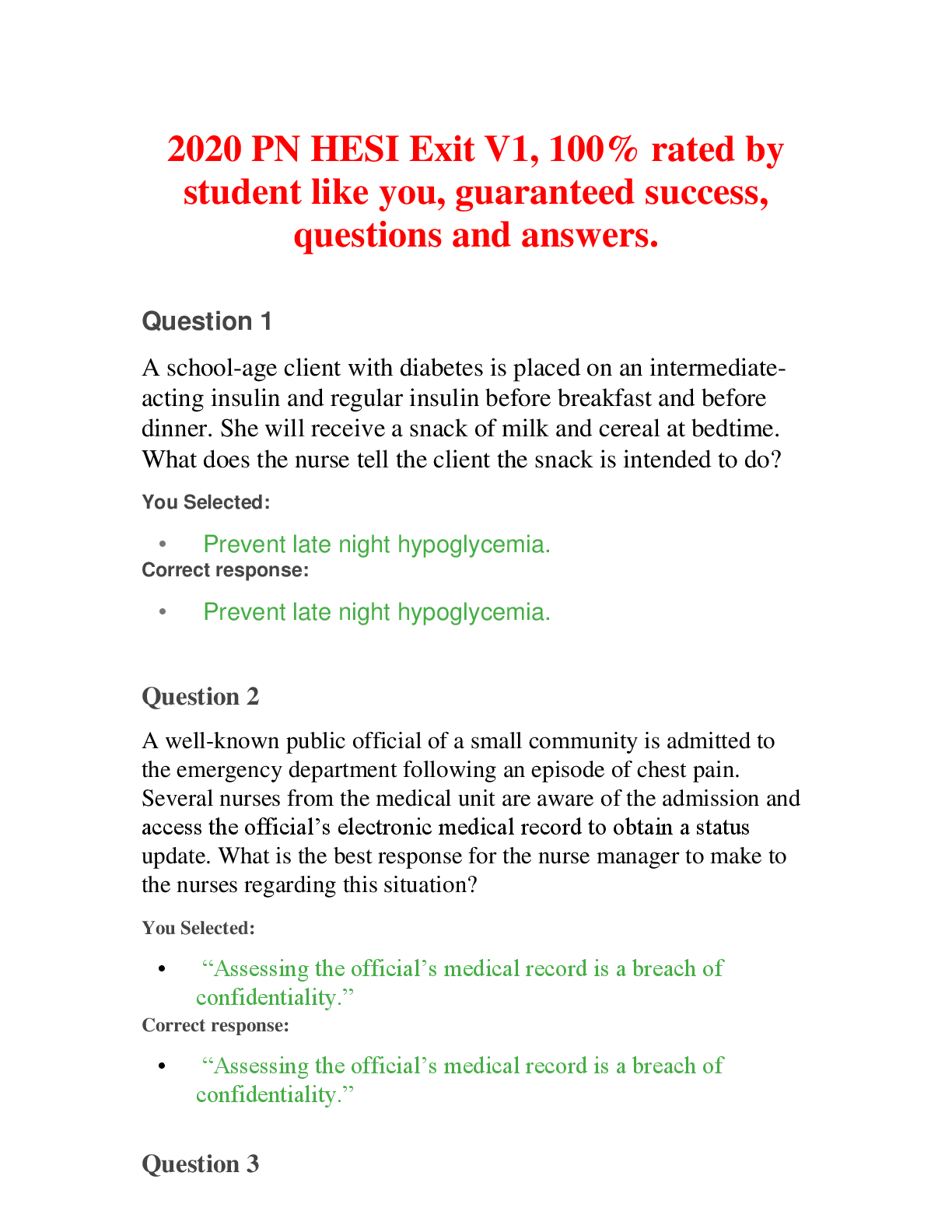
PN HESI Exit V1, 100% rated by student like you, guaranteed su...

Solutions Manual to Accompany Statistics for Business and Econ...

Solutions Manual to accompany THEORY OF MACHINES AND MECHANISM...