NURSING 1 EXAM 1
Osteomalacia
- Metabolic disease that causes poor & delayed mineralization of the bone cells in mature bones
- Main cause is a Vitamin D deficiency
Diagnostic procedures for Osteomalacia
Serum c
...
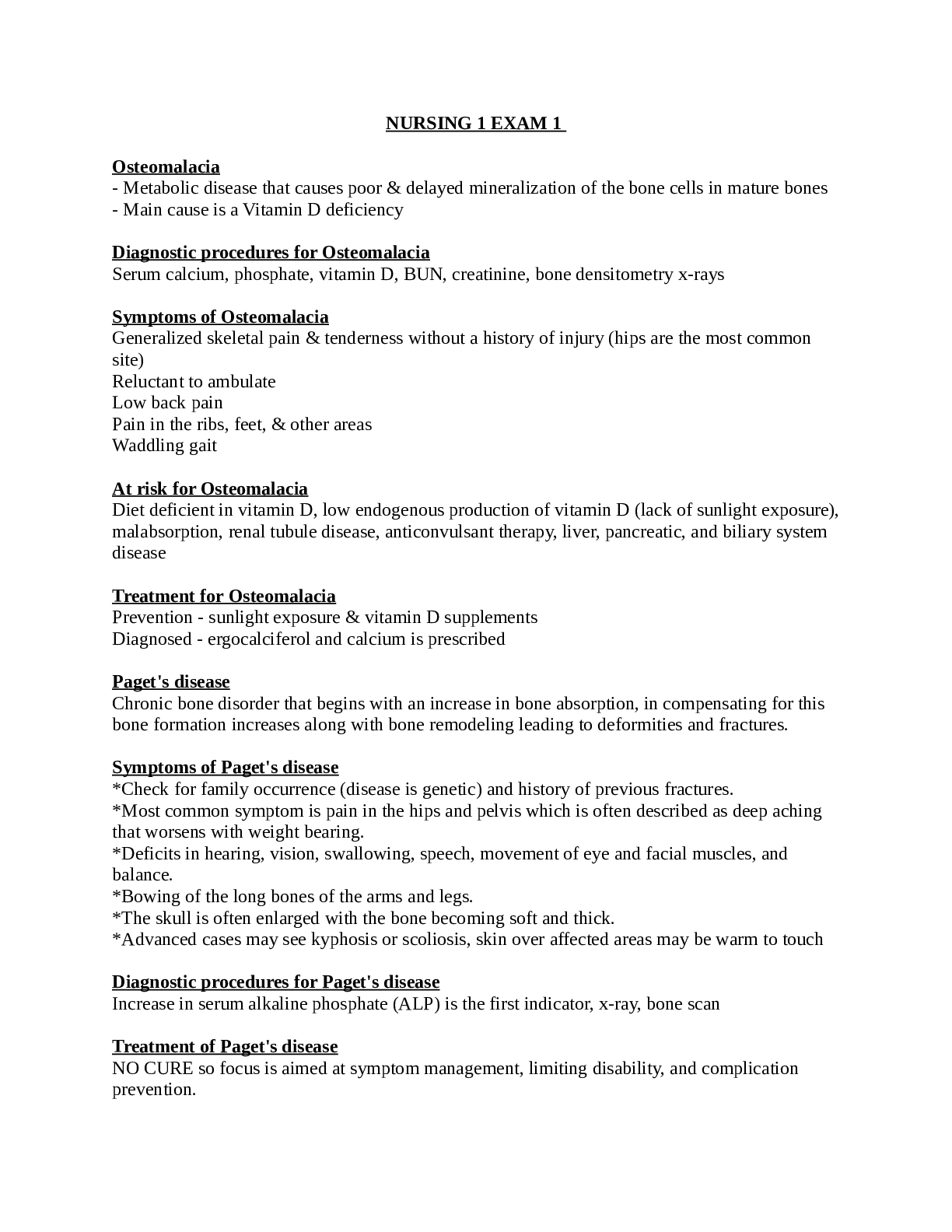
NURSING 1 EXAM 1
Osteomalacia
- Metabolic disease that causes poor & delayed mineralization of the bone cells in mature bones
- Main cause is a Vitamin D deficiency
Diagnostic procedures for Osteomalacia
Serum calcium, phosphate, vitamin D, BUN, creatinine, bone densitometry x-rays
Symptoms of Osteomalacia
Generalized skeletal pain & tenderness without a history of injury (hips are the most common site)
Reluctant to ambulate Low back pain
Pain in the ribs, feet, & other areas Waddling gait
At risk for Osteomalacia
Diet deficient in vitamin D, low endogenous production of vitamin D (lack of sunlight exposure), malabsorption, renal tubule disease, anticonvulsant therapy, liver, pancreatic, and biliary system disease
Treatment for Osteomalacia
Prevention - sunlight exposure & vitamin D supplements Diagnosed - ergocalciferol and calcium is prescribed
Paget's disease
Chronic bone disorder that begins with an increase in bone absorption, in compensating for this bone formation increases along with bone remodeling leading to deformities and fractures.
Symptoms of Paget's disease
*Check for family occurrence (disease is genetic) and history of previous fractures.
*Most common symptom is pain in the hips and pelvis which is often described as deep aching that worsens with weight bearing.
*Deficits in hearing, vision, swallowing, speech, movement of eye and facial muscles, and balance.
*Bowing of the long bones of the arms and legs.
*The skull is often enlarged with the bone becoming soft and thick.
*Advanced cases may see kyphosis or scoliosis, skin over affected areas may be warm to touch
Diagnostic procedures for Paget's disease
Increase in serum alkaline phosphate (ALP) is the first indicator, x-ray, bone scan
Treatment of Paget's disease
NO CURE so focus is aimed at symptom management, limiting disability, and complication prevention.
Physical therapy (weight-bearing exercises)
Pharmacological therapy: Calcitonin, Bisphosphonates, Analgesics Worst cases: Surgery
Bisphosphonates: Alendronate (Fosamax)
Dose: 40 mg once daily for three months
MUST be taken on an empty stomach with 6 to 8 ounces of water
It may be taken any time during the day, as long as there is no food for 30 minutes after the dose Do NOT lie down for at least 30 minutes after taking the dose
Osteomyelitis
Occurs when an infection has invaded the bone area.
Patients at risk for Osteomyelitis
Associated with patients who have diabetes of vascular problems Malnourished patients
Elderly patients Overweight patients Injection/needle drug use Poor blood supply Recent trauma
Symptoms of Osteomyelitis
Fever Malaise
Swelling &/or tenderness of the affected area Purulent drainage from the affected site
Pain in the affected area Tachycardia
Diagnostic procedures for Osteomyelitis
Bone biopsy/culture Bone scan
Bone x-ray
CBC (elevated WBC's)
Needle aspiration of the affected area CRP (will be elevated)
ESR (will be elevated)
Treatment of Osteomyelitis
IV antibiotic therapy for 4-6 weeks (type depends on culture) Pain medication
Wound debridement
Nursing Interventions with Osteomyelitis
Immobilize the body part Administer pain medications Perform checks
Perform sterile dressing changes Provide diet high in protein & vitamin C
Initiate antibiotics ASAP & teach the importance of completion
Prevention of Osteomyelitis
Prophylactic antibiotics and sterile technique during and after surgery
Osteoporosis
Chronic disease in which bone loss causes decreased density & possible fracture Wrist, hip & vertebral column are the most commonly affected.
Patients at risk for Osteoporosis
A = alcohol use
C = corticosteroid use C = calcium is low
E = estrogen is low (especially postmenopausal) S = smoking
S = sedentary lifestyle
Symptoms of Osteoporosis
Back pain Constipation
Decrease in height accompanied by kyphosis Fractures (forearm, femur, ribs, and spine) Decreased bone mass
Treatment of Osteoporosis
Exercise - walking, swimming, and water aerobics are recommended to increase muscle Diet - high in calcium, Vitamin D, fiber, & protein
Examples of foods high in calcium: breads cereals, apricots, molasses, milk, dairy products (especially yogurt), spinach, sardines, beans, carrots, asparagus, & collard greens
Avoid alcohol & smoking
NOTE: excessive caffeine can cause excretion of calcium through the urine
Safety in regards to Osteoporosis
Avoid the use of throw rugs, use of appropriate assistive devices, keep areas free of clutter, adequate lighting, non-skid socks/shoes
Treatment of Osteoporosis
Biophosphonates (i.e. Fosamax, Boniva, Didronel)
Calcium supplements (1000 mg to 1500 mg daily) Vitamin D supplements Estrogen (for post menopausal women)
Muscle relaxers
NSAIDS for discomfort/pain
Scoliosis
Spinal deformity that is characterized by a lateral curve, spinal rotation causing asymmetry, and less than normal curvature in the thoracic spine.
Symptoms of Scoliosis
Uneven pant or shirt length
Pain is usually not an issue until the deformity has progressed
Adams Bending Forward Test (test used in the schools to help with observing curvature in the spine)
Diagnostic tests for Scoliosis
Standing spinal x-rays where the curvature is measured
Treatment of Scoliosis
Mild curvature - exercises and braces
Surgery is recommended for curves greater than 40 degrees
Kyphosis
Posterior curvature of the thoracic spine greater than 45 degrees. Often results from osteoporosis
Treatment for Kyphosis
Accomplished with bracing or spinal fusion should the defect cause cardiopulmonary problems or pain.
Fibromyalgia
A clinical syndrome or condition involving chronic widespread diffuse musculoskeletal pain, stiffness, and tenderness.
Usually affects women of childbearing age & has no known cause.
Symptoms of Fibromyalgia
Pain (increases with stress &/or weather conditions) Fatigue
Insomnia
Treatment of Fibromyalgia
Focuses on client's symptoms: physical therapy, exercise with swimming & water exercises being recommended
Medications: antidepressants, NSAIDS (ibuprofen), skeletal muscle relaxants (cyclobenzaprine), for fibromyalgia treatment - pregabalin (Lyrica)
Gout
Accumulation of uric acid deposits in the joints, results from the body's inability to metabolize purine foods .
Causes of Attacks: fad starvation diets, stress, illness
Symptoms of Gout
Rapid development (within hours) of pain and edema in the one affected joint. Swelling, pain, and decreased range of motion in the affected joint
Kidney stones &/or renal abnormalities
Tophi may be seen on external ears, hands, feet, the olecranon process, and prepatellar bursas. Progressive joint damage and deformities
Diagnostic tests for Gout
Serum uric acid greater than 7mg/dL ESR will be elevated
**Synovial fluid analysis (will show uric acid crystals) Urine albumin will be greater than 100 mg/24 hours Urinary uric acid will be elevated
Leukocytosis will be present
Foods low in purine
cheese, eggs, fats, gelatin, milk, most vegetables, nuts, sugar, increase fluids, citrus juices, cherries
Foods high in purine
dried beans, fish (especially sardines), liver, lobster, oatmeal, oyster, peas, asparagus, poultry, spinach
Medications for Gout
Used to decrease the production of uric acid and promote the production of it Acute - Colchicine (Colsalide) and an NSAID, such as ibuprofen (Motrin) Chronic - Allopurinol (Zyloprim)
Osteoarthritis
Nonsystemic, noninflammatory progressive disorder of movable joints Changes are associated with increasing age and/or trauma
Risk factors for Osteoarthritis
Aging Obesity Genetics Joint injury
Occupation (those that use same joints continuously, for example athletes)
Symptoms of Osteoarthritis
Pain & stiffness (especially in the morning)
Immobility of the joint or limb due to the pain or joint changes
Enlarged and/or nodular joints Crepitation
Deformities Heberden's nodules Bouchard's nodules
Treatment of Osteoarthritis
*2 main goals are to provide comfort & maintain function/mobility Application of heat
TENS unit Weight loss
Nonpharmacological techniques for pain relief
Medications: acetaminophen, meloxicam, cortisone (orally, injections), arthritic rubs
If function is completely lost joint replacement surgery would then have to be explored
Lyme disease
Multisystem inflammatory process with devastating long-term effects if left untreated Bacterial infection that occurs after the bite of an infected deer tick, once the tick is attached it must remain in place for 36-48 hours for the infection to be transmitted
Symptoms of Lyme disease
Symptoms can begin as soon as three days after the bite or as late as 30 days:
Flu-like symptoms (low grade fever, fatigue, muscle and bone aches, chills, and malaise) Patient usually reports being active outside in the last 2-4 weeks, may or may not report bite 90% of cases report erythema migrans (which is the circular rash that continues to grow often resembling a bullseye)
Diagnostic test for Lyme disease
Western blot test to confirm positive ELISA (enzyme-linked immunosorbent assay) test
Treatment for Lyme disease
Antibiotics (type and route are dependent on the stage). Early illness is usually treated with oral drugs, for example, doxycycline (Vibramycin), amoxicillin, or cefuroxime axetil. Later illness such as nervous-system disease might require intravenous drugs such as ceftriaxone (Rocephin) and penicillin G.
Vaccine is no longer available
When synovitis accompanies arthritic symptoms, a synovectomy may be used to reduce edema and pain in the joint.
Patient teaching regarding Lyme disease
Dress appropriately for the outdoors when going out in a tick-laden environment: Light-colored clothing should be worn to make ticks more visible.
Tuck pants into socks when going into areas that could harbor ticks. Properly using insect repellents containing DEET is recommended
Daily tick checks should be conducted after coming in from the outdoors. If found, the tick should be properly removed by using fine-point tweezers to take away the ENTIRE creature.
Accident prevention - Infant
1. Place infants on back after eating and while sleeping; this will not increase risk of aspiration and will reduce risk of sudden infant death syndrome (SIDS)
2. Rapid changes in development and acquisition of new motor skills put infants at increased risk for injury from falls from tables, beds, high chairs, infant seats, and so on
3. Place infants riding in a car in a rear-facing car restraint system in back seat; use a rear-facing restraint system until child is 1 year old and at least 20 pounds
4. Because of their inability to communicate, infants are at risk for burns from applications of heat to skin, such as from a hot water bottle or other heated device
5. Carefully select infant furniture paying special attention to current safety standards
a) Infants may be trapped by crib slats spaced too far apart
b) Infants or young children may be poisoned by lead paint on antique furniture (normal serum lead
level is < 10 mg/dL; lead level 10-19 mg requires environmental history; higher levels require treatment to reduce or prevent neurological deficits)
Accident prevention - Toddler
1. Store medications, cleaning supplies, and poisons in locked cabinets to prevent poisonings in a curious toddler; have poison control phone number readily available to caregiver and posted on telephone
2. Place car restraint systems for toddlers in back seat and may be forward-facing after toddler has reached 1 year of age and 20 pounds; use car restraint systems until child's shoulders are above the harness or ears have reached top of seat; when a child outgrows system, a booster seat with a lap/shoulder belt is required
3. Toddlers explore all objects with their mouths; assess toys for small parts; avoid giving foods such as hard candy, peanuts, and chewing gum to prevent choking and aspiration
4. Burns in toddlers occur because of chewing on electrical wires, pulling hot liquids from tables or stove tops, and touching space heaters; keep electrical outlets covered and keep handles of pans on stove tops facing inward
5. Drowning is a leading cause of death in toddlers; accompany children at all times when in and around water in bathtub, wading pool, or swimming pool
Accident prevention - School age child
1. Teach and model pedestrian and bicycle safety to children at this age; bicycle helmets can prevent
head injury to children biking, rollerblading, or skateboarding
2. Place children under 12 years or under 4 feet 9 inches in rear seat of a car with a lap/shoulder seatbelt; never allow children to ride in the bed of a pickup truck or open, unsecured area of an automobile, such as a station wagon or van
3. Teach school-age children principles of fire safety; injury can occur from experimentation with
matches, lighters, and fireworks; school-age children can participate in implementing a school or home fire escape plan; teach to "stop, drop, and roll" if clothing catches fire
4. Teach school-age children principles of water safety; swimming lessons and life jackets are necessary for boating and swimming; children should never swim without adult supervision
5. Include in safety education for children to play in safe areas, avoid strangers, recognize unwelcome touch, and obey traffic signals
Accident prevention - Adolescent
1. Encourage courses in driver's education; seatbelt regulations should be role modeled and enforced by parents
2. Teach adolescents dangers of alcohol and substance use
3. Adolescents may be injured in sports-related accidents; encourage protective sporting gear for organized and impromptu sporting events
4. Review water safety principles because adolescents can overestimate endurance when swimming
5. Adolescents benefit from information about sexual health, including information about sexually transmitted infections and pregnancy prevention (birth control)
Accident prevention - Adult
1. Encourage working adults to participate in occupational health programs offered in workplace; musculoskeletal injury is most frequent workplace injury
2. Hazardous conditions and toxic substances may occur in workplace; educate employees in OSHA
(Occupational Safety and Health Administration) regulations and guidelines for use of safety devices and handling hazardous substances
3. Alcohol consumption is involved in 40% of deaths from motor vehicle accidents, making it important to have community education programs about hazards of drinking and driving; other drugs that cause impairment pose similar risks
4. Firearms in the home may lead to accidental injury or death; encourage owners to attend firearm safety class and store all firearms and ammunition in a locked cabinet
5. Residents in some neighborhoods may be at risk for crime and injury; assess for access to police and fire services
6. Residential fires account for the majority of fire-related injuries; teach that homes should have smoke detectors, a fire extinguisher in the kitchen, and a fire evacuation plan
Accident prevention - Older adult
1. Falls are the leading cause of accidents in older adults
a) Stairwells should be well lit
b) Small scatter rugs, runners, and mats should not be used
c) Bathroomsandshowers/tubsshouldhavegrabbars d) Furniture, floors, and passageways should be free of clutter
2. Home modifications may be necessary to accommodate safe use of wheelchairs or walkers
3. Clients taking some medications may have decreased cognitive abilities or impaired judgment; encourage them to ask for assistance with activities of daily living
4. Neighbors, police, and fire officials should be made aware of older adults with disabilities living alone
5. Decrease in temperature regulation may increase risk of hyperthermia or hypothermia; be careful with use of space heaters; be sure older adults have fans or air conditioners in summer
heat
6. Older adults may be prey to strangers and criminals who can inflict physical and financial injury; caution them against letting strangers into their homes or responding to telephone calls, e- mails, or letters asking for money or personal information
7. Motor vehicle accidents are of concern for older adults; frequent assessment of driving ability is required; loss of driving privileges can mean a loss of independence
Adverse reaction
An undesired effect of a prescribed medication, whether a severe side effect or a toxicity
Toxic reaction
Occurs when prescribed medication dosage is excessive or poisonous to an individual; this may be due to client's size, health condition, or other medications being taken
Idiosyncratic reaction
An unusual response to a drug that may be unrelated to dose; reaction may or may not reoccur if medication is given again
Nursing interventions to prevent medication errors
a. Know agency medication administration system; follow protocols
b. Be familiar with medication resources at agency c. ensure client information (e.g., height, weight, allergies) is accessible to healthcare providers,
clinicians, and pharmacists
d. Verify medication orders; do not transcribe orders that contain unapproved or nonstandard abbreviations until clarified
e. Use standard hours and times for medication administration
f. Be familiar with side effects or possible adverse reactions; observe for these on an ongoing basis
g. Ask a nurse colleague to double-check complex dosage calculations
h. Do not interrupt nurses giving medications, which can lead to errors during administration
i. Check client identification bracelet before administering medication; ask client to verbalize name and date of birth (or other method of checking two unique identifiers) according to agency policy; additional measures are needed with blood administration
j. Stop and double- check medication if client questions appearance or dose
k. Report and document any error or variation in medication administration process
Implementing a fall prevention program after fall assessment
1. Assess ability to ambulate and transfer
2. Orient client to nurse call system and encourage use
3. Be sure nurse call system is within reach
4. Keep bedside table and chair near bed
5. Keep hospital bed in low position
6. Do not use full-length bed side-rails for confused clients; do not leave confused client alone— evaluate need for constant companion
7. Keep crib side-rails up when child is unattended
8. Encourage use of nonskid footwear
9. Keep room tidy and free of clutter
10. Use a bed or chair monitoring device if necessary for clients at risk for falls
Restraints
Devices used to limit client mobility. Can be physical or chemical.
Purpose of restraints
a.Reduce risk of client injury from falls
b. Prevent interruption of therapy such as traction or IV infusions
c. Prevent a confused or agitated client from removing life support
d. Reduce risk of injury to others by an agitated client
Legal implications related to use of restraints
(Because they limit client freedom)
a. A written order is needed to restrain a client
b. Order must include reason for restraint and time period; PRN restraint orders are prohibited
c. A nurse may apply physical restraints; however, healthcare provider must evaluate client within an
agency-prescribed time period, which may be as soon as 1 hour; verify hospital policy and follow it
d. Assess and document condition of a restrained client HOURLY; remove restraints, assess skin, and allow or assist client to re position per agency policy
Bedside rails
Are considered restraints
Half or three-quarter rails may be better than full length rails for confused or agitated clients who may be injured climbing over rail
Keep bed in lowest position
Jacket, belt, or extremity restraints
a.Apply only as specified by manufacturer; never tie them to a movable part of a bed or chair
b. Use a half-bow knot for easy release when attaching restraint to bed or chair
c. Check for adequate circulation when using restraints; maintain TWO finger widths between client and restraint
Creative nursing measures to prevent use of physical or chemical restraints
a. Orient client to surroundings
b. Encourage family, friends, or a sitter to stay with client
c. Keep confused clients near nursing station
d. Provide confused clients with diversionary activities e. Maintain frequent toileting routine
f. Reposition or ambulate frequently if appropriate
g. Evaluate client medications for undesirable effects
h. Use relaxation techniques such as music, aroma therapy, and books on tape
Seizure precautions
1. Explain purpose of seizure precautions
2. Pad head and side-rails of bed with blankets and linens to prevent injury
3. Keep suction and oxygen equipment near bed; use oxygen mask after seizure activity has ceased4. Do not attempt to insert anything into mouth of client during seizure (bite-stick, airway)
5. Do not attempt to restrain or limit movement during a seizure; instead remove objects that could lead to client harm
Fire safety - R.A.C.E.
(R)emove clients from danger. (A)ctivate the fire alarm. (C)ontain the fire.
(E)vacuate the area (horizontal evacuation should be done before vertical evacuation if possible)
Incident report
Is an agency record of an accident or other event in the health care agency that is not consistent with hospital policy; may also be called unusual occurrence report or variance report.
Procedure for incident reporting
1. Prevent further injury and provide care for client, visitor, or employee
2. Notify health care provider immediately and take orders for interventions that may limit further harm
3. File report as soon as possible; person filing report may or may not be the person responsible for or involved in incident
4. Identify client, visitor, employee, and all witnesses to event
5. State objective facts of incident; do not draw conclusions or lay blame
6. Be specific—list name of medication or equipment involved
7. Document facts of incident in client's record also; do not document in client record that an incident report was filed
8. The policy for incident reporting is unique to each health care agency; review and follow specific agency policy for incident reporting
Emergency response - Primary survey
Primary survey focuses on airway, breathing, circulation, and neurological disability/deficits (ABCD)
a. Clear and open the airway
b. Assess for respiratory distress
c. Assess quality of ventilation (rate, color, auscultate lungs)
d. Check pulses for quality and rate
e. Assess for external bleeding
f. Take blood pressure
g. Assess level of consciousness and pupillary response, weakness or paralysis of extremities
Emergency response - Secondary survey
The secondary survey is initiated after initiating lifesaving interventions
a. Measure and record a full set of vital signs
b. Remove all of client's clothing
c. Do a complete health history and physical examination
d. Identify family members
e. Administer comfort measures or pain medication if appropriate
Identifying and labeling patients according to triage acuity system
a. Priority I or emergent care is needed for victims who need immediate treatment, such as those with cardiac or respiratory distress, trauma and bleeding, or neurological deficits; these victims should be labeled with a RED TAG
b. Priority II or urgent care is needed for victims who need treatment within 2 hours, such as clients with simple fractures, lacerations, or fevers; these victims should be labeled with a YELLOW TAG and reevaluated every 30 to 60 minutes
c. Priority III or non-urgent care is needed for victims who need treatment that can wait for hours; those with sprains, rashes, and minor pain should be labeled with a GREEN TAG and reevaluated every 1 to 2 hours; an ORANGE TAG indicates a client who has a non-emergent PSYCHIATRIC condition
Developmental factors affecting safety - Infants/toddlers
Cannot recognize danger
Tactile exploration of environment Totally dependent
Developmental factors affecting safety - Preschoolers
Play extends to outdoors More adventurous
Developmental factors affecting safety - School-age children
Try new activities without practice More time outside the home Stranger danger
Developmental factors affecting safety - Adolescents
False confidence; feel indestructible Risk-taking behaviors
Most lack adult judgment
Developmental factors affecting safety - Adults May be exposed to injury in the workplace Lifestyle choices impact health
Some decline in strength and stamina; others maintain fitness
Developmental factors affecting safety - Older adults
Loss of muscle strength, joint mobility; slowing reflexes; sensory losses Individual factors affecting safety
Lifestyle Cognition
Balance, gait, and mobility Ability to communicate Visual acuity
Emotional health Safety awareness
Safety hazards at home - Poisoning
Household chemicals, lead, medicines, cosmetics
Prevention: cabinet locks, store poison high, keep Poison Control telephone number available Treat: depends on type of poison ingested; antidotes, charcoal, etc.
Safety hazards in the home - Carbon Monoxide poisoning Produced by burning fuel: gas, wood, oil, kerosene Prevention: carbon monoxide detector
Treat: 100% humidified oxygen
Safety hazards in the home - Scalds & burns Hot water, grease, sunburn, cigarettes Prevention:
-Guardrails by fireplace
-Turning pot handles
-Care with candles
-Sunscreen
-Care when warming food in microwave
Safety hazards in the home - Fires
Cooking fires, smoke inhalation, home heating equipment Prevention:
-Smoke alarms
-Caution with cigarettes
-Fire extinguisher
-No candles unattended
-Safety with holiday lights
-Care with electrical cords
Safety hazards in the home - Falls
Prevalent in those older than 65 years
Slippery floors, stairs, tubs; low toilet seat; high bed Prevention:
-nonskid shoes
-tidy clothes
-proper lighting
-grab bars/rails
-no scatter rugs
Safety hazards in the home - Firearm injuries
Youth suicides; domestic violence Prevention:
-firearms safety education for parents and children
-proper locked storage, keep ammunition separate
Safety hazards in the home - Suffocation/Asphyxiation
Drowning, choking, smoke/gas inhalation Children 0-4 years old high risk Prevention
-Watch for small, removable parts
-Cut food into tiny pieces
-Pay attention to mobiles, strings, cords, plastic bags
-Apply a barrier to pool
-Know Heimlich maneuver
Safety hazards in the home - Take-home toxins Pathogenic microorganisms, asbestos, lead, mercury, arsenic Prevention:
-Be aware of workplace preventive measures
-Remove work clothing
-Shower if appropriate
-Use gloves
Safety hazards in the community - Motor vehicle injury
Causes: failure to use seatbelts; use of alcohol; pedestrian accidents; non-deployment of airbag Prevention:
-avoid distractions in car (cell phone, text messages, loud music)
-use designated driver
-use seat belts, proper age-dependent restraints for children
Safety hazards in the community - Community-acquired pathogens
Food-borne
Prevention: proper storage, cleaning, and cooking of foods; clean cooking surfaces; attention to folk remedies
Vector-borne
Prevention: drain standing water; insect repellents; protect skin contact with insects; wipe out breeding areas
Safety hazards in the community - Pollution
Air, water, noise, soil Prevention:
-Proper disposal and recycling of solid wastes
-Environmentally safe products
-Car pool, public transport
-Ear plugs
Safety hazards in the community - Electrical storms
Prevention:
-During a storm, seek lowest spot possible
-Seek shelter in large building, away from water
-Do not use metal objects
Safety hazards in the healthcare facility
-Falls
Prevention: fall risk assessment; environmental safety; clean, dry floors; client education
-Equipment-related accidents
-Fires/electrical hazards
-Restraints
-Side rails
-Mercury poisoning
Prevention: yearly facility training; following facility policy
Safety hazards for healthcare workers
-Back injury
-Needle stick injury
-Radiation injury
-Workplace violence Prevention:
-Body mechanics
-Sharps awareness; proper disposal
-Radiation precautions
-Environmental awareness of personal safety
Safety
Refers to the prevention of health care errors and the elimination or mitigation of patient injury caused by health care errors.
Adverse event
Unintended harm by an act of commission or omission rather than as a result of disease process
Near miss
Error of commission or omission that could have harmed a patient, but harm did not occur as a result of chance
Sentinel event
Unexpected occurrence involving death or serious injury
**Always signals the need for immediate investigation and response
Categories of errors Diagnostic Treatment Preventive Communication
Scope of errors - Latent Care coordination Documentation Electronic records
Scope of errors - Active
-Prevention of decubitus ulcers
-Medication administration:
*10 Rights
*Components of an physician med order
-Fall prevention
-Invasive procedures
-Diagnostic workup
-Recognition of/action on adverse events
-Communication
Organizational culture of safety
-Historically, a culture of blame has existed; identify the clinical at fault, followed by disciplinary measures.
-Now: the focus is on what when wrong rather than who to blame.
-Culture of safety is needed to address errors and to prevent a re-occurrence.
Concept Attributes of Safety
*Knowledge
Focus of safety is on the execution of skills, as well as on technology and systems level.
*Skills
Nurses need to use tools to contribute to safer systems.
*Attitudes
Nurses and other health care professionals need to value their roles in safety and collaboration.
Reason's Swiss Cheese Model of Accident Causation
The model shows how errors occur when situational factors align, despite multiple layers of safeguards for the prevention of errors.
Human factors
Study of the interrelationships among people, technology, and the work environment. Consider the ability or inability to perform tasks while attending to multiple things at once. Work of nurses in acute care environment is very complex.
Focus is on supporting health professionals and eliminating hazards.
Crew Resource Management
Six critical components:
1. Situational awareness
2. Problem identification
3. Decision making by generating alternative acceptable solutions
4. Appropriate workload distribution
5. Time management
6. Conflict resolution
Just Culture
A health care system's value is in reporting errors without punishment.
Seeks to find a balance between the need to learn from mistakes and the need for disciplinary action against employees.
Nursing diagnoses related to safety
Risk for falls Risk for injury
Risk for falls - Nursing interventions
-Provide signs or secure a wristband identification to remind healthcare providers to implement fall precaution behaviors.
-Transfer the patient to a room near the nurses' station.
-Move items used by the patient within easy reach, such as call light, urinal, water, and telephone.
-See to it that the beds are at the lowest possible position. If needed, set the patient's sleeping surface as adjacent to the floor as possible.
-Use side rails on beds, as needed. For beds with split side rails, leave at least one of the rails at the foot of the bed down.
-Guarantee appropriate room lighting, especially during the night.
-Encourage the patient to don shoes or slippers with nonskid soles when walking.
-Make the primary path for walking clear and as straight as possible. Avoid clutter on the floor surface.
-Bed and chair alarms must be secured when patient gets up without support or assistance.
-Teach client how to safely ambulate at home, including using safety measures such as handrails in bathroom.
Risk for falls - Goals/outcomes
Long term:
-Client will not experience any falls during stay
-Client will make necessary physical changes in environment to ensure increased safety within first week of returning home
Short term:
-Client will identify factors that increase potential for injury by the end of the day
-Client will remain free of falls per shift
Major complications of immobility
1. Psychological effects: powerlessness and possible reduced self-esteem
2. Atrophy and contractures
3. Disuse osteoporosis
4. Pressure ulcers
5. Orthostatic intolerance (postural or orthostatic hypotension)
6. Deep vein thrombosis
7. Pneumonia
8. Decrease in peristalsis (paralytic ileus)
9. Kidney stones
Nursing interventions for impaired mobility - Explain benefits of exercise
a. Improves tone and strength of muscles, joint flexibility and range of motion (ROM)
b. Promotes good pulmonary ventilation, which prevents pooling of secretions in lungs and reduces risk of pneumonia
c. Improves GI motility and tone, thereby improving digestion and elimination
d. Decreases bone loss of calcium, which helps maintain acidic urine to decrease risk for renal calculi
Mobility
Mobility is a state or quality of being mobile or movable.
Other terms: Immobility Disuse syndrome Deconditioned
Isometric exercise
Muscle tension or resistance is produced without a change in muscle length Strengthen muscle groups that will be used later in ambulation
Teach client to push or pull against a stationary object Muscles exercised are abdominal, gluteus, and quadriceps
Isotonic exercise
Exercises that shorten muscle to produce contraction and active movement with no significant change in resistance, so force of contraction stays stable
Increase muscle tone and maintain joint flexibility
Examples are using a trapeze to lift body or pushing body into a sitting position
Passive ROM
Accomplished with assistance of a caregiver who supports client's body part while moving it
Active ROM
Isotonic exercises of each joint in body that are performed by client Can maintain or improve muscle strength
Prevent deterioration of joint movement and subsequent contractures
Skeleton
Consists of bones, joints, and cartilage; provides framework for body and protects soft tissue and vital organs
Composed primarily of calcium (as Ca++ phosphate and Ca++ carbonate) Provides points of attachment for muscles
Classifications of bones
1. Two major classifications are based on structure: -compact bone (dense)
-cancellous bone (spongy)
2. A central shaft (diaphysis) and two end portions (epiphyseals) characterize long bones (e.g., humerus and radius)
3. Short bones are characterized by cancellous bone covered by a thin layer of compact bone (examples: carpals and tarsals)
4. Flat bones are characterized by two layers of compact bone separated by a layer of cancellous bone (e.g., skull, ribs, scapula, and sternum)
Bone marrow
1. Soft, spongy, highly cellular blood-forming tissue that fills cavities of bones and is site for hematopoiesis (red blood cell or RBC production) and storage of RBCs.
2. Responsible also for production of white blood cells (WBCs), and platelets.
3. Becomes predominantly fatty with age, particularly in long bones of limb.
Axial section
1. Each vertebra is constructed like a ring, one on top of another, with a padding of cartilage between; vertebral rings are studded with bony projections called processes, which function as attachments for muscles and points of articulation with bones
2. Twelve pairs of ribs attach to thoracic vertebrae; upper 7 opposing pairs attach at front to sternum; 3 of remaining 5 pairs attach to rib immediately above by cartilage, and lowest 2 pairs are unattached
Appendicular section
Connected to axial skeleton by bones of upper and lower extremities
a. Shoulder girdle supports arms; humerus is located in upper arm and ulna and radius in forearm
b. Each innominate bone (hip bone) consists of 3 parts—ileum, ischium, and pubis; innominate bones unite with sacrum and coccyx of vertebral column to form pelvic girdle, which supports legs
Joint articulations
1. Result when two bones are joined together; categorized according to type of motion
2. Composed of fibrous connective tissue and cartilage (dense avascular connective tissue) that covers ends of bones making movement smooth
3. Joint cavity secretes synovial fluid, which lubricates joint and reduces friction
Synarthroses
Immovable joints (e.g. the sutures between cranial bones).
In youth these joints have some flexibility to allow growth but they gradually become rigid.
Amphiarthroses
Allow for limited movement. (e.g. the joints between the vertebrae and the pubic bones
Diarthroses
Or synovial joints.
Freely movable because of the amount of space between the articulating bones.
Ligaments
Bands of rigid connective tissue that HOLD JOINTS TOGETHER, allowing for movement and stability
Have a relatively poor blood supply, which significantly prolongs healing process after injury
Tendons
Fibrous tissue that attaches muscle to bone
Muscles
1. Primarily function as a source of power and pull against bones to move body
2. Three primary types of muscle:
-skeletal muscle (striated, voluntary) moves extremities and external areas of body
-cardiac muscle (striated, involuntary) is found in heart
-smooth muscle (nonstriated, involuntary) is found in walls of arteries and bowel
Principles of body mechanics
To move your body without causing injury Proper alignment
Wide base of support Avoid bending and twisting Squat to lift
Keep objects close when lifting Raise beds
Push vs. lift Get help
Factors affecting mobility and activity
Lifespan Nutrition Lifestyle Stress
External environment Disease
Consequences of immobility
Muscle atrophy Joint dysfunction
Atelectasis/pneumonia Venous stasis Increased coagulability
Orthostatic hypotension Glucose intolerance Pressure ulcers Constipation
Paralytic ileus Urinary tract infection Renal calculi Depression
Sleep disturbances Disorientation
Elements of a musculoskeletal assessment
Past medical history Family history Current medications Lifestyle behaviors Occupation
Social environment Problem-based history Pain (0-10)
Reduced joint movement
Reduced sensation or loss of sensation Falls (history of)
Fatigue
Altered gait or imbalance Reduced functional ability
X-ray
Assist in the diagnosis of fractures, abnormal fracture healing, tumors, arthritic conditions, and osteomyelitis.
MRI
Used to confirm the diagnosis of musculoskeletal dysfunction and infection by orthopedic surgeons.
Similar to CT images; however, the ability to see tissue is much clearer, without contrast
Bone, joint, and soft tissue injuries can be seen without bone artifact and can distinguish whether a tumor is within or adjacent to a bone
CT Scan
100 times more sensitive than normal x-rays. Can be done with or without contrast dye.
For long bones and joints CT scans can allow for a detailed examination of cross-sections of the areas examined.
Defects that can be seen include tumors and fractures. Contrast may be used in joint examination.
Bone scan
A nuclear scan used to detect early bone disease, bone metastasis, and bone response to therapeutic regimens.
Bone scans assist in detecting fractures, abnormal healing of fractures, and degenerative bone diseases.
The patient is injected with the isotope because radioisotopes are used in the bone scan. One the isotope is injected and has been distributed in the body, the body is scanned for hot spots. Dark spots on the scan indicate areas where the radioisotope uptake is greater, usually indicating an abnormality in that region.
Dual Energy X-ray Absorptiometry Scan (DEXA)
Dual energy X-ray absorptiometry (DEXA) scans measure bone density. These scans assist in the early diagnosis of osteoporosis.
Using a computer analysis, the scans can determine size, thickness, and mineral content of the bone.
Myelogram
Used to diagnose defects in and around the spinal column.
Using fluoroscopy and radiography, a contrast dye is injected into the subarachnoid space of the spinal canal.
Defects are revealed when a smooth flow of contrast is not seen.
Herniated discs, tumors, and spinal nerve root injury are examples of defects that may be seen.
Angiogram
Most often used after trauma or surgery to confirm a diagnosis of a DVT or a pulmonary embolism or to assess damage in an injured area.
An angiogram is an invasive procedure that can have serious complications. A catheter is inserted in the femoral, brachial, subclavian, or carotid artery and a contrast dye is injected to visualize the vessels.
The most serious complication is an embolus forming due to catheter clot formation.
Athrography
The injection of air, contrast medium, or both into a joint, which is then examined either by plain X-rays, CT scan, or MRI.
Once considered the mainstay in diagnosing cartilage lesions or damage, plain radiographic (X- ray) arthrography is SELDOM used today.
Arthrometry
Measures and documents cruciate ligament laxity of the knee both passively and actively. The arthrometer measures the distraction forces on the knee. These measurements are taken passively, actively, and manually.
Measurements are taken on both the noninjured and injured knee.
**Surgeons use this method to diagnose anterior cruciate ligament (ACL) or posterior cruciate ligament (PCL) tears.
Arthroscopy
Most often performed in the knee joint, an arthroscopy is an endoscopic procedure used to diagnose and repair meniscal, patellar, extrasynovial, and synovial diseases. Biopsies can also be performed.
Bone marrow aspiration and biopsy
Used to examine the bone marrow for abnormal tissue growth or to monitor the progress of bone marrow disease.
This procedure is performed under local anesthesia.
The aspiration can be performed at the ILIAC CREST or the STERNUM.
Bone marrow is aspirated using a needle that is inserted into the cancellous bone.
Joint aspiration
Performed to examine the synovial fluid in the joint cavity. It is also used to relieve pain in the joint resulting from edema and effusion.
The procedure involves inserting a needle into the joint space and withdrawing fluid using a syringe. The fluid is then sent to the laboratory to be analyzed for infection or abnormal cells. The procedure is generally done under local anesthetic in the health care provider's office.
Nerve conduction studies
Electromyography (EMG) measures electrical activity of skeletal muscle at rest and during voluntary muscle contraction.
EMG is used to diagnose neuromuscular diseases and nerve damage. The EMG can be used to differentiate between muscle and nerve damage.
Needles are inserted into the muscle to detect electrical activity, which is printed as a graph. This procedure may be uncomfortable for the patient.
Somatosensory Evoked Potentials (Evoked Potentials)
Used to measure time in meters per second from the stimulation of a peripheral nerve through the response.
**It is used when EMG is not appropriate.
This measurement documents axonal continuity when sensory potential cannot be measured due to nerve trauma.
It is useful in the evaluation of radiculopathies and peripheral nerve function and the diagnosis of Charcot-Marie-Tooth disease.
Electrodes are placed on the skin, stimulus is applied, and time intervals are calculated based on the time it takes from the stimulus to be given at one electrode and reach the next electrode along the peripheral nerve pathway.
Clinical Management: Primary Prevention
-Regular physical activity
-Protection against injury
-Optimal nutrition
-Fall prevention measures
Osteoporosis screening
-What are the national guidelines for screening?
**Women 65 and older AND/OR younger who have increased fracture risk
-Test?
DEXA of the hip and spine
Mobility screening
-What are common screening tools?
**Timed Get-up-and Go test
**Performance-oriented mobility assessment
Fall assessment screening
-What are common screening tools?
**Morse Falls Score sheet When should these be used?
**Upon admission - meeting
**Ongoing as conditions change
General care guidelines for immobilized patient
-Frequent turning, positioning, alignment
-Skin assessment and skin care
-Range of motion
-Deep breathing
-Weight bearing (if possible)
-Measures to optimize elimination
-Nutrition
Exercise therapy
Ambulation Joint mobility Stretching Balance
Pharmacologic agents Anti-inflammatory agents Analgesics
Nutrition supplementation
Surgical interventions
Curative versus palliative
Immobilization Casts and splints Braces
Traction Slings
Shoulder immobilizers Pillows
Assistive devices
Crutches Canes Walkers Wheelchairs Prostheses
Nursing Process - Nursing diagnoses (related to immobility)
Impaired physical mobility Activity intolerance
Risk for ineffective peripheral tissue perfusion Risk for disuse syndrome
Acute pain / chronic pain Ineffective health maintenance Risk for injury / risk for falls
Self-care deficit (bathing/hygiene, feeding, dressing/grooming, toileting
Nursing Process - Goals and outcomes (related to immobility)
-Increase ambulation (distance and time) daily
-Report more control over muscles during ambulation
-Identify ways to conserve energy to manage fatigue
-Describe modifications to daily routine to reduce stress and promote relaxation
Immobility Interventions - Assessment (immobility)
Asses ability to walk - assess environment at home
Assess ability to carry out functional activities (ADLS, etc) Complete assessment of ROM, muscle strength, pain, fatigue, etc.
Immobility Interventions - Decreases stressors / strengthen lines of defense
Set up consultation with multidisciplinary care team (PT, OT, social worker, etc) Perform ROM (passive, passive/active, active, etc)
Work with PT to recover body rhythm and muscle control (lessens fatigue)
Immobility Interventions - Teach
Teach family to modify home environment (rugs, steps, shower, etc.) Explore regular activity schedule (work, volunteer, etc)
Explore energy conservation techniques (activity during energetic time and scheduled rest periods, etc)
Immobility related to impairments in gas exchange
May become immobile because of excessive fatigue.
Are at risk of developing complications associated with gas exchange, such as stasis pneumonia.
Immobility related to impairments in intracranial regulation
May become immobile as a result of unsteadiness and imbalance
Immobility related to nutrition
May be unable to purchase, prepare, and/or consume adequate nutrition
Individuals with inadequate nutrition may have decreased mobility because of excessive fatigue
Immobility related to pain
Acute or chronic pain may interfere with mobility Some conditions associated with immobility cause pain
Immobility related to perfusion
Are less able to be mobile because of reduced oxygenated blood reaching peripheral tissues Those who are immobile, perfusion is less effective because of reduced venous return
Immobility related to tissue integrity
May have diminished tissue integrity or be at risk of experiencing such problems, like skin breakdown
Immobility related to elimination
May have changes to their pattern of elimination or be at risk of experiencing problems such as constipation or urinary retention.
[Show More]