1. Peripheral Vascular System and Lymphatic System – Chapter 20
- Anatomy and Physiology
a. Arteries - The heart oxygenated blood through the arteries to all body tissues. The pumping heart
makes this a high-pressure
...
1. Peripheral Vascular System and Lymphatic System – Chapter 20
- Anatomy and Physiology
a. Arteries - The heart oxygenated blood through the arteries to all body tissues. The pumping heart
makes this a high-pressure system. The artery walls are strong, tough, and tense to withstand pressure
demand. Arteries contain elastic fibers, which allow their walls to stretch with systole and recoil with
diastole. They also contain muscle fibers (vascular smooth muscle), which control the amount of blood
delivered to the tissues. The vascular smooth muscle contracts or dilates, which changes the diameter of
the arteries to control de rate of blood flow. Each heartbeat creates a pressure wave, which makes the
arteries expand and then recoil. It is the recoil that propels blood through like a wave. All arteries have
this pressure wave, or pulse, throughout their length, but you can feel it only at body sites where the artery
lies close to the skin and over a bone.
Temporal Artery – is palpable in front of the ear.
Carotid Artery – The carotid artery is palpated n the groove between the sternomastoid muscle and the
trachea.
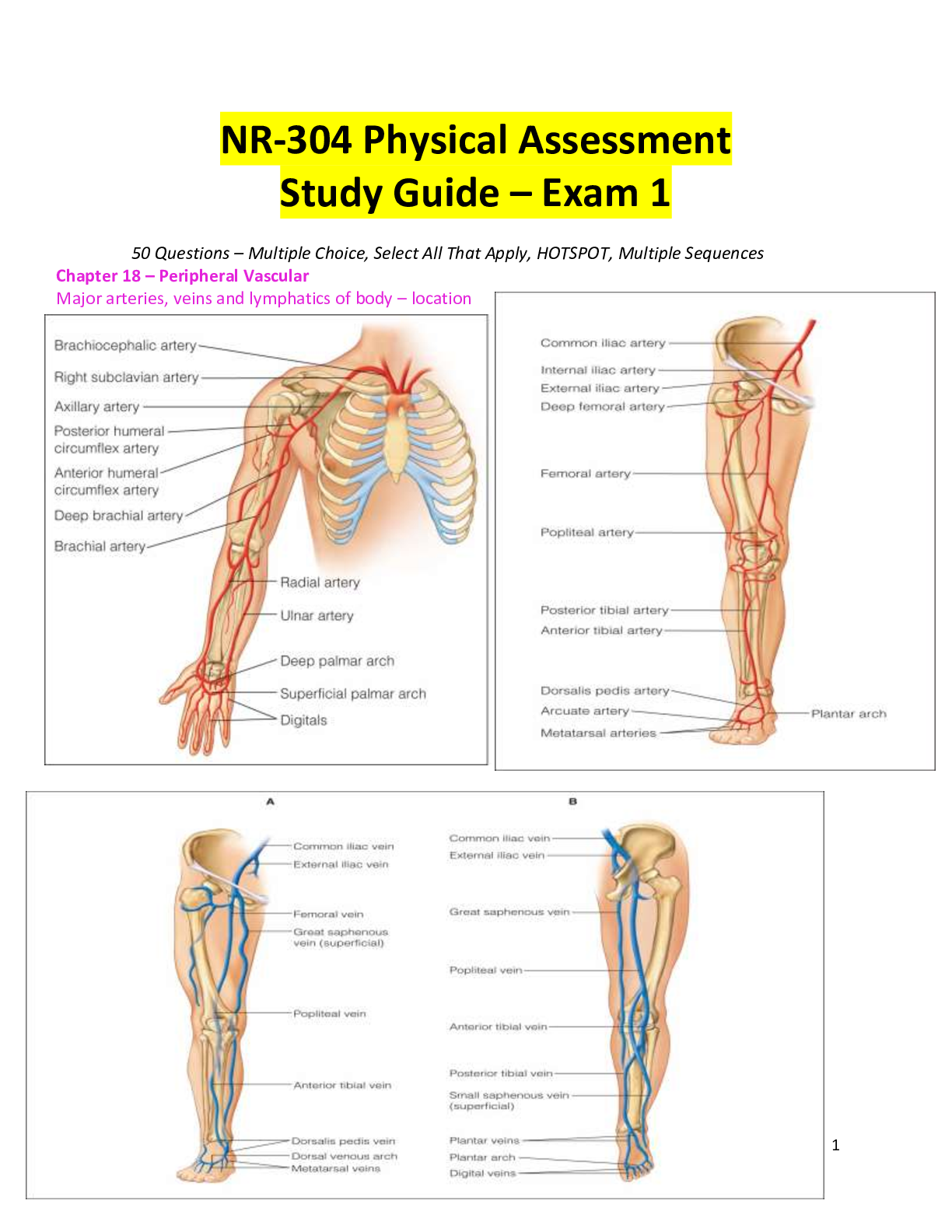
Structure and Function – Arteries in the Arm: the major artery supplying the arm is the brachial
artery, which runs in the biceps-triceps furrow of the upper arm and surfaces at the antecubital fossa in the
elbow medial to the biceps tendon. Immediately below the elbow the brachial artery bifurcates into the
ulnar and radial arteries. These run distally and form two arches supplying the hand; these are called the
superficial and deep palmar arches. The radial pulse lies just medial to the radius at the wrist; the ulnar
artery is in the same relation to the ulna, but it is deeper and often difficult to feel.
The function of arteries is to supply oxygen and essential nutrients to tissues
Complete blockage leads to death of distal tissue
Partial blockage creates an insufficient supply, and ischemia may be apparent only at exercise when
oxygen needs increase
Structure and Function: Arteries in the Leg – the major artery to the leg is the femoral artery, which
passes under the inguinal ligament. The femoral artery travels down the thing. At the lower thigh it
courses posteriorly; then it is termed the popliteal artery. Below the knee the popliteal artery divides. The
anterior labial artery travels down the front of the leg to the dorsum of the foot, where it becomes the
dorsalis pedis. In back of the leg the posterior tibial artery travels down behind the medial malleolus and
forms the plantar arteries in the food.
The function of the arteries is to supply oxygen and essential nutrients to the tissues. Ischemia is a
deficient supply of oxygenated arterial blood to a tissue caused by obstruction of the blood vessel. A
complete blockage leads to death of the distal tissue. A partial blockage creates an insufficient
supply, and the ischemia may be apparent only at exercise when oxygen needs increase. Peripheral
artery disease PAD affect noncoronary arteries and usually refers to arteries supplying the limbs.
*** Please note that this is meant to serve as an augmentation tool. Students ARE subject to be tested any material
related to readings, simulations, labs, assignments and activities within the course. ***
1
Exam Study Guide
It usually is caused by atherosclerosis, and less commonly by embolism, hypercoagulable states, or
arterial dissection.
b. Veins – The course of the veins is parallel to the arteries, but the direction of flow is opposite; the veins
absorb CO2 and waste products from the periphery and carry them back to the heart. The body has more
veins, and they lie closer to the skin surface.
Jugular Veins – the jugular veins empty unoxygenated blood directly into the superior vena cava.
Because no cardiac valve exists to separate the superior vena from the right atrium, the jugular veins give
information about activity on the right side of the heart. Specifically they reflect filling pressure and
volume changes. Because volume and pressure increase when the right side of the heart fails to pump
efficiently, the jugular veins expose this.
Veins in the Arm – each arm has two sets of veins: superficial and deep. The superficial veins are in the
subcutaneous tissue and are responsible for most of the venous return.
Vein in the Leg – the legs has 3 types of veins
1. The deep veins run alongside the deep arteries and conduct most of the venous return from the legs.
These are femoral and popliteal veins. As long as these veins remain intact, the superficial veins can be
excised without haring he circulation.
2. The superficial veins are the great and small saphenous veins. The great saphenous veins, inside the
leg, starts at the medial side of the dorsum of the foot. You can see it ascend in front of the medial
malleolus; then it crosses the tibia obliquely and ascends along the medial side of the thigh. The small
saphenous veins, outside the leg, starts on the lateral side of the dorsum of the foot and ascends behind the
lateral malleolus, up the back of the leg, where it joins the popliteal vein. Blood flows from the superficial
veins into the deep leg veins.
3. Perforators are connecting veins that join the two sets. They also have one-way valves that route blood
from the superficial into the deep veins and prevent reflux to the superficial veins.
Structure and Function: Venous Flow Veins drain the deoxygenated blood with its waste products from
the tissues and return it to he heart. Unlike the arteries, veins are a low-pressure system. Because they do
not have pump to generate their blood flow, they need a mechanism to keep blood moving. This is
accomplished by:
1. the contracting skeletal muscles that milk the blood proximally, back toward the heart.
2. the pressure gradient caused by breathing, in which inspiration makes the thoracic pressure decrease
and the abdominal pressure increase.
3. the intraluminal valves, which ensure unidirectional flow.
Each valve is a paired semilunar pocket that opens toward the heart and closes tightly when filled to
prevent back flow of blood.
[Show More]


 Exam 1 Concept Study Guide MY21.png)
.png)