Unit 1
Visual
• Eye accommodation – pupils will constrict when moving an object towards the nose
• Light – pupils constrict
• Dark – pupils dilate
• Cataract development
o Cataract = opacity within the lens
o Risk
...
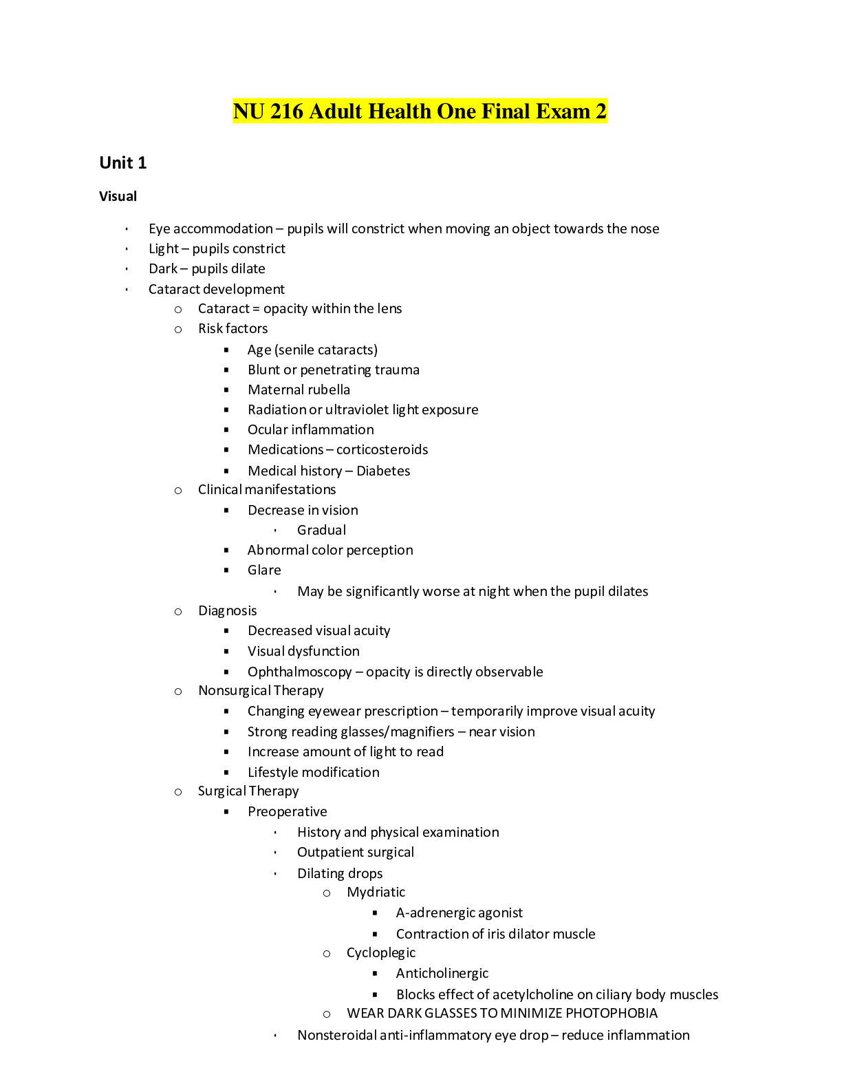
Unit 1
Visual
• Eye accommodation – pupils will constrict when moving an object towards the nose
• Light – pupils constrict
• Dark – pupils dilate
• Cataract development
o Cataract = opacity within the lens
o Risk factors
▪ Age (senile cataracts)
▪ Blunt or penetrating trauma
▪ Maternal rubella
▪ Radiation or ultraviolet light exposure
▪ Ocular inflammation
▪ Medications – corticosteroids
▪ Medical history – Diabetes
o Clinical manifestations
▪ Decrease in vision
• Gradual
▪ Abnormal color perception
▪ Glare
• May be significantly worse at night when the pupil dilates
o Diagnosis
▪ Decreased visual acuity
▪ Visual dysfunction
▪ Ophthalmoscopy – opacity is directly observable
o Nonsurgical Therapy
▪ Changing eyewear prescription – temporarily improve visual acuity
▪ Strong reading glasses/magnifiers – near vision
▪ Increase amount of light to read
▪ Lifestyle modification
o Surgical Therapy
▪ Preoperative
• History and physical examination
• Outpatient surgical
• Dilating drops
o Mydriatic
▪ A-adrenergic agonist
▪ Contraction of iris dilator muscle
o Cycloplegic
▪ Anticholinergic
▪ Blocks effect of acetylcholine on ciliary body muscles
o WEAR DARK GLASSES TO MINIMIZE PHOTOPHOBIA
• Nonsteroidal anti-inflammatory eye drop – reduce inflammation
▪ Intraoperative
• Phacoemulsification – small incision is made in the surface of the eye in or near the cornea, ultrasonic vibrations dissolve the clouded lens,
particles are suctioned out
• Extracapsular cataract extraction procedure – cataract is removed in one piece
o Advanced cataracts when lens is too dense
• IOL lens – posterior chamber lens implanted in capsular bag behind iris
▪ Postoperative
• Antibiotic drops – prevent infection
• Corticosteroid drops – decrease postoperative inflammatory response
• Activity restrictions – bending, stooping, coughing, lifting
o Health Promotion
▪ Wear sunglasses
▪ Avoid extraneous or unnecessary radiation
▪ Maintain appropriate intake of antioxidant vitamins – C and E
• Macular degeneration development
o Most common cause of irreversible central vision loss
o 2 forms
▪ Dry (nonexudative)
• Most common
• Close vision tasks difficult
• Macular cells atrophy – slowly progressive and painless vision loss
• Abnormal accumulation of yellowish extracellular deposits (drusen) in retinal pigment epithelium
• Acute vision loss
▪ Wet (exudative)
• More severe
• Accounts for AMD-related blindness
• More rapid onset of vision loss
• Development of abnormal blood vessels in or near macula
o May leak fluid and bleed – scar tissue
• Acute vision loss
o Risk factors
▪ Retinal aging
▪ Genetics and family history
▪ White ethnicity
▪ Chronic inflammation conditions
▪ Smoking
▪ Hypertension
o Reduce risk
▪ Increase intake of dark green, leafy vegetables with lutein (kale/spinach)
▪ Vitamin and mineral supplements
▪ Smoking cessation
o Clinical manifestations
▪ Blurred and darkened vision
▪ Scotomas – blind spots in visual field
▪ Metamorphopsia – distortion of vision
o Interprofessional Care
▪ Limited treatment options – wet AMD
• Medications injected into vitreous cavity to help slow vision loss
o Side effects
▪ Blurred vision
▪ Eye irritation
▪ Eye pain
▪ Photosensitivity
o Given at 4- or 6-week intervals
• Photodynamic therapy – damages abnormal blood vessels
o Avoid direct exposure to sunlight and other intense forms of light for 5 days after treatment
Pressure Injuries and Wound Care
• Pressure Injury
o Localized injury to skin and/or underlying tissue
o Result of pressure with or without shear
o Development factors
▪ Amount of pressure (intensity)
▪ Length of time the pressure is exerted (duration)
▪ Ability of patient’s tissue to tolerate the pressure
▪ Shearing force – pressure on skin when it adheres to bed and skin layers slide in direction of body movement
▪ Excessive moisture
o Risk factors
▪ Increased age
▪ Incontinent
▪ Unable to reposition
▪ Bed- or wheelchair-bound
• Staging
o Stage 1
▪ Intact skin with nonblanchable redness
▪ Usually over a bony prominence
▪ Area may be painful, firm, soft, warmer/cooler compared to adjacent tissue
o Stage 2: Partial Thickness
▪ Shallow open ulcer with red-pink wound bed – no slough
▪ Intact/open/ruptured serum-filled or serosanguineous-filled blister
▪ Shiny or dry shallow ulcer without slough or bruising
o Stage 3: Full-Thickness Skin Loss
▪ Subcutaneous fat may be visible – no bone/tendon/muscle
▪ Slough, undermining, tunneling may be present
o Stage 4: Full-Thickness Tissue Loss
▪ Exposed bone/tendon/muscle
▪ Slough, eschar, undermining, tunneling may be present
o Unstageable
▪ Full-thickness tissue loss
▪ Depth is obscured by slough/eschar
o Suspected Deep Tissue Injury
▪ Purple/maroon localized area intact skin
▪ Blood-filled blister
▪ Due to damage of underlying soft tissue from pressure and/or shear
• Undermining
o Tissue under the wound edges becomes eroded, results in a pocket beneath the skin at the wound’s edge
• Slough
o Consequence of inflammatory phase of wound healing
o Comprises dead white blood cells, fibrin, cellular debris, and liquefied devitalized tissue
• Tunneling
o Wound channels that extend from a wound into and through subcutaneous tissue/muscle
• Eschar
o Slough/piece of dead tissue that is cast off from the surface of the skin
• Maceration
o Softening and breakdown of skin from prolonged exposure to moisture
• Dehiscence
o Separation and disruption of previously joined wound edges
• Evisceration
o Wound edges separate to the extent that intestines protrude through wound
• Wound healing
o Primary intention – wound margins neatly approximated
▪ 3 phases
• Initial – 3 to 5 days
o Edges of incision are aligned and sutured/stapled in place
o Incision area fills with blood from cut blood vessels, blood clots form, platelets release growth factors
o Acute inflammatory reaction occurs
• Granulation phase – 5 days to 4 weeks
o Proliferating fibroblasts, proliferating capillary sprouts (angioblasts), WBCs, exudate, loose/semifluid/ground substance
o Wound is pink and vascular, at risk for dehiscence
• Maturation and Scar Contractions – 7 days to several months
o Secondary intention
▪ Wounds that have edges that cannot be approximated
▪ Debris may have to be cleaned away before healing can take place
▪ Greater defect and gaping wound edges
▪ Healing and granulation take place from edges inward and from bottom upward until defect is filled
▪ More granulation tissue and larger resulting scar
o Tertiary Intention
▪ Delayed suturing of a wound
▪ Two layers of granulation tissue are sutured together
▪ Contaminated wound is left open and sutured closed after infection is controlled
▪ Primary wound becomes infected, opens, granulates, sutured
▪ Results in larger/deeper scar
• Impediments to wound healing
o Malnourished
o Lack of protein – decreases supply of amino acids for tissue repair, albumin
o Diabetics – decreases collagen synthesis, delays capillary growth, impairs phagocytosis, reduces supply of oxygen and nutrients secondary to vascular disease
........................................CONTINUED......................................
[Show More]