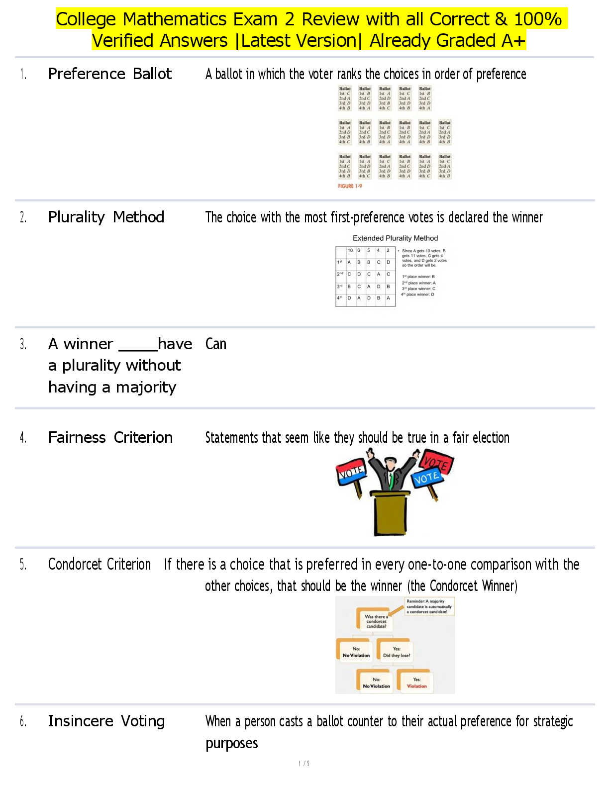
HESI Exit RN Exam Over 700 Questions, Answers Rationale New 2021-2022 latest 100%.
Biology > EXAM REVIEW > APII Module 5 (All)
Kidney Anatomy An adult kidney is bean-shaped, about the size of a can of soup and weighs about 5 ounces. The right kidney (because of the liver's location just above it) lies slightly lower than t ... he left and both are protected by the rib cage since they are located in the lumbar region between the T-12 and L-3 vertebra. The kidney's medial surface is concave and has a cleft called the renal hilus leading to a space within the kidney called the renal sinus. The ureters, blood vessels, and nerves are located in the sinus and entering the kidney at the hilus. On top of each kidney is an adrenal gland. The kidney surface is protected by three layers of specialized tissue. The renal capsule is a tough fibrous outer skin of the kidney which protects it from injury and infection. Outside of the renal capsule is a fatty layer that protects the kidney from trauma which is called the adipose capsule. The outer renal fascia is dense fibrous connective tissue which keeps the kidney in place inside the abdominal cavity. The three separate regions evident in a vertical section of the kidney are the cortex, the medulla, and the pelvis. The outer renal cortex, just inside the renal capsule, is a continuous outer region with a number of projections (cortical columns) that extend down between the renal medulla pyramids. Located within the cortex are the glomerular capsule and the distal and convoluted tubule sections of the nephrons along with associated blood vessels. Deeper within the kidney lies the renal medulla which is divided into sections called pyramids that point toward the center of the kidney. Located within the medulla are the Loop of Henle and the collecting duct sections of the nephrons along with associated blood vessels. The centermost section of the kidney near the renal hilus is the renal pelvis which constitutes a funnel- shaped tube that connects to the ureter as it leaves the hilus. Several extensions of the pelvis called calyces collect urine which drains continuously into the renal pelvis and subsequently into the ureter, which transports the urine to the bladder to be stored. Blood and Nerve Supp ly The kidneys are innervated by many blood vessels so that they can filter the blood to regulate its composition with the renal arteries delivering about 1200 ml of blood per minute of blood directly from the abdominal aorta which amounts to 20% of the cardiac output. The renal arteries branch into 5 segmental arteries which divide further into lobar arteries then further into interlobar arteries which pass between the renal pyramids. The interlobar arteries then divide into the arcuate arteries which branch into several interlobular arteries that feed into the afferent arterioles that supply the glomeruli. After filtration occurs the blood moves into the efferent arterioles and either the peritubular or vasta recta capillaries and then drain into interlobular veins which converge sequentially into arcuate then interlobar veins then to the renal vein which exits the kidney. ....................................................................CONTINUED............................................................... [Show More]
Last updated: 3 years ago
Preview 1 out of 25 pages

Buy this document to get the full access instantly
Instant Download Access after purchase
Buy NowInstant download
We Accept:

Can't find what you want? Try our AI powered Search
Connected school, study & course
About the document
Uploaded On
Jul 01, 2021
Number of pages
25
Written in
All
This document has been written for:
Uploaded
Jul 01, 2021
Downloads
0
Views
75
Scholarfriends.com Online Platform by Browsegrades Inc. 651N South Broad St, Middletown DE. United States.
We're available through e-mail, Twitter, Facebook, and live chat.
FAQ
Questions? Leave a message!
Copyright © Scholarfriends · High quality services·

.png)