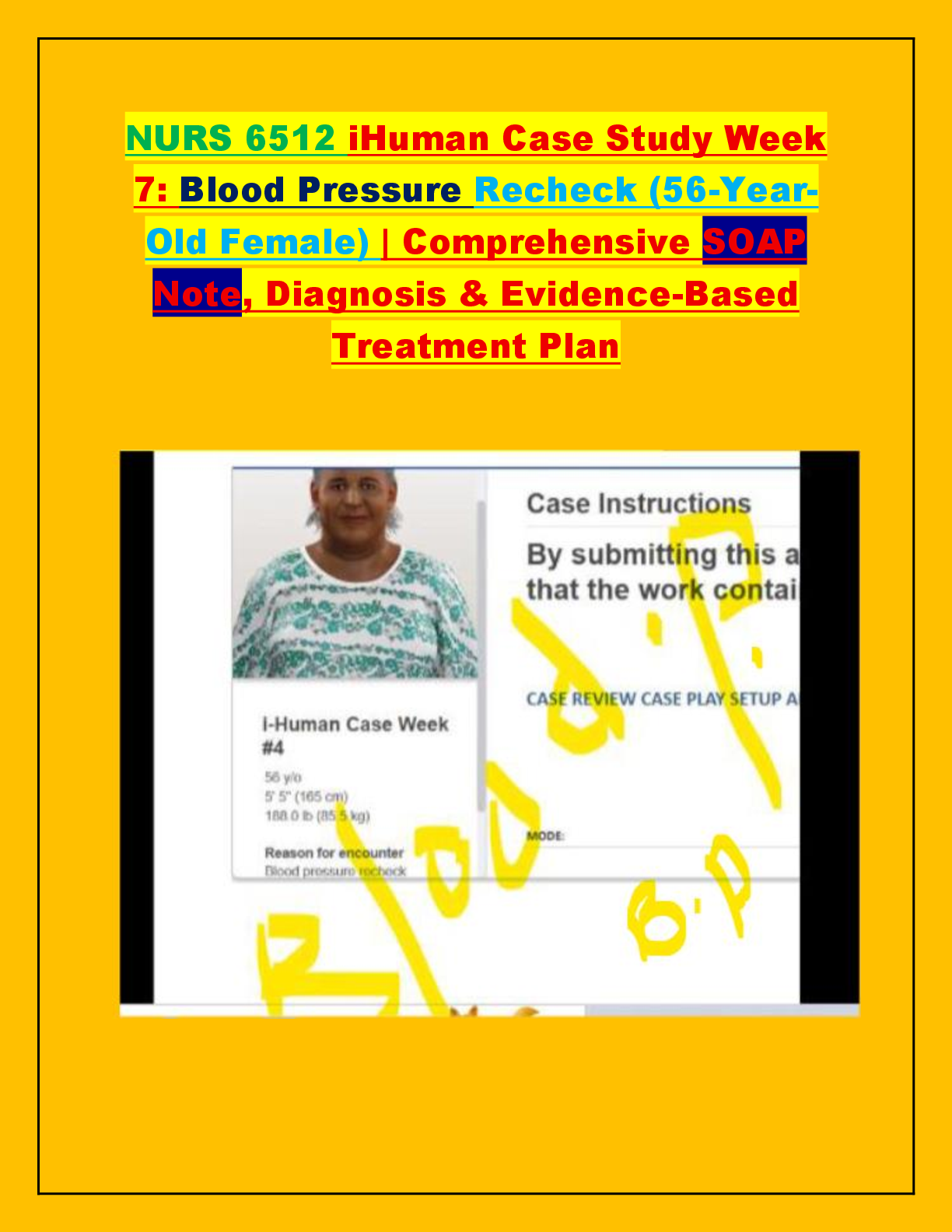
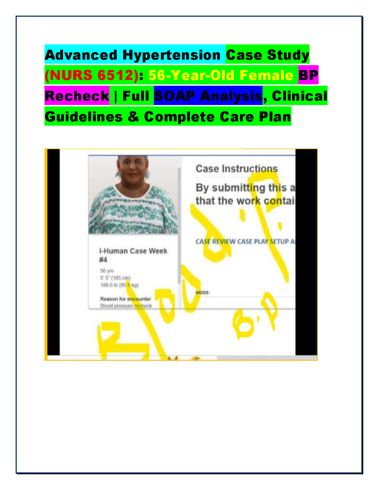
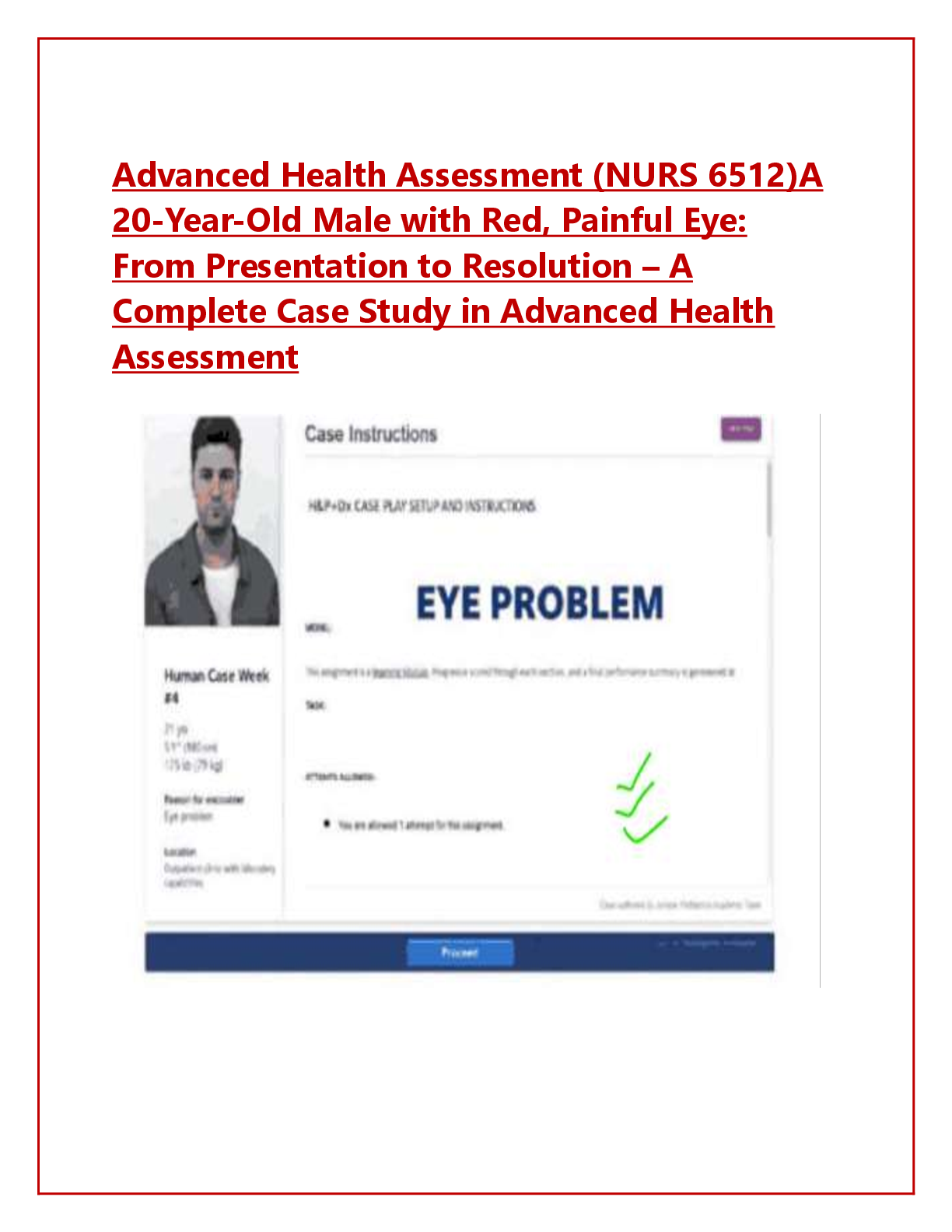
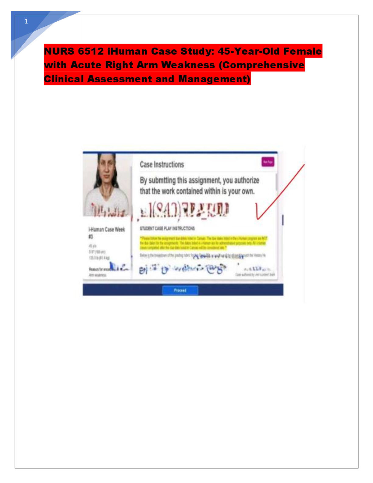
NURS 6512N; Week 8 - Case Study Guide (Latest 2019) Already Graded A.
Document Content and Description Below
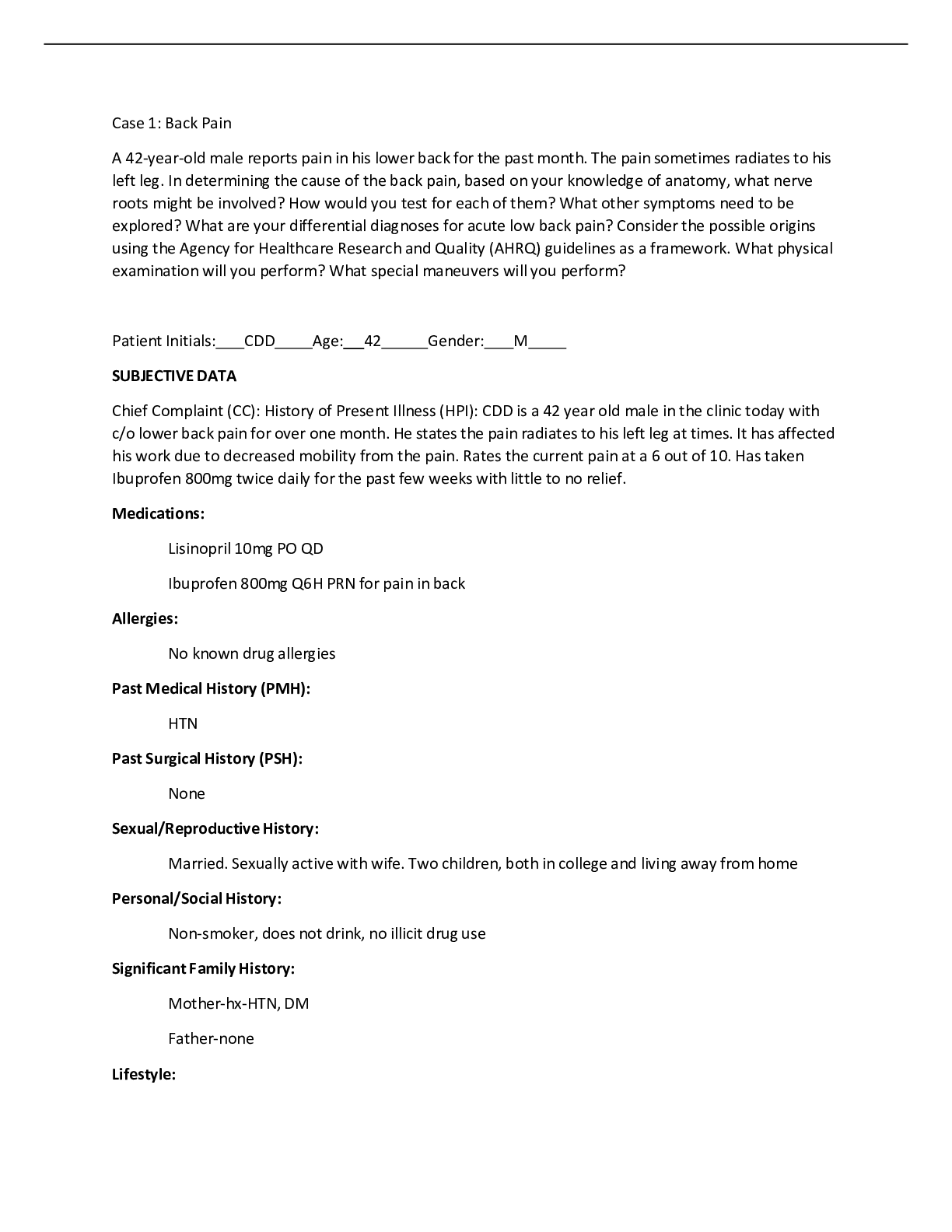
Case 1: Back Pain
A 42-year-old male reports pain in his lower back for the past month. The pain sometimes radiates to his left leg. In determining the cause of the back pain, based on your knowledge of anatomy, what ne
...
[Show More]
Last updated: 3 years ago
Preview 1 out of 4 pages
Instant download
Buy this Document to get the Full Access Instantly
Provided by Students Who Aced it
We Verify Document Content to Gurantee Accuracy
Reviews( 0 )
Document information
Connected school, study & course
About the document
Uploaded On
Sep 23, 2021
Number of pages
4
Written in
All

Additional information
This document has been written for:
Uploaded
Sep 23, 2021
Downloads
0
Views
315
Document Keyword Tags
Recommended For You
Get more on Summary »
$10
4 Pages
NURS-6512N / NURS 6512 / NURS6512, Advanced Health Assessment...

$11.5
26 Pages
NURS 6512 / NURS6512 Midterm Study Guide (Latest): Advanced He...

$13
65 Pages
NURS 6512 / NURS6512 Exam #1 Study Guide (Latest): Advanced He...
$18.5
23 Pages
NURS 6512 / NURS6512: Advanced Health Assessment and Diagnosti...

$15
15 Pages
NURS-6512N-53, Advanced Health Assessment.2020 FINAL EXAM
$10
16 Pages
NURS-6512N / NURS 6512 / NURS6512, Advanced Health Assessment...
$10
26 Pages
NURS-6512N / NURS 6512 / NURS6512, Advanced Health Assessment...

$10
17 Pages
NURS-6512N-8/NURS-6512C-8-Advanced Health Assessment Winter 20...
$9
20 Pages
NURS-6512N-8/NURS-6512C-8-Advanced Health Assessment2019 Winte...

$15
5 Pages
Week 5 Discussion NURS 6512 Case # 2 Lily 20-year-old Caucasia...

$10
5 Pages
NURS 6512 Week 5 Episodic/Focused SOAP Note Template > 2021

$3
3 Pages
Test Bank Lehnes Pharmacotherapeutics for Advanced Practice Nu...

$10
49 Pages
Summary Pathophysiology Final Exam Study Guide_all chapters (1...

$10
10 Pages
NR 509 Week 4 Chest Pain SOAP Note (version 1) Complete Spring...
More related documents below