Chapter25 Assessment of Cardiovascular Function
Overview of Anatomy and Physiology
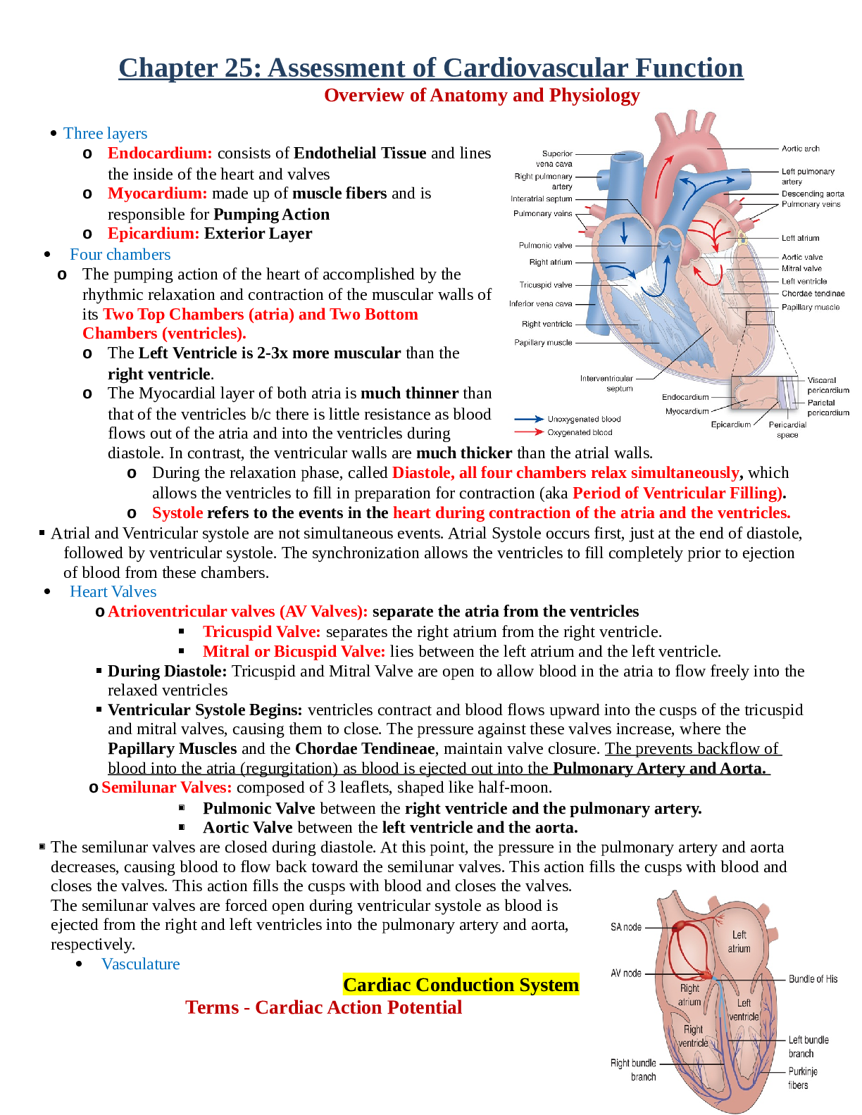
• Three layers
o Endocardium: inside layers
o Myocardium: made up of muscle fibers and is responsible for the pumping actions
o Ep
...
Chapter25 Assessment of Cardiovascular Function
Overview of Anatomy and Physiology
• Three layers
o Endocardium: inside layers
o Myocardium: made up of muscle fibers and is responsible for the pumping actions
o Epicardium: outer layers
o Fluids can be accumulate in these layers
• Four chambers
o 2 atria and 2 ventricles
o Hemodynamic: the use of pressure monitoring devices to directly measure cardiovascular function
• Heart valves
• Chambers
• Heart Valves
o Atrioventricular valves
o Semilunar valves
• Vasculature
• Perfusion: oxygenation and flow
Cardiac Conduction System
Cardiac conduction system generates and transmits electrical impulses that stimulate contraction of the myocardium. Under normal circumstance, the conduction system first stimulates contraction of the atria and then the ventricles.
SA node (peacemaker) AV node Bundles of His Left & right bundle branch Purkinje fibers
Terms - Cardiac Action Potential
• Depolarization: electrical activation of cell caused by influx of sodium into cell while potassium exits cell
• Repolarization: return of cell to resting state caused by re-entry of potassium into cell while sodium exits
• Refractory periods
o Myocardial cells must completely repolarize before they can depolarize again. During the repolarization process. Two phases
o Effective refractory period: the cells is completely unresponsive to any electrical stimulus; phase in
which cells are incapable of depolarizing
▪ Corresponds with the time in phase 0 to the middle of phase 3 of the action potential
o Relative refractory period: phase in which cells require stronger-than-normal stimulus to depolarize
▪ Corresponds with the short time at the end of phase 3
Cardiac Action Potential
23.4: Cardiac action potential of a fasting-response Purkinje fiber. The arrows indicate the approximate time and direction of movement of each ion influencing membrane potential. Ca++ movement out of the cell is not well defined but is thought to occur during phase 4.
Great Vessel and Heart Chamber Pressures
25.2 : great vessel and chamber pressures. Pressures are identified in millimeters of mercury (mm Hg) as mean pressure or systolic or systolic over diastolic pressure.
Cardiac Hemodynamics CO= HR X SV
Hemodynamics
Terms - Cardiac Output
• Stroke volume: amount of blood ejected with each heartbeat
o Average resting stoke volume is about 60 to 130 mL
• Cardiac output: amount of blood pumped by ventricle in liters per minute
o In a resting adult is 4 to 6 L/min normally
• Preload: degree of stretch of cardiac muscle fibers at end of diastole
o Preload is referred to as left ventricular
o End of diastole is the period when filling volume in the ventricles is the highest and the degree of stretch of the muscle fibers is the greatest
o Less blood return to the heart reduced preload
o IV fluids will increase preload
• Contractility: ability of cardiac muscle to shorten in response to electrical impulse
o Increased contractility result in increased stroke volume
o Contractility is depressed by acidosis, hypoxemia, and certain medication such as beta blockers
o Enhanced SNS, such as digoxin, epinephrine increase contractility
• Afterload: resistance to ejection of blood from ventricle
o Arterial vasodilation decrease afterload increase stroke volume
o Arterial vasoconstriction increase afterload decrease stoke volume
• Ejection fraction: percent of end diastolic volume ejected with each heartbeat
o Ejection fraction of normal left ventricle is 55% to 65%
o Used as a measure of myocardial contractility
o Measured by echocardiogram
*Low heart rate will affect the cardiac output
*Stroke volume, if anything is affecting contractility the heart is not contracting, or heart is not stretching/pumping will affect the cardiac output heart is not perfuse b/c CO is low
Age-Related Changes of the cardiac System
• Atria
• Left ventricles
o Myocardial thickening (hypertrophy) stiff and less compliant, and progressive decline cardiac
output = fatigue, decrease exercise tolerance, s4 may be present
o Left side heart failure- auscultating lungs sounds is cracks. Possible wheeze and gurgles
• Valves
o Thickening or rigidly of the AV valves abnormal blood flow across the valves during cardiac
cycle = murmurs may be present
• Conduction system
o Loss function slow heart rate
• Sympathetic nervous system
• Aorta and arteries
• Baroreceptor response
Health History
• Chief complaint
• History of Present Illness
• Past medical, surgical history
• Past family history
• Past social history
• Home medications
• Nutrition
• Allergies
Physical Assessment
• General appearance
o Evaluate the patient’s LOC & mental status
o Changes may be attributed to inadequate perfusion of the brain from a compromised cardiac output of thromboembolic event (stroke)
o Observe for signs of distress (pain, discomfort, SOB, anxiety)
o Measure height and weight
• Skin and extremities
o Skin color, temperature, and texture for problems with arterial or venous circulation
• Blood pressure
• Arterial pulses
• Jugular venous pulsations (JVD)
o Right side heart failure fluid overload
o What does patient taking to improve fluid overload?
• Heart inspection and auscultation
• General assessment
• Any deviations 偏差 from the normal?
o Do you have any change in activity tolerance?
o Heart as a pump
o Atrial/ventricular filling volumes
o Cardiac output
o Compensatory mechanisms
▪ Did they change their shoes because their legs are swollen?
▪ What position are they are laying to sleep?
▪ Tripod position: patient falling a sleep on a chair - one sits or stands leaning forward and
supporting the upper body with hands on the knees or on another surface.
*right sided heart failure= Ascites, hepatojugular reflex, distention
Most Common Clinical Manifestations
• Chest pain
• Dyspnea
• Peripheral edema, weight gain, abdominal distention
• Palpitations
• Fatigue
• Dizziness, syncope, changes in level of consciousness
o Why would a patient be dizzy? Cardio output and perfusion issues
o LOC- due lack of oxygen to the brain
Chest Pain
• Chest pain and chest discomfort are common symptoms that may be caused by a number of cardiac and noncardiac problems summarizes the characteristics and patterns of common of chest pain or discomfort.
• Identify quantity of pain (0-10 SCALE)
• Identify location of pain
• Identify quality of pain
• Radiation of pain
o Chest pain can radiate to left side, jaw, epigastrium, back, shoulder
• Associated signs/symptoms (diaphoresis or nausea)
• Duration of pain
o Severity or duration of chest pain does not predict the seriousness of its cause. For example, when asked to rate pain using a 0 to 10 scale, patient with esophageal spasm may rate as 10. In contrast, patient with MI, which is a potentially life-threatening event, may report 4 to 6.
• Assess for other cardiac conditions
• Assess for other significant conditions (see p. 663-664)
o Pneumonia, pulmonary embolism
o Hiatal hernia, GERD
▪ Doctor will order labs or endoscopy on the patient to rules out GERD instead of heart attack
o Costochondritis
o Vascular
Assessment
• Medications
o Aspirin, a nonprescription medication is an important antithrombotic therapy for secondary prevention in patients recovering from ACS (acute chest syndrome)
• Nutrition
o Dietary modification, exercise, weight loss, and careful monitoring are important strategies for managing three major cardiovascular risk factors: hyperlipidemia, hypertension, and diabetes.
o Diet restriction: sodium, fat, cholesterol, or calories.
o Height + weight
o BMI (assessment for obesity)
o Lab result: glucose, glycosylated Hb (diabetes), cholesterol, HDL, LDL, triglycerides
o Patient food habits and culture preference
• Elimination
o Typical bowel and bladder habits need to be identified
o Nocturia (awakening at night to urinate) is common with HF patient
o Vagal maneuvers: try to slow down the fast heart rate by stimulate the vagal nerve
increase pressure to the baroreceptor to slow down HR
▪ Bearing down means that you try to breathe out with your stomach muscles but you don't let air out of your nose or mouth.
• Activity, exercise
o Changes in the patient’s activity tolerance are often gradual and may go unnoticed.
o Compare the recent changes in the patient current activity level with that performed in the past 6 to 12 months.
o Activity-induced angina or shortness of breath may indicated CAD.
▪ Symptoms occur when myocardial ischemia is present, due to an inadequate arterial blood supply to the myocardium, in the setting of increased demand (exercise, stress or anemia)
o Fatigue: associated with a low left ventricular ejection fraction (less than 40%)
o Medication (beta-adrenergic blocking agents) can result in activity intolerance.
• Sleep, rest
o Clues to worsening cardiac disease, especially HF, can be revealed by sleep-related events.
o Patient with worsening HF often experience orthopnea (need to sit upright or stand to avoid feeling short of breath).
▪ Need to sleep upright in chair or add extra pillows to bed
o Paroxysmal nocturnal dyspnea- sudden awakening with shortness of breath = symptom of worsening HF
• Self-perception, self-concept
o Quit smoking to reduce risk of future cardiovascular related health problems
• Roles, relationships
o assess the pts support system (esp bc many invasive cardiac procedures like cardiac catheterization and percutaneous coronary intervention (PCI) are being performed as outpatient procedures.
• Sexuality, reproduction
o Erectile dysfunction may develop in men as a side effect of cardiac meds (beta blockers)- may cause some men to stop taking it!
• Coping, stress tolerance
o Anxiety, depression, and stress are known to influence both development of and recovery from CAD and HF
o High levels of anxiety are associated with an increased incidence of CAD
• Prevention strategies
Health Promotion, Perception, and Management Questions
• Ask regarding health promotion, preventive practices
o What type of health issues do you have? Are you able to identify any family history or behaviors that put you at risk of this health problem?
o What are your risk factors for heart disease? What do you do to stay healthy?
o How is your health? Have you noticed any changes?
• Ask regarding health promotion, preventive practices
o Do you have a cardiologist or primary health care provider? How often do you go for check-ups?
o Do you use tobacco or alcohol?
o What medications do you take?
Cardiac Specific
• General appearance
• Assessment of skin/extremities
• BP
o Pulse pressure = systolic – diastolic normally 30 – 40
▪ Decreased pulse pressure reflects reduced stroke volume and ejection velocity
(shock, HF, hypovolemia, mitral regurgitation) or obstruction to blow flow during systole (mitral or aortic stenosis)
o Postural blood pressure changes postural/orthostatic hypotension in pts with CVD most
often due to sig. reduction in preload, which compromises cardio output.
▪ Decrease BP within 3 minutes of moving from a lying or sitting to a standing position. Accompanied by dizziness, lightheadedness, or syncope.
▪ Take BP laying, sitting, standing!
• Arterial pulses
o Pulse rate
▪ Normal 60- 100 bpm
o Pulse rhythm
▪ Regular or irregular changes in the EKG
o Pulse amplitude
▪ How much forces do the pulse, tells about volume fluid overload or dehydration
▪ indicative of the BP in the artery, is used to assess peripheral arterial circulation
▪ 0: Not palpable or absent (use a doppler)
▪ +1: Diminished—weak, thready pulse; difficult to palpate; obliterated with pressure
▪ +2: Normal—cannot be obliterated
▪ +3: Moderately increased—easy to palpate, full pulse; cannot be obliterated
▪ +4: Markedly increased—strong, bounding pulse; may be abnormal
• Jugular venous distention
o Indicative of fluid overload, an assessment done with heart failure specifically
o Right side heart function can be estimated by observing the pulsation of the jugular veins of the neck, which reflects central venous pressure (CVP).
▪ CVP is the pressure in the right atria of the right ventricle at the end of diastole.
o The abnormality is observed is patients with right-sided HF, due to hypervolemia, pulmonary hypertension, and pulmonary stenosis (pulmonary valve between the right ventricle & pulmonary artery is too small and narrowed)
• Heart inspection/palpation
• Heart auscultation
o Normal heart sounds
▪ S1, S2 (AV closure, semilunar closure)
o Abnormal heart sounds
▪ S3, S4 gallops
▪ Opening snaps, systolic clocks, and murmurs
▪ S3, S4 gallops are heard during diastole
▪ S3 (DUB) indicated with heart failure or fluid overload is heard early in diastole during the period of rapid ventricular filling as blood flows from the atrium into a
complain ventricle
▪ S4 (LUB) specific for coronary artery disease & hypertension
• Assessment of lungs
o Hemoptysis 咳血 pink/frothy = pulmonary edema
o Coughs, crackles, wheezes
▪ Crackles is the first heard at the base of the lung
▪ Wheezes can cause by nonselective beta blockers narrowing airway
• Assessment of abdomen
o Abdominal distention indicative of ascites
o Bladder distention- reduced urine output may indicate inadequate renal perfusion
o Hepatojugular reflux- this test is performed when right ventricular or biventricular HF is suspected
Diagnostic Interpretation
Laboratory Evaluation
• Assists in making a diagnosis
• Screens for risk factors associated with CAD
• Establishes baseline values
• Monitor responses to therapy
• Assess for abnormalities that affect prognosis
Laboratory Tests: Cardiac Biomarkers
• Myocardial cells that become necrotic from prolonged ischemia or trauma release specific enzymes:
o Creatinine Kinase (CK)
o Creatinine Kinase Isoenzymes (CK-MB)
▪ Heart muscle
▪ Cardiac-specific isoenzyme; it found mainly in cardiac cell and therefore increase when there has been damage to these cells
▪ Indicator of acute MI
▪ The levels begin to increase within a few hours and peaks within 24 hours of an infarct
▪ Returns to normal in 3 to 4 days
▪ CK–MB range from 3 to 5% (percentage of total CK) or 5 to 25 IU/L
o Proteins
▪ Myoglobin
• Increase within 1 to 3 hours and peaks within 12 hours after the onset of symptoms
• Increase myoglobin (early as 1 hour) is not very specific in indicating an acute cardiac event; however, negative result can be used to rule out an acute MI
• Normal range is 25 to 72 ng/mL (1.28 to 3.67 nmol/L) OR 5-70???
▪ Troponin T and Troponin I
• Biomarkers are currently recognized as reliable and critical markers of myocardial injury.
• An increase in the level of troponin in the serum can be detected within a few hours during ACUTE MI.
• It REMAINs elevated for a long period, often as long as 2 weeks, and it therefore can be used to detect recent myocardial damage
• Normal range: 0 and 0.4 ng/mL
o Abnormally high levels of these substances can be detected in serum blood samples
Laboratory Tests: Hematology, Chemistry, Coagulation Studies pg. 693
• BUN and Creatinine
o BUN: 10- 20
o Creatinine: 0.7 -1.3
o BUN and creatinine were flush the dye for the test
• Potassium: 3.6 -5.2
o Medication can reduce potassium hypokalemia dysrhythmia
• Sodium: 135-145
• Lipid profile
o Total cholesterol 100-199
o Triglycerides 0-149
o LDL value 0-100 mg/dL
o HDL value > 49
• Brain (b-type) natriuretic peptide (BNP)
o Neurohormone that helps regulate BP and fluid volume
o Secreted from the ventricles in response to increase preload resulting elevated ventricular pressure
o BNP level increase as ventricle walls expands from pressure increase makes helpful for diagnostic, monitoring, and prognostic tool in the setting of HF.
o BNP level > 100 suggests HF
o BNP more specific to CHF**
• C-reactive protein
o Protein produced by liver in response to systemic inflammation
o People with high CRP (3 m/L or greater) may be at greatest risk for CVD
• Homocysteine
o An amino acid linked to development of atherosclerosis because it can damage the endothelial lining of arteries and promote thrombus formation.
o Elevated level = high risk CAD, stoke, PVD
o Test results are interpreted as optimal (less than 12 mcmol/L), borderline (12 to 15 mcmol/L), and high risk (greater than 15 mcmol/L)
• Blood chemistries
o BNP
• Coagulation studies
o PTT, PT, INR
o PT: 9.5- 12 seconds
o PTT: 60- 70 seconds
o INR: 1 (therapeutic range is 2- 3.5)
▪ generally, an effective therapeutic range for people taking warfarin for disorders such as atrial fibrillation or a blood clot in the leg or lung
▪ The determine if the anticoagulation given is working or not
o Also, patient goes for cardiac catheterization testing or PTCH testing, they may get medication in the procedure that might affect the function of PT, PTT and INR
• Hematologic studies
o CBC (WBC, Hgb, hematocrit)
o Hematocrit
▪ Male: 42 -52%; Female: 35- 47%
o Hemoglobin
▪ Male: 13- 18 g/dL; Female: 12- 16 g/dL
o Platelets: 150,000 – 45,000 (mm3)
Diagnostic Evaluation
Imaging
• Chest-x-ray
o Size, position, contour of heart
o Reveals cardiac and pericardial calcifications
o Assists in diagnosing other conditions
o Does not help diagnose acute MI, it can help diagnose some complication (HF)
Electrocardiography
• Electrical activity of the heart (12 leads, looks at 12 different views)
• Diagnoses
o Dysrhythmias
o Conduction abnormalities
o Chamber enlargement
o Myocardial ischemia, infarction, injury
o Electrolyte disturbances
Electrocardiography: Continuous ECG monitoring
• Standard of care for patient who at high risk for dysrhythmias
• Monitors more than one lead at a time
o Can have continuous monitoring with the 5 leads or 12 leads monitoring for patient are in emergency situation
• Monitors for ST segment changes
o Which are used to identify the presence of MI or injury (ST Depression)
o ST elevation provide evidence often evolving MI
• Provides visual and audible alarms
o Interprets and stores alarms
o Trends data over time
o Prints copy of rhythms from one or more specific ECG leads over a set time (called a rhythm strop)
Apply electrodes for cardiac monitoring( from PrepU)
1. Clip (do not shave) hair from around the electrode site
2. If patient is diaphoretic (sweaty), apply small amount of benzoin on the skin, avoiding the area the center of the electrode
3. Debride the skin surface of dead cell and water and dry well
4. Change electrode every 24- 48 hr
5. Examine the skin irritation and apply the electrode to different location
Example of 12-lead EKG (You do not need to memorize this!)
Cardiac Stress Testing
• Normally: cardiac arteries dilate to four times their usual diameter in response to increased metabolic demands for oxygen and nutrients
o Atherosclerotic vessels dilate less ischemia
o Abnormalities are likely to be detected during times of increased demand (stress)
• Cardiac stress test procedures are noninvasive ways to evaluate response to stress
o The stress test helps determine:
“To determine coronary arteries disease, to look at the cause of chest pain, and determine the medication prescribed are working”
Stress test determines the following: 1. Presence of CAD
• S4
2. Cause of chest pain
3. Functional capacity of the heart after an MI or surgery
4. Effectiveness of antianginal or antiarrhythmic medications
• To know if the drugs are working stress the heart at what point the patient has pain on the medication
5. Occurrence of dysrhythmias
6. Specific goals for physical fitness program
• Contraindications
o Severe aortic stenosis
o Acute myocarditis or pericarditis
o Severe HTN
o Suspected left main disease
o Heart failure
o Unstable angina
• Complications
o Acute MI (within 48 hours)
o Cardiac arrest
o Heart failure
o Unstable angina
• ACLS training required
Nursing Interventions BEFORE the stress testing
• NPO x 3-4 hours
• Avoid stimulants (caffeine, tobacco)
• Meds with sips of water
• May instruct to hold beta blocking agents, theophylline, aminophylline before test
o Beta blocker will lower the heart rate
• Appropriate attire 服装
• Sensations with medications
• If patient is nicotine pack on, try to avoid it
1. Exercise Stress Testing
o Walks on a treadmill (most common), bike, arm crank
o Exercise intensity increases the demands of the heart rate
o During the test, patient monitoring (EKG, VS, HR, ischemic changes, or symptoms including chest pain, dizziness, leg cramping and fatigue)
o This test is terminated when the target HR is achieved or patient experiences signs of myocardial ischemia
o Nurses need to monitor the patient and stop the exercise if patient experiences any signs and symptoms
2. Pharmacologic Stress Testing
o Patient who are cognitively impaired and unable to follow directions or physically disabled/deconditioned
1. Persantine and Adenocard given as an IV infusion
a. Used to mimic effects of stress by maximally dilating normal coronary arteries
2. Dobutamine is another option for use during a pharmacologic stress test.
a. Medications increases HR, myocardial contractility, and BP, thereby increasing the metabolic demands of the heart.
b. if no tolerance to exercise
“The GOAL of both stress test is increasing demand of the heart”
Cardiac Evaluations
• Transthoracic echocardiography
o Noninvasive ultrasound is used to measure ejection fraction and examine the size, shape and motion of cardiac structure.
• Transesophageal echocardiography
o NPO 6 hours
o Obtain Consent Form
o Ultrasound provides clear images of the heart
o The transducer is inserted into the mouth and the patient is asked to swallow several times until it is positioned in the esophagus
▪ Patient’s throat will be anesthetized, will withhold fluid and food until gag reflex return
o Invasive- internal visualization
o Ejection fraction
▪ Determine the amount of volume that’s left after the pump in the left ventricular
o Size, shape and motion of cardiac structures
o Pericardial effusions
▪ Collection of fluids
o Determines chamber size
o Etiology of heart murmurs
o Function of heart valves
o Ventricular wall motion
• Myocardial perfusion imaging
o It can show areas of the heart muscle that aren’t getting blood flow
o Commonly performed after an acute MI to determine if arterial perfusion to the heart is compromised during activity and to evaluate the extent of myocardial damage.
o Also elevate if MI from CAD is the cause of chest pain or CAD- related symptoms
o Perfusion: tissues are receiving blood
• Computerized tomography (CT)
o Form of cardiac imaging that uses x-rays to provide accurate cross-sectional “virtual” slices of specific area of the heart and surrounding structures
• MRI
o Looking at different lens of the ability to see different structures
• PET scan
o How much uptake of the dye is happening and where
o Used to determine muscle injury
Cardiac Catheterization
• Invasive diagnostic procedure
o To determine oxygen level and pressure within the heart chamber
o Used to diagnostic structural or functional disease of the heart and great vessels (sclerosis or blockage)
• The catheters are inserted either the femoral or the radial artery catheter will be placed catheter going to the subclavian. Once the catheter is determined to be in the right anatomical location, will do a x-ray (called fluoroscopy). Give dye in the catheter go through the vessels able to see if there is a blockage.
• Patient will go to a cath lab (diagnostic area of the hospital), they’re going to have an arterial line inserted inject dye take pictures see blockages once they see it, what we going to do? PTCA (plan) depending on the vessels on what we’re going to do, patient could get a stent, or clean out the plaque in the vessels
• Radiopaque arterial and venous catheters advanced into right and left heart
• Guided by fluoroscopy (x-ray)
• Gold standard for diagnosis=
o CAD
o Coronary artery patency
o Determines extent of atherosclerosis
o Determines whether revascularization may be necessary
o Pulmonary artery HTN
o Valvular hear disease
Cardiac Catheterization: Complications
• Anaphylaxis (iodine)
o Make sure patient have IV fluids, increase fluids to get rid of the iodine
o Need to check lab values before and after
o Contrast MUST excreted by the kidney! If not, lead to CIN
• Contrast induced nephropathy [CIN] (increase of baseline creatinine by 25% within 2 days of procedure)
o If you can not excrete the dye
• Hematoma: artery become damaged, it can cut the circulation or patient can loss their extremities
o Make sure to assess the patient affected extremities: check for pulses
• Patient back from cath lab, need to be on bedrest, line straight if the catheter inserted into the femoral artery, can’t move right leg, apply pressure to stop the bleeding
• Bleeding
o Manual pressure: if hematoma present, apply pressure
o FemoSTop
o QuickSeal/AngioSeal
o Sutures
o 以上都是:apply pressure on the artery when taken the catheter out
Nursing Interventions
Before Procedure
• Fast 8-12 hours prior to procedure
• Transportation home
• Explanation of procedure
• Reassuring the patient that IV medication are given to maintain comfort
• Informing patient about sensation that will be experience during the catheterization.
During a cardiac catheterization:
• The patient has one or more IV catheters for administration of fluids, sedatives, heparin, and other medications.
• Monitor BP, ECG to observe for hemodynamic instability or dysrhythmias
• Resuscitation equipment must be readily available
After procedure
• Observe catheter access site for bleeding/hematoma formation
o Assessing peripheral pulses in the affected extremity (dorsalis pedis and posterior tibial pulses in the lower extremity, radial pulse in the upper extremity) every 15 minutes for 1 hr, 30 min for next 1 hr, 4 hours or until discharge.
• Neurovascular assessment of extremity
o Assess for circulation by checking peripheral pulses, color, sensation
• Screening for dysrhythmias
o Assess for hypotension/hypertension, respiratory difficulty
• Bedrest for 2-6 hours, positioning
o Keep extremities of the insertion site straight “full spine”
o Avoid bending the leg, strain or lift heavy object if groin was used for 24 hours
• Monitor cardiovascular status/chest pain/VS
• Monitor for Contrast Induced Nephropathy (CIN)
o Observing for elevation in serum creatinine levels
o Oral and IV hydration is used to increase output and flus the contrast (dye) agent from the urinary tract
Hemodynamic Monitoring
Central Venous Pressure (CVP)
• IV line enter the subclavian and going directly to the core
• Central venous pressure not just to administer fluid or medication. It’s able to monitor through pressure volume status that hook it up to a transducer, which is pressure reading monitor get a waveform (CVP numbers) how much volume patient have centrally
• Measures filling pressure in right atrium/ventricle at end of diastole (PRELOAD)
▪ Hypervolemia
▪ Hypovolemia
o CVP 2-6 mm Hg
▪ Measure the values how much volume status patient have centrally
▪ CVP greater than 6 mm Hg indicated an elevated right ventricular preload.
▪ Ex: CVP 10 mm Hg = too much (hypervolemia) or right-side HF
• Give diuretic to get rid of fluids
▪ Ex: CVP less than 2 mm Hg = hypovolemia dehydration, excessive blood loss, vomiting or diarrhea and over diuresis
Pulmonary Artery Pressure Monitoring
• Central monitoring of give readings about different areas of the heart
• Assess left ventricular function
o You can administer medications, elevate to therapy and monitor chamber pressure.. etc
• Diagnose ethology of shock
• Evaluate response to therapy (fluid administration vasoactive medications)
• Administer medications
• Place pacemaker
• Measures cardiac chamber pressures
Red: 129/58: arterial line – cord blood pressure reading
• Anytime you have a catheter connected to a waveform, you need to level it
Phlebostatic Level is the reference point for the atrium when the patient is positioned supine. It is the intersection of two line on the chest wall
o SPECFIFIC POINT that you level the monitor
Pulmonary Artery Catheter and Pressure Monitoring System
• Patient will have a different type of IV connection because you need pressure monitoring tubing. Very specific tubing that goes to the transducer that gets hooked up to the computer component.
• Monitoring is used in critical care for assessing left ventricular function diagnosing the etiology of shock, and evaluating the patient’s response to medical intervention
Intra-arterial Blood Pressure Monitoring
• An “A” line is cord blood pressure reading
o EX: if you have a cuff pressure and A line, which one is more accurate? A line, because it’s a core value
• It is an arterial access, nurses are NOT inserting the arterial catheter, done by physician. But nurses need
to be monitoring
• Obtains direct and continuous BP measurement in critically ill patients with severe hypertension or hypotension
• Obtain arterial blood gas specimen
o Taken the arterial specimen for ABG values
• Radial artery is the usual site selected
“Measurement of arterial pressure by inserting a cannula needle in the suitable artery”
Heart Failure
How do you know patient have good perfusion?
Capillary refill, skin warm, pink, normal vital signs, SpO2, urine output at least 30 ml/hr, mucous membrane.
Chronic Heart Failure
• 6 million people diagnosed with heart failure
• > 75 years of age
• Most common reason for hospitalization for those over 65
• especially during holiday time
• 25% of patients discharged with CHF are readmitted within 30 days
• Need to provide education to the patient
• Economic burden: $30 billion annually
Types of Heart Failure
• Diagnosed by echocardiogram
• Normal Ejection fraction (EF 55-65%)
• Left ventricular ejecting
• ECG will tell you the ejection fraction
• Systolic heart failure
• Alteration in ventricular function (when ventricular cannot contract properly 血 pump 不出去)
• Weakened heart muscle
• Severely reduced EF
• Left ventricular weaken can affect EF, can lead to backup of blood or accumulation
• Diastolic Heart failure
• Right side of the heart
• Less common
• Stiff and noncompliant heart muscle difficult for ventricle to fill (when ventricular cannot relax properly)
• Normal EF
*Assessment for both HF: decreased of cardio output, congestion, edema will occur
Etiology
• Coronary Artery Disease
• Cardiomyopathy
o Disease of the myocardium
• Valvular disorders
o Valvular dysfunction it become increasingly difficult for blood to move forward, increasing pressure within the heart and increasing cardiac workload HF
• Renal dysfunction with volume overload
• Systemic or pulmonary hypertension
o Increase afterload, which increase cardiac workload and leads to hypertrophy of myocardial muscle fibers
• DM are high risk for HF
• Atherosclerosis of the coronary arteries is a primary cause of HF
Pathophysiology
• Significant myocardial dysfunction occurs
• Neurohumoral compensatory mechanisms represent the body’s attempt to cope with the HF
• Responsible for signs and symptoms
• Such as shortness of breath, edema or fatigue
• Treatment is aimed at opposing the causes, relieving symptoms
Clinical manifestations
• S/S are related to congestion and poor perfusion
• s/s related to ventricle which is most affected
• Left-side heart failure (left ventricular)
• Right-side heart failure (right ventricular)
• May have s/s of both
• Right-sided failure
• RV cannot eject sufficient amounts of blood blood backs up in the venous system This results in peripheral edema, hepatomegaly (enlargement of liver), ascites, anorexia, nausea, weakness, and weight gain
• Increase pressure leads to Jugular vein distention
• Left-sided failure
• LV cannot pump blood effectively to the systemic circulation. Pulmonary venous pressures increase and result pulmonary congestion with dyspnea, cough, crackles, and impaired oxygen exchange & shortness of breath, decrease SpO2, fatigue, crackles lung sound (start at the base of the lung),
mucous membrane.
o Pulmonary edema will occur
o Decrease of renal function
o Legs: will be cold if they are swollen
o Patient will report orthopnea, difficulty breathing when lying flat
▪ Require sitting on the chair to sleep
o Monitor daily weight for edema: same time, same clothes everyday
▪ Sodium and water retention
o Decrease of the cardio output confusion, restlessness, tired, fatigue and nocturia need to
monitor oxygen
o S3 gallop or ventricular gallop may be detected on auscultation
o BNP (enzyme) will increase
▪ *CHF is ventricular is not functioning in both side
o Patient will be anxiety, fearful
• Left ventricular failure can cause also right ventricle failure back up from the lungs into the right side of the heart.
• Chronic heart failure is frequently biventricular.
General Manifestations Congestion
• Dyspnea/orthopnea/PND (paroxysmal nocturnal dyspnea)
• Require sitting on the chair to sleep
• PND: patient have sudden attacks of dyspnea at night
• Cough
• Dry or nonproductive
• Pulmonary crackles
• Weight gain
• Dependent edema
• Ascites
• JVD
• Fatigue
Low cardiac output
• Muscle wasting/weakness
• Lightheadedness/dizzy
• Unexplained confusion/AMS
• Resting tachycardia
• b/c heart cannot pump properly. The heart will try to compensate by increasing heart rate
• Pallor/cyanosis
Diagnostic studies
• Assessment of ventricular function
• Echocardiogram is usually determine the EF, identify structural abnormalities and value malfunction, and confirm the diagnosis of HF
• Chest X-ray
• 12-lead EKG
• Serum electrolytes [CMP], Liver function tests, CBC
• BNP (HR specific)
• Key diagnostic indicator of HF; high levels are a sign of high cardiac filling pressure and can aid in both the diagnosis and management of HF
Goal in the Management of Heart Failure
• Relieve symptoms
• Improve functional status and quality of lift (QOL)
• Extend survival
• Improve cardiac function by reducing preload and afterload
• Delay progression of HF
• Promote healthy lifestyle
Pharmacologic therapy (refer to page 824 table 29-3)
• Angiotensin-converting enzyme inhibitors (decrease BP & afterload)
• Angiotensin receptor blockers (decrease BP & afterload)
• Hydralazine (vasodilator)
• Nitrates (Vasodilator)- nitroglycerin venous dilation = decrease preload
• Beta-blockers (decrease afterload)- decrease the workload of the heart
• Diuretics (refer to page 803)
• To decrease preload
• Ex: laxis- excrete too much potassium hypokalemia
• Patient might need potassium in their diet
• Digitalis (increases contractility)
• Slow down the conduction of AV node = slow down heart rate
• Digoxin- if not enough potassium will enhance digoxin toxicity
• IV infusion
• Milrinone
• Dobutamine
• Antiarrhythmic
HOPE
H: head of the bed O: oxygen
P: Push furosemide or laxi, morphine used in reduce workload of the heart E: end sodium and no excess fluid
Other Management Strategies
• Oxygen therapy
• PCI/CABG
• Biventricular pacemaker
• Help them to pump to improve their cardio output
• Ultrafiltration for volume overload
• Nutritional therapy
• Less sodium
Nursing Process
• Assessment/health history
• Physical examination
• Mental status: confusion and dizziness
• Lung sounds: crackles and wheezes
• Heart sounds: S3
• Fluid status/signs of fluid overload
• Daily weight and I&O
• Assess for JVD and edema
• Diagnosis
• Activity intolerance r/t decreased CO
• Lack of oxygen and fluids in the lung
• Excess fluid volume r/t HF syndrome
• Anxiety
• Powerlessness
• b/c of muscle wasting, they will be fatigue
• intervention: break things up, and do things in slow motion
Nursing Interventions
• Promote activity tolerance
• Manage fluid volume
• Control anxiety
• Monitoring for complications
• See page 835
Activity Intolerance
• Bed rest for acute exacerbations
• Encourage regular physical activity; 30–45 minutes daily
• Exercise training
• Pacing of activities
• Wait 2 hours after eating for physical activity
• Avoid activities in extreme hot, cold, or humid weather
• Modify activities to conserve energy
• Positioning; elevation of the Head of the bed to facilitate breathing and rest, support of arms
Fluid Volume Excess
• Assessment for symptoms of fluid overload
• Daily weight
• I&O
• Diuretic therapy; timing of meds
• Fluid intake; fluid restriction
• Maintenance of sodium restriction
Patient Teaching
• Medications
• Diet: low-sodium diet and fluid restriction
• Monitoring for signs of excess fluid, hypotension, and symptoms of disease exacerbation, including daily weight
• Exercise and activity program
• Stress management
• Prevention of infection
• Know how and when to contact health care provider
• Include family in teaching
Complications
• Hypotension
• Cardiogenic shock
• Dysrhythmias
• Thromboembolism
• Pericardial effusion
• Cardiac tamponade: fluid and clot accumulation in the pericardial sac, which compresses the heart, preventing the blood from filling the ventricles.
Which one is normal?
Chapter 31 Hypertension
Epidemiology
• 32.6% of adults have HTN
• Prevalence: Hispanics, African Americans
• “Silent Killer”
• Accompanies other risk factors for atherosclerotic heart disease (plaque buildup in the lumen)
o Dyslipidemia
o Obesity
o DM, metabolic syndrome
o OSA (obstructive sleep apnea)
Risk Factors
• Contributes to rate at which atherosclerotic plaque accumulates
• Major contributor to death related to cardiac, cerebrovascular, renal and peripheral vascular disease.
• Outcomes=>
o AMI, heart failure, renal failure, CVA, impaired vision, LVH
• “CAKE” (Affected from HTN)
• Cardiovascular lar disease (CHF)
• A brain (stroke)
• Kidney
• Eyes
Macro-organs:
• Heart
• Brain
Micro- organs:
• Eyes: increase intracranial pressure blindness
o Educated the patient to get eye exam
• Kidney: pressure increase, decrease filtration
Primary Hypertension
• “Essential Hypertension”
• High blood pressure from an unidentified cause
• 95% of cases
Secondary Hypertension
• Cause for HTN is identified
o Renal disease
o Hyperaldosteronism
o Medications (prednisone)
o Pregnancy
o 5% of cases
What is Blood Pressure?
• Blood pressure is the product of:
o Cardiac Output x Peripheral Resistance
▪ Cardiac Output= HR x Stroke Volume (SV)
▪ HTN results in
• ↑ CO – related to an expansion in vascular volume
• ↑ peripheral resistance (constriction of blood vessels)
o Amount of the pressure against blood vessel wall
Pathophysiology
• Genetic= 40 single gene mutations identified
• Many causes of HTN have been suggested:
o Increased sympathetic nervous system activity related to dysfunction of the Autonomic nervous system
o Increased renal reabsorption of sodium, chloride and water
o Increased activity of renin-angiotensin-aldosterone (RAA) system expansion of extracellular fluid volume and increased systemic vascular resistance
o ↓ vasodilation of the arterioles related to dysfunction of the endothelium BP
o Resistance to insulin action
o Activation of immune system contributing to renal inflammation
Clinical Manifestations
• May reveal no abnormalities other than elevated BP
• Retinal changes/hemorrhages
• Papilledema (swelling of optic disc)
• Specific s/s may indicate vascular damage:
o Coronary artery disease (CAD), angina, AMI
o Left ventricular hypertrophy (LVH) in response to the increased workload
o Heart failure, when damage is extensive
o ↑Creatinine, BUN (nocturia)
▪ Kidney damaged
o Cerebrovascular (CVA) may lead to a transient ischemia attack (TIA) or stroke.
▪ Manifested by alteration in vision or speech, dizziness, weakness, a sudden fall
Assessment and Diagnostic Findings
• Health history
• HPI
• Physical examination
o Retinal examination
• Laboratory
o Urinalysis
o Blood chemistry (sodium, potassium creatinine, fasting glucose, and total HDL level)
o Lipid profile
o 12-lead EKG
o Echocardiography to assess left ventricular hypertrophy
• Risk factor assessment as per Joint National Committee of Prevention, Detection, Evaluation, and Treatment of High Blood Pressure (JNC) 7
o Classifies HTN
o Guides treatment
Blood Pressure Classifications
Management
JNC Medical Management
• Prevent complications and death
• Achieve and maintain arterial BP at 140/90 mm Hg or lower
o 130/80 mm Hg for people with DM or chronic kidney disease (CKD)
Pharmacologic Management
• Lifestyle change: exercise, decrease sodium, stress reduction, reduce caffeine intake
• Patient can be on more than one blood pressure medications.
o Diuretics to remove sodium and water
▪ For renal patient: blood pressure medication ace- inhibitors good for patient, prevent vasocontraction blood flow better
o If not control, then other medication will be keep adding depending on the patient condition
• Uncomplicated HTN
o Diuretics, beta blockers or both
• Promote adherence
o Prescribe simplest treatment schedule
o Ideally one pill/day
• Diuretics
o Thiazide diuretics
o Loop diuretics
o Potassium-sparing diuretics
o Aldosterone receptor blockers
• Central Alpha² Agonists
o Reserpine
o Methyldopa
o Clonidine
• Beta blockers (记住 “lol”)
o Atenolol
o Metoprolol
o Propranolol
o Beta blockers are used to decrease workload of the heart
o Used treatment of CAD to decrease the myocardial oxygen by reducing heart rate and workload of the heart
• Vasodilators
o Hydralazine
o Nitroglycerin: use for vasodilation
o Minoxidil
• ACE Inhibitors
o Captopril
o Enalapril
o Lisinopril
• Angiotensin II Receptor Blockers (ARB)
o Losartan
o Valsartan
• Calcium Channel Blockers
o Diltiazem
o Verapamil
o Norvasc
o Clevidipine
o Decrease SA and AV node conduction, resulting in a slower heart rate and decrease in the strength of myocardial contraction (negative inotropic effect)
• Direct Renin Inhibitors
o Tekturna
Primary HTN: Medications
Lifestyle Modifications
• Weight reduction
• DASH diet
• Dietary sodium restriction
• Regular physical activity
• Moderate consumption of alcohol
DASH
• Dietary approaches to stop hypertension:
o Grains: 7-8 servings per day
o Vegetables 4-5 servings per day
o Fruits 4-5 servings per day
o Low fat/Fat free dairy 2-3 servings per day
o Meat 2 or fewer servings per day
o Nuts/seeds 4-5 servings per week
Nursing Process
• Assessment
o Measuring BP (Chart 31-2; p. 866)
• Nursing Diagnosis
o Knowledge deficit regarding the relation of the treatment regimen and control of the disease process
o Noncompliance with therapeutic regimen related to side effects of prescribed therapy
• Collaborative Problems/Complications
o Left Ventricular Hypertrophy (LVH)
o AMI/Heart failure/TIA
o Cerebrovascular disease (CVA)- stoke or brain attack
o TIA
o Chronic Kidney Disease (CKD)
• Nursing Interventions
o The objective of nursing care for patients with hypertension focuses on lowering and controlling the blood pressure without adverse effects and without undue cost.
o Increasing knowledge
▪ Lifestyle changes, taking meds as prescribed, and scheduling regular follow-up appt
o Promoting adherence to therapy
▪ Adherence to the therapeutic regimen increases when patient actively participate in self-care, including self-monitoring of BP and diet.
o Promoting home and community-based care
• Evaluation
o Maintains adequate tissue perfusion
o Adheres to self-care program
▪ Reduces calories, sodium, and fat intake; increase fruit and vegetable intake
▪ Exercise regularly
▪ Abstains from tobacco and excessive alcohol intake
o Has no complications
▪ Report no changes in vision
▪ Exhibits no retinal damage on vision testing
▪ Reports nod dyspnea or edema …etc
Hypertensive Crisis Classification Hypertensive Emergency
• Clinical dysfunction of target organ
• Extremely elevated BP and must be lowered immediately to prevent damage to target organs
o HTN of pregnancy
o Acute Myocardial Infarction
o Dissecting Aortic Aneurysm
o Intracranial hemorrhage
• Goal reduce BP by 20-25% within first hour of treatment (160/100 mm HG) over a periods of up to 6 hours, and then more gradual reduction in pressure over a period of days
• IV vasodilators
Hypertensive Urgency
• Elevated BP but no evidence of progressive target organ damage
• HA, epistaxis, anxiety
• PO agents with goal of normalizing BP in 24-48 hours
Chapter 26 Cardio Dysrhythmia
*Ischemia, infarction, electrolyte disturbances, drug toxicity
Management of Patients with Dysrhythmias
• Regular rate and rhythm required to circulate oxygenated blood and life-sustaining nutrients to body organs
Dysrhythmias
• Disorder of the formation or conduction of the electrical impulse within the heart
• Irregularity causes disturbances in:
o Heart Rate
▪ Normal: 60 to 10 bpm
▪ Slow heart beats are less than 60 bpm
o Heart Rhythm
o Both
• Hemodynamic effects 血液动力学
o Cause change in conduction may change the pumping action of the heart and cause decreased blood pressure
• Diagnosed by analyzing the electrocardiogram
• Named according to site of origin
Review of Cardiac Conduction System
Electrical impulses cause and is followed by the mechanical contraction of the heart:
**Electrical Stimulation: Depolarization
**Mechanical Stimulation: Systole (contraction)
**Electrical Relaxation: Repolarization
**Mechanical Relaxation: Diastole (relaxation)
Influences on HR and Contractility
• Stimulation of sympathetic system increases HR (positive chronotropy)
o conduction through AV node (positive dromotropy) and the
o force of myocardial contraction (positive inotropy).
• Parasympathetic system reduces HR, AV conduction and force of myocardial contraction.
Cardiac Assessment
• Effectiveness of the heart as a pump
• Filling volumes and pressures
• Cardiac output
• Compensatory Mechanisms
• “Atrial Kick”
o phenomenon of increased force generated by the atria during contraction
The Electrocardiogram
• EKG/ECG is a graph tracing of the electrical activity of the heart
o Not the mechanical activity!
• Information obtainable from an EKG rhythm strip analysis:
o Heart rate? Yes
o Rhythm/regularity? Yes
▪ Rhythm is regular or irregular
o Impulse conduction time intervals? Yes
▪ Impulse conduction is where is the impulse starting from
o Pumping action? No
o Blood pressure? No
o Cardiac Output? No
• What else does the EKG reflect?
o Ischemia
o Infarction
o Electrolyte disturbances
o Drug toxicity
o Enlarged cardiac chambers
o Primary electrolyte is potassium, magnesium and drug toxicity
EKG Waveforms/Segments/Intervals
• P wave
• QRS complex
• T wave
• PR interval: prolonged would indicate a first-degree heart block
• ST segment
Normal EKG
Total 6 leads in the EKG. This is 12 leads EKG
+ in the upper position; - is downward position
P wave
• Represents the electrical impulse starting in the SA node and spreading through the atria
• Atrial depolarization
• <0.11 seconds in length
• Range Normal: 0.04 -0.12
o About 3 boxes
QRS Complex
• Represents ventricular depolarization
• 1st negative deflection: Q wave
o Negative means pointing downward
• 1st positive deflection: R wave
o Pointing upward
• 1st negative deflection after the R wave is S wave
• <0.12 seconds in length
o 3 little boxes
T wave
• Represents ventricular repolarization
• Resting phase
• Follows the QRS complex
PR Interval
• Measured from the beginning of the P wave to the beginning of the QRS complex
o Represents time needed for sinus node stimulation, atrial depolarization and conduction through the AV node before ventricular depolarization.
• 0.12-0.20 seconds in length
• If one box is 0.12 ( 3-5 boxes)
ST Segment
• Represents early ventricular repolarization
• Lasts from the end of the QRS complex to the beginning of the T wave
• Normally isoelectric
• Analyzed above or below the baseline the isoelectric line
o Which may among other signs and symptoms, a sign of cardia ischemia
EKG Waveforms
• A waveform recorded on the EKG strip refers to movement away from the baseline or isoelectric line and is represented in the following manner:
o A positive deflection is above the isoelectric line
o A negative deflection is below the isoelectric line
• As the electrical impulse leaves the SA node, waveforms are produced on the graph paper
Each large box are 0.20 seconds Each smaller box are 0.04 seconds
How many boxes of 5 to get you in one second?
6 seconds in one ekg strip = 30 boxes 15 boxes = 3 seconds
EKG Graph Paper
► Measures time and amplitude or voltage:
1. Time is measured on the horizontal line
2. Voltage 电压 is measure on the vertical line
General Rules in Identifying Heart Rhythms
• Look at your patient
• Read every strip from left to right, starting at the beginning of the strip
• Apply the five-step systematic approach
• Avoid shortcuts and assumptions
• Ask and answer each question in the five-step approach…this is important for consistency!
Five-Step Approach for EKG Interpretation
• Step 1: Heart rate
• Step 2: Heart rhythm/regularity
• Step 3: P wave
• Step 4: PR interval
• Step 5: QRS complex
• At completion, evaluate the overall appearance
Step 1: Heart rate
• Normal: 60-100 bpm
o Bradycardia
▪ < 60 BPM
o Tachycardia
• Two ways to determine ventricular heart rate from an EKG
1. Evaluation of 1-minute strip/ 1500 Method (Regular Rhythm)
o Most accurate
o Cannot be used with irregular rhythms
o Count number of small boxes within an RR interval and divide 1500 by that number
▪ If all the R to R distance is the same, then the rhythms is normal
▪ Ex: 1500 divide 22 (little boxes from R to R) = 68 bpm
Example 2 for 1500 Methods
2. Six-Second Method (Irregular Rhythm)
o Quick and easy
o Not as accurate as other methods
o Multiply by 10 the number of QRS complexes found in a six second portion of ECG tracing
o Count number of RR intervals in 6 seconds and multiply by 10
o Top of EKG paper marked in 3-second intervals which contains 15 large boxes horizontally
o RR intervals counted rather than QRS complex, because HR based on QRS may be inaccurately high
Heart Rate Determination
• 30 big boxes = 6 seconds
• Now, count the how many R wave within 30 big boxes
► 7 x 10 = 70 bpm
Step 2: Heart Rhythm
• Determines whether the heart rate is regular
o Measure the intervals between R-to-R (Ventricular rhythm)
o Measure the intervals between P-to-P waves (Atrial rhythm)
• Ask the following questions:
• Are P waves present?
• Are the P waves occurring regularly?
• Is there one P wave for each QRS complex?
• Are the P waves smooth, rounded, and upright in appearance?
• Do all the P waves look similar?
• P waves tell the SA nodes
• Little wave too long, the PR will be too much time
• Count how many little boxes within the p wave: About 3 little boxes
Step 4: The PR Interval
• Ask the following questions:
• Are PR intervals greater the 0.20 seconds?
• Are the PR intervals constant across the EKG strip?
o Count the small boxes for the PR waves
o each smaller box are 0.04 x 5 = 0.20
Step 5: The QRS Complex
• Ask the following questions:
• Is the QRS complex greater or less than 0.12 seconds?
• Are the QRS complexes similar in appearance across the EKG strip?
o Count the smaller box within the QRS
o Ex: 3 boxes x 0.04 = 0.12 seconds
Look at the isoelectric line for:
ST depression common at ischemia. ST elevated is the isoeletrical line
ST Segment Elevation
• Usually you see ST = injury
• ST depression= ischemia
• Also have T wave invertion (frap)- usually upward positon become downward postion 相反
T wave is inverted/ ST depression– patient had a heart attack = zone of ischemia
Elevated ST segment- injury occurred in the heart = zone of injury
Pronounce Q waves- zone of infarction
Dysrhythmias
• Dysrhythmias: disorders of the formation or conduction (or both) of the electrical impulses in the heart
• These disorders can cause disturbances of:
o Rate
o Rhythm
o Both rate and rhythm
• Potentially can alter blood flow & cause hemodynamic changes
• Diagnosed by analysis of ECG waveform
• Step 1 heat rate: using 1500 Methods
o 1500 divide by 20 = 75 bpm
• Step 2 heart rhythm: R-R intervals and P-P
o About one little box within R-R
o About three little boxes within P-P
o The heart rhythm is regular
• Step 3: P wave is normal and consistent shape; always in front of the QRS
• Step 4: PR wave consistent interval between 0.12 to 0.20 seconds
• Step 5: QRS complex normal less than 0.12 duration
Sinus Bradycardia (Sinus Node Dysrhythmia)
• HR<60 bpm
• Sinus node creates impulse at slower than normal rate
R-R distance is regular
Step 1: 1500 divide by 45 = bpm (less than 60 bmp in adult)
Step 2: heart rhythms is regular
Step 3: p wave present, smooth, round; always in front of the QRS
Step 4: PT interval is consistent interval between 0.12 and 0.20 seconds
• 5 little boxes: 0.04 x 5 = 0.20 seconds Step 5: QRS shape and duration is normal
Etiology of Sinus Bradycardia • Vomiting
• Hypoxia
• Hypothermia
• Medications: beta blockers
• Well-conditioned athletes
• Coughing/Straining
• Acute MI
• Carotid sinus pressure
• Increase intracranial pressure (IICP)
• H’s & T’s
o Hypovolemia
o Hydrogen ion
o Hypo/hyperkalemia
o Hypoglycemia
o Hypothermia
o Toxins
o Tamponade 心包堵塞- pressure surrounding the heart, fluid pressing down of your heart
o Tension pneumothorax (PTX)
o Thrombosis
o Trauma
Clinical Manifestations: Slow heart rate causes significant hemodynamic changes resulting:
• SOB
• Decreased LOC
• Angina
• Hypotension
• EKG changes (ST segment changes or premature ventricular complexes (PVC’s)
Management: • Treat symptoms
o such as acute alteration in mental status, chest discomfort, or hypotension)
• Withhold medications
o Ex: beta-blockers
• Decrease Vagal stimulation Avoid Bearing down
• Atropine 0.5 mg IV (vagolytic)
o IV bolus and repeated every 3 to 5 minutes until a maximum dosage of 3
mg is given
• Pacemaker
o If unresponsive to atropine, then emergency transcutaneous packing can be instituted or catecholamines such as dopamine or epinephrine are given
Sinus Tachycardia
• HR: 100-160 bpm
• Sinus node creates impulse at faster than normal rate
• Does not start or stop suddenly
Step 1: heart rhythm is regular
Step 2: 1500 divided by 13 = 115 bpm (fast heart rate greater than 100 bmp)
Step 3: p wave is normal and consistent shape, always in front of the QRS Step 4: PT intervals consistent between 0.12 to 0.20 seconds
Step 5: QRS shape and duration- usually are normal
Etiology of Sinus Tachycardia • Exercise
• Fear/Anxiety
• MI
• CHF
• Fever
• Stimulants (caffeine, cocaine)
• Hypovolemia
• Medications
• Pain
• Infection
Clinical Manifestations: • Decreased filling time of heart
o Reduces cardiac output
▪ Syncope 晕厥
▪ Hypotension
Management • Abolish the cause 1st!
• Beta-blockers
• Calcium-channel blockers
• Increased fluid/sodium
• vagal maneuvers, such as carotid sinus massage, gagging, bearing down against a closed glottis (as if having a bowel movement), forceful and sustained coughing,
and applying a cold stimulus to the face (such as immersing the face in ice water), or administration of adenosine (Adenocard) may be considered to interrupt the
tachycardia.
Atrial Flutter (Atrial Dysrhythmia)
• Occurs in the atrium and cause a rapid
• Creates atrial rate between 250-400 times/minute (Ventricular rate 75-150 bpm)
• Not all atrial impulses can conducted into the ventricle, causing therapeutic block at AV node
o This is important feature of this dysrhythmia. If all atrial impulses were conducted to the ventricle, the ventricular rate would be 250- 400 bpm ventricular fibrillation (life-threatening dysrhythmia)
o So atrial flutter not so much life-threantening compare to a-fib
• P: QRS ratio: 2:1, 3:1, 4:1
o 2 p waves to 1 QRS
o 3 p wave to 1 QRS
• Regular atrial activity with a saw tooth appearance
• HR > 100 bpm
o “uncontrolled”
• HR > 150 bpm
o “rapid ventricular rate”
Step 1: Look at the R-R distance: regular (heart rhythms)
Instead of p wave, we have these F waves or flutter waves, which also equal distance apart.
• If we count the number of small boxes between these F wave, about 5 boxes each
wave.
• Step 2:1500 divided by 5 = 300 bpm
o For Atrial flutter heart rate are very fast: atrial rate ranges between 250 to 400 bpm; ventricular range between 75 -150 bpm
• Step 3: P wave saw-toothed shape; these waves are referred to as F waves
o The p waves more than one, the rate will be regular because is even in between each QRS, if there is four than is irregular
• Step 4: PR interval multiple F waves may make it difficult to determine the PR
interval
• Step 5: QRS usually are normal
• QRS ratio: 2:1, 3:1, 4:1
o 2 p waves to 1 QRS
o 3 p wave to 1 QRS
P wave normally look rounded, here it looks sharp.
There are 2 p-waves between each QRS complex. 2,2,2,2, regular (if the flutter waves are equal between each QRS complex regular)
=P wave is atrial
Etiology of Atrial Flutter
• Hypoxia
• Pulmonary embolism
• Pneumonia
• MI
• Hyperthyroidism
o increases the heart rate and can cause palpitations as well as abnormal heart
rhythms
• Cardiac surgery
“atrial flutter and a- fib is the same risk factors!!!
Clinical Manifestations • Chest pain
• Dyspnea
• Hypotension
• Diaphoresis (sweating)
Management • Unstable
o Cardioversion
• Stable
o Beta blockers
o Calcium channel blockers
o Digitalis
o Adenosine (CCB)
▪ Causes sympathetic block and slowing of conduction through the AV node
Atrial Fibrillation
• Rapid, disorganized and uncoordinated twitching of atrial muscle
• Paroxysmal or chronic
• Rapid and irregular ventricular response reduce the time for ventricular filling result
in smaller stroke volume
• Loss of atrial kick (the last part of diastole and ventricular filling, which accounts for 25- 30% of cardiac output)
• Ventricular rate: 120-200 bpm
Instead of P wave before the QRS complex, there are many fib wave or little bumps between the
QRS complexes.
Step 1: distance between each R wave is irregular! irregular ventricular rhythms
Step 2: ventricular and atrial rate: atrial rate is 300 – 600 bmp; ventricular 120 -200 bmp in untreated atrial fibrillation.
Step 3: No discernible P waves; irregular undulating waves that vary in amplitude and shape are seen and referred to as fibrillatory or F waves
Step 4: PR interval CANNOT measure
Step 5: QRS complex are nice and normal and under three small boxes in duration or under 0.12 seconds in duration.
Etiology of Atrial Fibrillation • Advanced age
• Valvular disease
• CAD
• Hypertension
• s/p open heart surgery
• Diabetes Mellitus (DM)
• Obesity
• Hyperthyroidism
• ETOH
• Obstructive sleep apnea (OSA)
• Excessive caffeine
• Hypoxia
• Stress
• Electrolyte imbalance
• Pulmonary embolism
Clinical Manifestations: • Palpitations
• Fatigue
• Malaise
• SOB
• Exercise intolerance
• Chest pain
• Pulmonary
Management • Treatment of atrial fibrillation depends on cause and duration, patient age, symptoms, and
co-morbidities
In some cases, atrial fibrillation spontaneously converts to sinus rhythm within 24 to 48 hours and without treatment
• Stable? (记住 AABCD)
o Adenosine
o Amiodarone
o Beta blockers
o Calcium channel blockers
o Digoxin
Electrical cardioversion is indicated for patients with atrial fibrillation that is hemodynamically
unstable (acute alteration in mental status, chest discomfort, hypotension) does not response to medication
Unstable?
• Cardioversion for AF < 48 hours old (Acute onset)
• Avoid Cardioversion if AF > 48 hours old (needs Coumadin)
• Shorter diastole time reduces coronary artery perfusion; can cause myocardial ischemia
• Erratic atrial contraction promotes formation of thrombus within atria
o Increases risk of thrombotic event
o 2-5-fold increase in risk of stroke
▪ Coumadin (3-4 weeks prior to cardioversion)
▪ Transthoracic echocardiogram (TEE): check for mural thrombus/Heparin gtt prior to cardioversion if no thrombus, cardioversion can proceed
▪ 如果有 tee 差出有 check 出有 thrombus, 不能做 cardioversion
*If a cardioversion elective and the dysrhythmia has longer than 48 hours, anticoagulation performed for a few weeks before cardioversion may be indicated
*anticoagulant should be prescribed, to reduce the risk of emboli formation associated with
ineffective circulation
Premature Ventricular Complexes (PVCs)
• Impulse that starts in ventricle and is conducted through the ventricle before the next
normal sinus impulse
• Bigeminy – every other complex is a PVC
• Trigeminy - every third complex is a PVC
• Quadrigemy – every fourth complex is a PVC
Step 1: ventricular and atrial rhythm: irregular due to early QRS, creating one RR interval that is shorter than the others. The PP interval may be regular, indicating that the PVC did not depolarize the sinus node
Step 2: heart rate depends on the underlying rhythm (sinus rhythm)
Step 3: P wave visibility of the p wave depends on the timing of the PVC; may be absent (hidden in the QRS or T wave) or in front of the QRS. If the P wave follows the QRS, the shape of the P wave may be different
Step 4 PR intervals: if the P wave is in front of the QRS, the PR interval is less than 0.12
seconds
Step 5 QRS: duration is 0.12 seconds or longer; shape is bizarre and abnormal
Etiology of PVC’s • Can occur in healthy patients (intake of caffeine, alcohol, nicotine)
• Cardiac ischemia or infarction
• Digitalis toxicity- too much digoxin or digitoxin, a medication used to treat heart
conditions
• Hypoxia
• Acidosis
• Electrolyte imbalance
Clinical Manifestation: • Patient may be asymptomatic
• Patient may state “my heart skipped a beat”
• The effect of a PVC depends on its timing in the cardiac cycle and how much blood
was in the ventricles when they contracted. Initial treatment is aimed at correcting
the cause
Management: • Stable:
o Correct the cause
• Unstable:
o Patient are frequent and persistent may treat with
o Medications
▪ Amiodarone
▪ Sotalol
PVCs are not considered a warning for ensuring VT. However, a recent study reported a
higher frequency of PVCs preceded the development of cardiomyopathy, with an increased incidence of heart failure and increase mortality.
Atrioventricular Nodal Reentry Tachycardia
(aka Paroxysmal atrial tachycardia)
Common dysrhythmia that occurs when an impulse is conducted in the AV node that causes the impulse to be rerouted back into the same area over and over again at a very fast rate
• Conducted down into the ventricular causing:
• Causes fast ventricular rate
• Paroxysmal (突发)
• Atrial rate150-250 bpm (P wave)
• Ventricular rate 120-200 bpm
Step 1: regular rhythm
Step 2: heart rate 150 -250; ventricular 120-200 Step 3 p wave very difficult to discern
Step 4 PR interval: if the p wave is in front of the QRS, the PR interval is less than 0.12 seconds (indicate that the electric impulse has originated in an ectopic pacemaker in the lower part of the atria or in the AV junction)
Step 5 QRS: usually are normal
Etiology of AV nodal reentry tachycardia • Caffeine
• Nicotine
• Hypoxemia
• Stress
• Coronary artery disease
• Cardiomyopathy
Clinical Manifestations: • The clinical symptoms vary with the rate and duration of the tachycardia and the
patient’s underlying condition. The tachycardia usually is of short duration, resulting only in palpitations.
• A fast rate may reduce cardiac output:
o Restlessness
o Chest pain
o SOB
o Pallor
o Hypotension
o Loss of consciousness
Management • Treatment aimed at breaking reentry of impulse
o Vagal maneuvers (“VAGUS” NERVE TO SLOW DOWN HR)
▪ Carotid sinus massage
▪ Gag reflex
▪ Breath holding against a closed glottis (as if having a bowel movement)
▪ Forceful and sustained coughing, and applying a cold stimulus tot the face (such as immersing the face in ice water) or administration of adenosine
(Adenocard) may be consided to interrrupt the tachycardia
▪ These techniques increase parasympathetic stimulation, causing slower conduction through the AV node and blocking the reentry of the rerouted impulse.
▪ Some patient use some of these methods to terminate the episode on their
own. Because of the risk of a cerebral embolic events, carotid sinus massage, which may be performed by physician, is contraindicated in patient with carotid artery disease.
Pharmacologic Therapy
• Adenosine
o If the vagal maneuvers are ineffective, the patient may then receive a bolus of adenosine to correct the rhythm, 100% effective in terminating AVNRT
o The effect of adenosine is so short, AVNRT may recur.
• Cardioversion
o If patient does not respond to the medication, then cardioversion
• If P-waves not identified may be called Supraventricular Tachycardia (SVT)
o Indicates only that it is NOT ventricular tachycardia
o Abnormal fast heart beats. Any forms of heart rhythm problems (heart arrhythmias) that originate above the ventricles (supraventricular) in the atria or AV node
• SVT can be
o Atrial fibrillation
o Atrial flutter
o AV nodal reentry tachycardia
o Vagal maneuvers/Adenosine help slow rhythm to allow visualization of P waves
Nursing Management
• Acquire Health history
o Presence of coexisting conditions
o Indications of previous occurrence
• Assessment:
o Indicators of cardiac output
o Oxygenation
o Changes in level of consciousness.
• Physical assessment includes:
o Rate and rhythm of apical and peripheral pulses
o Assess heart sounds
o Blood pressure and pulse pressure
o Signs of fluid retention
o Continuous ECG monitoring
• Administration of medications and assessment of medication effects
• Provide education to patient regarding dysrhythmias
• Provide support to decrease anxiety
• Obtain consent for adjunct therapy (cardioversion, pacemakers)
Take home point!!!
• It is IMPERATIVE, AT ALL TIMES, to observe and treat the patient based on his or her clinical presentation regardless of the rhythm being observed on the EKG monitor.
• Ask yourself: “How is this rhythm clinically significant to my patient?”
Atherosclerosis
CH. 27 Coronary Vascular Disorders
• Leading cause of CAD
An abnormal accumulation of fatty substances, and fibrous tissue in the lining of the arterial blood vessels.
* These substances block and narrow the lumen of the coronary vessels and reduces blood flow to the heart.
* Repetitious inflammatory response to the injury of the vessel wall
Coronary Arteries
Decrease blood supply coronary arteries might cause the heart stop beating
cardiac arrest”
Angina
• Syndrome characterized by episodes or paroxysmal 突发 pain or pressure in the anterior chest secondary to insufficient coronary blood flow decreased oxygen supply
• Physical exertion or emotional stress increases myocardial demand for oxygen and the coronary vessels are decreased to supply the increased heart's needs - patient will experience chest pain
Clinical Manifestations
• Ischemia of the heart muscle may produce pain or other symptoms
• Pain varies - mild to severe
• Tightness, choking, or a heavy sensation-chest, frequently retrosternal and may radiate to the neck, jaw, shoulders, back or arms (usually left)
• Anxiety frequently accompanies the pain
• Possible other symptoms: dyspnea/SOB, dizziness, N/V
• With stable Angina the pain subsides with rest or nitroglycerin
o Predicable
o Patient stop the activity the pain will go away
o Given three nitroglycerin every fiver fives. After taken the first pill, start call 911
• The symptoms and complications are due to the myocardial ischemia, this is related to the location and degree of vessel obstruction
Unstable Angina
• Characterized by increased frequency and severity and is NOT relieved by rest and nitroglycerin. Requires immediate Medical Intervention!
Nonmodifiable risk factor: race (African American), gender (men higher risk ), age, family history with CVD, Modifiable: diet, smoking exercise, alcohol, hypertension, obesity, depression, stress, oral contraceptive Nursing Intervention: smoking cessation, balance diet: limit sodium and fat intake, check blood pressure and cholesterol regularly and engage in physical activities
Disorders and Complications
• Angina Pectoris
• Myocardial Infarction
• Heart Failure
• Sudden Cardiac death
Angina Treatment
* If the patient report pain (cardiac ischemia is suggested by prodromal symptoms, which may include sensation of indigestion or nausea, choking, heaviness, weakness, or numbness in the upper extremities, dyspnea or dizziness), the nurse takes immediate action.
* Have patient stop all activity and rest
* In a semi-fowler positions to reduce the oxygen requirement of the ischemic myocardium
* Assess the patient while performing other necessary interventions
* Measuring vital signs and observing for signs of respiratory distress
* Attach 12-lead ECG
* Administer Oxygen & Meds per protocol
* 2 L/min by nasal cannula
* Nitroglycerin is given sublingually
Angina-Nursing Care Plan Assessment
• Assess all symptoms carefully and compare to previous and baseline data to detect any changes or
complications
• Monitor ECG
• Obtain 12 lead EKG may show change indicative of ischemia; ex: T-wave inversion, ST segment
elevation, or the development of an abnormal Q wave
• Labs:
o Cardiac biomarker
o Stress test
• Assesses the patient’s risk factors for CAD, the patient’s response to angina
Diagnosis
• Ineffective Cardiac Perfusion
• Risk for fluid imbalance
• Risk for ineffective peripheral tissue perfusion
• Death Anxiety
• Knowledge Deficit
Plan/Goal
• Pain relief and resolution of ischemic signs
• Prevention of further myocardial damage
• Absence of respiratory difficulties
• Adequate tissue perfusion
• Decrease Anxiety: taking deep breathe
• Self-care adherence
• Absence or early recognition of complications
Anxiety
• Use a calm manner
• Use stress reduction techniques
• Educate patient
o Provide information about the illness, treatment and methods to preventing its progression
• Assess and meet both patient and family needs
Patient Teaching
• Educate regarding when to seek emergency treatment
• Recognize and adapt behaviors that produce episodes
• Educate regarding disease process, meds, and stress reduction
Myocardium Infarction
* An area of the myocardium is permanently destroyed
* Usually caused by decreased blood flow to a coronary artery due to an atherosclerotic plaque rupture and subsequent occlusion
* The lumen builds up heart become necrotic and ischemic
* Unstable angina-plaque ruptures but the artery is not completely occluded--unstable angina and Acute MI are considered the same process, but at different points in time
* The term acute coronary syndrome includes unstable angina and MI
• Unable angina does NOT relieve can become a heart attack or MI
• Unbale angina artery is NOT completely occluded but MI is completely occluded!
Clinical Manifestations
• Chest pain w/ possible radiating pain arm, back pain, jaw (LEFT side!)
• Diagnostics:
o ECG: used to elevated ventricular function
• Assist in diagnosing an MI, especially when ECG is nondiagnostic
• Can detect hypokinetic and akinetic wall motion and can determine the ejection fraction
o CK- MB
• Cardiac-specific isoenzyme
• Found mainly in cardiac cell and therefore, increase when there has been damage to these cells
o Myoglobin
• Heme protein that helps transport oxygen.
• Found in the cardiac and skeletal muscle
• Increase myoglobin (early as 1 hour) is not very specific in indicating an acute cardiac event; however, negative result can be used to rule out an acute MI
• Other biomarker starts to increase in 2 to 4 hours
• 5-70?
o Troponin
• -Protein found in myocardial cells, regulates the myocardial contractile process.
• Most accurate serum determinant of an MI is troponin level (题里说的)
Treatment
• Diagnostic tests within 10 mins of ED admit
o Obtain 12-lead Electrocardiogram
o Obtain laboratory specimens of cardiac markers include troponin
• Oxygen, aspirin, nitro, morphine, and beta blockers
o Metoprolol increase cardiac life
o Morphine decrease the workload of the heart
▪ Monitor respiratory rate
o Nitro: vasodilation
• Ace inhibitor within 24 hours
o Ace inhibitor within 24 hours following PCI or thrombolytic therapy
• Evaluate for PCI or thrombolytic therapy
o After elevating the patient that having chest pain or heart attack. If they need it
o Within 90 minutes, the patient has to be in the Cath Lab (or may need a stent)
o PCI: used to open the occluded coronary artery and promote reperfusion to the area that has been deprived of oxygen.
o
o Thrombolytic therapy: to dissolve the thrombus in a coronary artery (thrombolysis), allowing blood to flow through the coronary artery again (reperfusion), minimizing the size of the infarction and preserving ventricular function.
• As indicated IV heparin, Low-molecular-weight heparin (LMWH), clopidogrel or ticlopidine
(blood thinners)
o LMWH: anticoagulant medications. They are used in the prevention of blood clots and treatment of venous thromboembolism
o Clopidogrel (Plavix) is an antiplatelet for the stent, to prevent inflammatory process on the stent
• Bedrest
o 12-24 hours
o To decrease the workload of the heart
Myocardial Infarction Treatment
Priority 1
• Decrease the hearts demand for oxygen.
• MONA
• Reduce and control risk factors and possible reperfusion
• Medications- Nitro, beta blockers, calcium channel blockers, antiplatelet and anticoagulant, clopidogrel
and ticlopidine.
INFARCT***
Ischemic myocardial:
• Tissue change cause elevation of the ST segment, an inverted T wave and a pathological Q wave.
T wave is inverted – patient had a heart attack = ischemia Elevated ST segment- injury occurred in the heart Pronounce Q waves- zone of infarction
Nursing Process- M.I.
Assessment
Patients symptoms & Activities that precede the attack Risk factors-lifestyle/family history
Nursing Diagnosis
Ineffective cardiac tissue perfusion Death Anxiety
Knowledge deficit
Noncompliance- ineffective management of therapeutic regimen
Planning/Goal
Immediate and appropriate angina treatment, prevention, decrease anxiety, awareness of the disease process, understanding of prescribed care, adherence to the self-care program, and absence of complications
Percutaneous Coronary Intervention (PCI)
Invasive interventional procedures to treat CAD
A balloon-topped catheter is used to open blocked coronary vessels and resolve ischemia. It is used in patients with angina and as an intervention for ACS. The purpose of PTCA is to improve blood flow within the coronary artery by compressing the atheroma.
• A:balloon-tipped catheter is passed into the affected coronary artery and placed across the area of the atheroma (plaque)
• B:the balloon is then rapidly inflated and delated with controlled pressure
• C:a stent is placed to maintain patency of the artery and the balloon is removed
CABG & Vessels Used
• Major indication for CABGS:
o Alleviation of angina that cannot be controlled with medication or PCI
o Treatment for left main coronary artery stenosis or multivessel CAD
o Prevention of and treatment for MI, dysrhythmias, or heart failure
o Treatment for complications from an unsuccessful PCI
Coronary Artery Bypass Graft: procedures are performed with the patient under general anesthesia. In the tradition CABG, the surgeon performs a median sternotomy and connects the patient to the cardiopulmonary bypass machine. Next, a blood vessel from another part of the patient’s body (saphenous vein & left internal mammary artery) is grafted distal to the coronary artery lesion, bypassing the obstruction. CPB (machine) is then discontinued, chest tubes and epicardia pacing wires are placed and the incision is closed. The patient is then admitted to an ICU
Nursing Intervention
• Respiratory complication that may occur include atelectasis.
• An incentive spirometer and the use of deep breathing exercise are necessary to prevent atelectasis and pneumonia
• CVP (2-6 mm hg), low indicated reduced right ventricular preload, commonly caused by hypovolemia, which is common cause of decreased cardiac output after cardiac surgery
• Replacement fluids such as colloids, packed red blood cells, or crystalloid solution may be prescribed.
• One or more procedure may be performed using various veins and arteries
• A: Left internal mammary artery, used frequently because of its function longevity
• B: Saphenous vein also used as bypass graft
Heart Bypass
CPB (cardiopulmonary bypass). The procedure mechanically circulates and oxygenates blood for the body while bypassing the heart and lungs.
o -Maintain perfusion to body organs and tissues and allows the surgeon to complete the anastomoses in a motionless, bloodless surgical field.
CPB: placing a cannula in the right atrium, vena cava or femoral vein to withdraw blood from the body.
1. Cannula is connected to the tubing filled with an isotonic crystalloid solution
2. Venous blood removed from the body by the cannula is filtered, oxygenated, cooled, or warmed by the machine and then returned to the body
3. The cannula used to returned the oxygenated blood I usually inserted in the ascending aorta or it may inserted in the femoral artery.
4. The patient received heparin to prevent clotting and thrombus formation in the bypass circuit when blood come in contact with the surface of the tubing.
5. At the end of procedure, patient is disconnected from the bypass machine, protamine sulfate is given to reverse the effects of heparin.
The cardiopulmonary bypass system, in which cannulas are placed through the right atrium into the superior and inferior vena cava to divert blood from the body and into the bypass system. The pump system creates a vacuum, pulling blood into the venous reservoir. The blood is cleared of air bubbles, clots, and particulates by the filter and then is passed through the oxygenator, releasing carbon dioxide, and obtaining oxygen. Next, the blood is pulled to the pump and pushed out to the heat exchanger, where its temperature is regulated. The blood is then returned to the body via the ascending aorta
IMPORTANT SIDE NOTES:
Ischemic myocardial:
• Tissue change cause elevation of the ST segment, an inverted T wave and a pathological Q wave.
A Prolonged PR interval occurs with first-degree heart block, the leas dangerous atrioventricular heart block; this disorders may arise in healthy people but sometime result form drug toxicity, electrolyte or metabolic disturbances, rheumatic fever, or chronic degenerative disease of the conduction system
An absent Q wave is normal; an MI may cause a significant Q wave
A widened QRS complex indicates a conduction delay in the His-Purkinje system
Students need to memorize the following unit conversions
• 2 lbs. (pounds) = 1 kg (kilograms)
• 1000 g (grams) = 1 kg (kilograms)
• 1000 ml (milliliters) = 1L (Liters)
• 30 ml (milliliters) = 1 oz. (ounces)
• 8 oz. (ounces) = 1 cup
• 30 ml (milliliters) = 2 tbsp. (tablespoons)
• 5 ml (milliliters) = 1 tsp. (teaspoon)
• 15 ml (milliliters) = 1 tbsp. (tablespoon)
• 240 ml (milliliters) = 1 cup, 8 oz. (ounces)
• 500 ml (milliliters) = 1 pint, 16 oz. (ounces)
• 1000 ml (milliliters) = 1 liter = 1 quart = 32 oz.(ounces)
• 4 L (liters) = 1 gallon = 4 qts (quarts)
• 1000 mcg (micrograms) = 1 mg (milligrams)
• 1000 mg (milligrams) = 1 g (gram)
• 1,000,000 mcg (micrograms) = 1 g (grams)
• 1 gm (gram) = 1000 mg (milligrams)
Implied decimal points
• Zeros that follow the last number after a decimal point are called trailing zeros.
o Do write 1.3; Do not write 1.30.
o Do write 3; Do not write 3.0
**Students must place a zero before point in order for the answer to be correct. (Example: .5 should be 0.5) **
[Show More]