MDC 3 – Exam 1 Blueprint
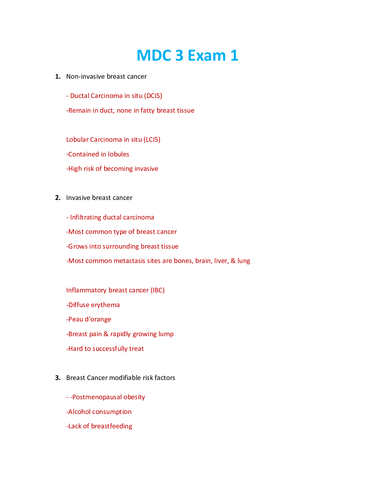
Breast Cancer
Patho:
- Impaired cellular regulation in the breast tissue. One single cell that grows and multiplies in epithelial cells in one or
more of the mammary ducts or lobules
Risk Fa
...
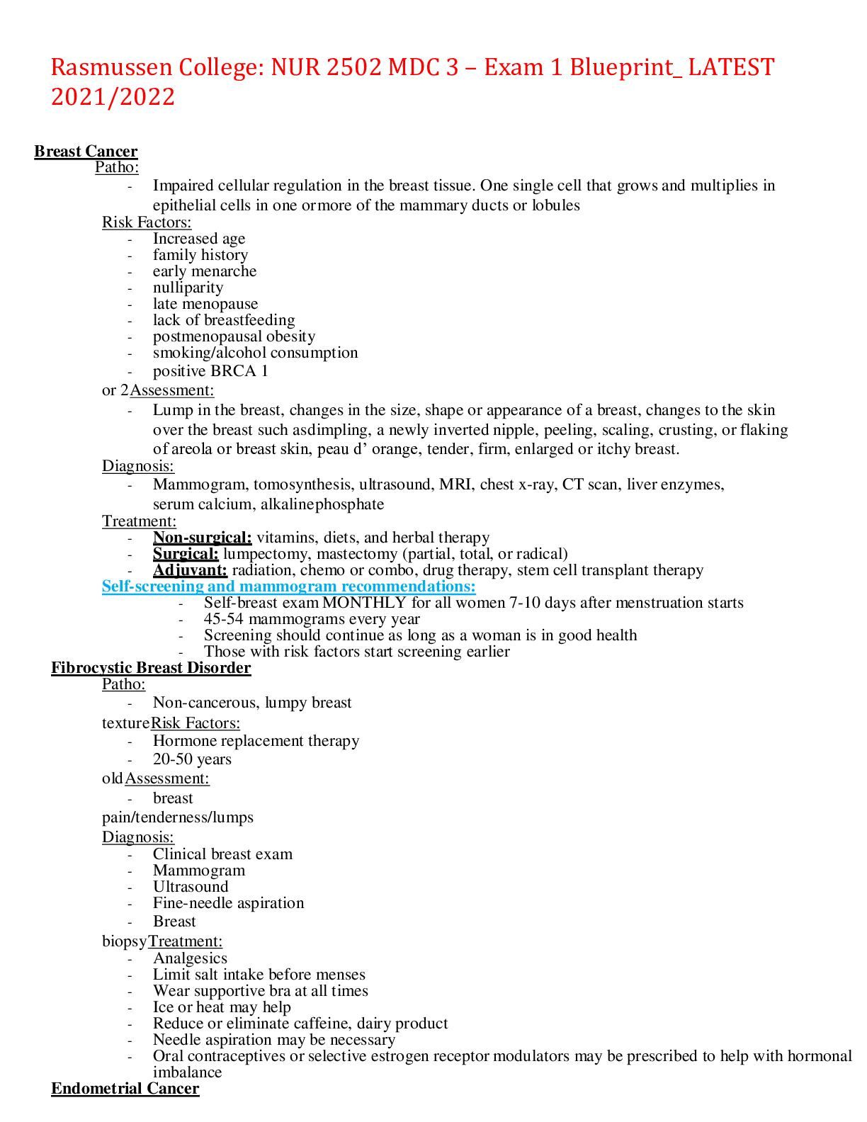
MDC 3 – Exam 1 Blueprint
Breast Cancer
Patho:
- Impaired cellular regulation in the breast tissue. One single cell that grows and multiplies in epithelial cells in one or
more of the mammary ducts or lobules
Risk Factors:
- Increased age
- family history
- early menarche
- nulliparity
- late menopause
- lack of breastfeeding
- postmenopausal obesity
- smoking/alcohol consumption
- positive BRCA 1 or 2
Assessment:
- Lump in the breast, changes in the size, shape or appearance of a breast, changes to the skin over the breast such as
dimpling, a newly inverted nipple, peeling, scaling, crusting, or flaking of areola or breast skin, peau d’ orange,
tender, firm, enlarged or itchy breast.
Diagnosis:
- Mammogram, tomosynthesis, ultrasound, MRI, chest x-ray, CT scan, liver enzymes, serum calcium, alkaline
phosphate
Treatment:
- Non-surgical: vitamins, diets, and herbal therapy
- Surgical: lumpectomy, mastectomy (partial, total, or radical)
- Adjuvant: radiation, chemo or combo, drug therapy, stem cell transplant therapy
Self-screening and mammogram recommendations:
- Self-breast exam MONTHLY for all women 7-10 days after menstruation starts
- 45-54 mammograms every year
- Screening should continue as long as a woman is in good health
- Those with risk factors start screening earlier
Fibrocystic Breast Disorder
Patho:
- Non-cancerous, lumpy breast texture
Risk Factors:
- Hormone replacement therapy
- 20-50 years old
Assessment:
- breast pain/tenderness/lumps
Diagnosis:
- Clinical breast exam
- Mammogram
- Ultrasound
- Fine-needle aspiration
- Breast biopsy
Treatment:
- Analgesics
- Limit salt intake before menses
- Wear supportive bra at all times
- Ice or heat may help
- Reduce or eliminate caffeine, dairy product
- Needle aspiration may be necessary
- Oral contraceptives or selective estrogen receptor modulators may be prescribed to help with hormonal imbalance
Endometrial Cancer
Patho:
- Most common reproductive cancer of the inner uterine lining. Adenocarcinoma is the most common tumor type. It
arises from the glandular part of the endometrium and usually follows endometrial hyperplasia (overgrowth)
- Stages of Endometrial Cancer:
Stage 1: endometrium only
Stage 2: endometrium, cervix
Stage 3: endometrium, cervix, vagina, lymph nodes
Stage 4: endometrium, cervix, vagina, lymph nodes, bowel and bladder
Risk Factors:
- women in reproductive years
- family history
- diabetes mellitus
- hypertension
- obesity
- uterine polyps
- late menopause
- nulliparity
- smoking
Assessment:
- The main symptom of endometrial cancer is postmenopausal bleeding. Ask the patient how many tampons or pads
they use each day.
- Other symptoms include low pelvic pain, watery and bloody vaginal discharge, low back/abdominal pain. Ask the
patient to describe where the pain is and perform a pelvic examination.
Diagnosis:
- CBC’s because of blood loss and serum tumor markers CA-125
- -transvaginal ultrasound and endometrial biopsy **GOLD STANDARD**
- Chest x-ray, abdominal US, CT of pelvis, MRI of the abdomen and pelvis.
Treatment:
- Surgical: total hysterectomy and bilateral salpingo-oophorectomy
- Nonsurgical: radiation, chemotherapy, drug therapy.
Cervical Cancer
Patho:
- The uterine cervix is covered with squamous cells on the outer cervix and columnar (glandular) cells that line the
endocervical canal. Most cervical cancers arise from the squamous cells on the outside of the cervix. The other
cancers arise from the mucus-secreting glandular cells (adenocarcinoma) in the endocervical canal.
Risk Factors:
- HPV or STI
- smoking
- having multiple full-term pregnancies
- age (late-teens-early 30’s most at risk)
- no access for screening
- oral contraceptives
Assessment:
- (hematuria) painless vaginal bleeding, unexplained weight loss, dysuria (painful urination), pelvic pain, chest pain,
rectal bleeding.
- Pap smear will be performed to visualize the tumor if it is visible.
Diagnosis:
- Pap smear
- colposcopy-visualizing the characteristics of cells by using a light.
- endocervical curettage (scraping of the uterine lining-may cause bleeding)
Treatment:
- LEEP (loop electrosurgical excision procedure)- a thin loop wire electrode that transmits a painless electrical current
is used to cut away affected tissue.
- Chemotherapy, radiation, cryotherapy, laser therapy, total hysterectomy, conization
Cervical ablation- post op care:
- No sexual intercourse
- No tampons / douche
- No baths (showers only)
- No fragrant soaps on vagina
- Avoid lifting heavy objects
- report vaginal bleeding
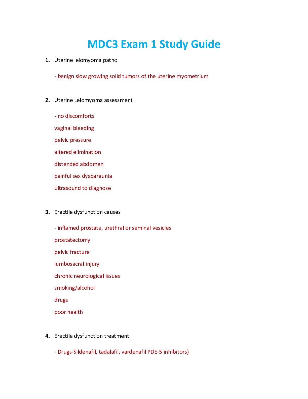
Uterine Leiomyoma/Fibroid
Patho:
- benign, slow-growing solid tumors of the uterine myometrium. Classified by the location. Intramural leiomyomas
are contained in the uterine wall. Submucosal leiomyomas protrude into the cavity of the uterus and can cause
bleeding and disrupt pregnancy. Subserosal leiomyomas protrude through the outer surface of the uterine wall and
may extend to the broad ligament pressing other organs.
Risk Factors:
- Cause is unknown but they think leiomyomas develop from excessive local growth of smooth muscle cells.
- May be a genetic error causing a lack of ability to halt growth.
- The growth of leiomyomas may also be stimulated by estrogen, progesterone, and growth hormone.
Assessment:
- Acute pain, usually seek medical attention because of heavy vaginal bleeding, depending on the location she can
have constipation, urinary frequency, or retention., abdominal pressure.
Diagnosis:
- Abdominal, vaginal, and rectal examinations usually reveal the presence of a uterine enlargement.
- CBC, pregnancy test, transvaginal US, laparoscopy, hysteroscopy
Treatment:
- Nonsurgical: oral contraception, MRI
- Surgical: MRI focused ultrasound- heat to tumor to kill it, Uterine artery embolization-starves tumor or circulation
allowing it to shrink, myomectomy- laser removal of tumor, hysterectomy-removal of ovaries
Hysterectomy- post op care:
- monitor patient for fluid overload, embolism, hemorrhage, and to make sure the patient reports any sudden
onset PAIN
Vulvovaginitis
Patho:
- inflammation of the lower genital tract / imbalance of hormones. Think yeast, bacterial vaginosis, pH changes.
Risk Factors:
- Multiple sexual partners
- Sexually transmitted diseases
- Spermicides
- Use of vaginal sprays or douching
- Diabetes
- Birth control use
- Wet clothing
- Antibiotics/Steroids
Assessment:
- Pruritus
- Painful sex
- abnormal vaginal discharge
- vaginal itching/odor
Diagnosis:
- Pelvic exam
- Vaginal discharge microscopy
- pH testing
Treatment:
- Antifungals
- Antibiotics
Education:
- Practice safe sex
- wear cotton underwear/dry clothing
- wipe front to back
- cleanse vagina with water NO soap
- Void after sex
- Vulvar self-exams monthly
Toxic Shock Syndrome (TSS)
Patho:
-Infection from STAPHYLOCOCCUS AUREUS & bacteria from menstruation and tampon usage which crosses
into the bloodstream.
Risk Factors:
-Immunosuppression
-use of highly absorbent tampons or contraceptive sponges
-deep skin infection
Assessment:
-fever
-rash
-hypotension
-GI upset
-neuromuscular disturbances
-increased liver enzymes
-THINK SEPSIS
Diagnosis:
-blood cultures
-assist provider with pelvic exam
Treatment:
- IV antibiotics
- fluid replacement
- Corticosteroids
Education:
- Teach about hand hygiene when changing pads/tampons
- se pads at night, no longer that 4 hours without changing tampon
- No intercourse during treatment
Prostate Cancer
Patho:
- Slow growing cancer with predictable metastasis. The 2nd leading cancer in men after lung cancer, but is almost
100% curable.
Risk Factors:
- over 50
- African Americans AT HIGHER RISK
- high fat diet
- no exercise
- consume alcohol or nicotine
Assessment:
- difficult starting urination/ dribbling after urination
- polyuria
- nocturia
- urinary retention
- Latesymptoms:
- blood in the urine or semen. (DIFFERENTIATES FROM BPH)
Labs:
- urinalysis, ultrasound, PSA, alkaline phosphate, biopsy
Treatment:
- Active surveillance (check up every 6 months) as long as there is no growth
- Prostatectomy: removal of whole prostate. The patient will be catheterized.
Education:
- monitor urine output, control pain, watch for bleeding. Erectile dysfunction is common after total or radical
prostatectomy
Benign Prostatic Hyperplasia (BPH)
Patho:
- Non-cancerous enlargement of the prostate resulting in compression of the urethra and urinary retention
Assessment:
- polyuria, nocturia, trouble starting urination, feeling that the bladder is not empty
Diagnosis:
- transabdominal ultrasound or MRI
- urinalysis and culture, CBC, BUN, PSA,
Treatment:
- Non-surgical
- meds to stop hyperplasia (growth of prostate)
- alpha blockers
- 5alpha reductase inhibitors (Finasteride or Dutasteride)
- Cialis
- Surgical
- prostatectomy (TURP) through the urethra
Education:
- TURP- post op care: monitor urine output, control pain, watch for bleeding
Testicular Cancer
Patho:
- very rare - usually affects men 20-35 years of age (reproductive years)
- Usually curable with early detection – self-examinations.
- Germ cell tumors: arise from sperm-producing cells
- Non germ cell tumors: arise from stromal, interstitial, or Leydig cells (more rare)
[Show More]