FINAL EXAM STUDY GUIDE – MICRO
INSTRUCTIONS:
It’s time to show your skills and knowledge of what you have learned!
This Final Exam contributes 230 points to your overall grade, so please do your best.
• There ar
...
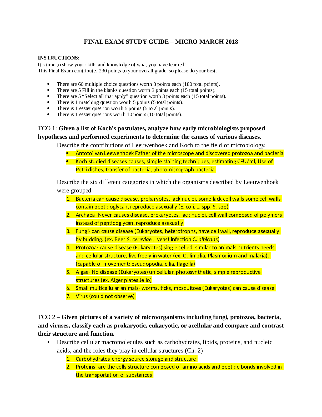
FINAL EXAM STUDY GUIDE – MICRO
INSTRUCTIONS:
It’s time to show your skills and knowledge of what you have learned!
This Final Exam contributes 230 points to your overall grade, so please do your best.
• There are 60 multiple choice questions worth 3 points each (180 total points).
• There are 5 Fill in the blanks question worth 3 points each (15 total points).
• There are 5 “Select all that apply” question worth 3 points each (15 total points).
• There is 1 matching question worth 5 points (5 total points).
• There is 1 essay question worth 5 points (5 total points).
• There is 1 essay questions worth 10 points (10 total points).
TCO 1: Given a list of Koch's postulates, analyze how early microbiologists proposed hypotheses and performed experiments to determine the causes of various diseases.
Describe the contributions of Leeuwenhoek and Koch to the field of microbiology.
• Antotoi van Leewenhoek Father of the microscope and discovered protozoa and bacteria
• Koch studied diseases causes, simple staining techniques, estimating CFU/ml, Use of
Petri dishes, transfer of bacteria, photomicrograph bacteria
Describe the six different categories in which the organisms described by Leeuwenhoek were grouped.
1. Bacteria can cause disease, prokaryotes, lack nuclei, some lack cell walls some cell walls contain peptidoglycan, reproduce asexually (E. coli, L. spp, S. spp)
2. Archaea- Never causes disease, prokaryotes, lack nuclei, cell wall composed of polymers instead of peptidoglycan, reproduce asexually
3. Fungi- can cause disease (Eukaryotes, heterotrophs, have cell wall, reproduce asexually by budding. (ex. Beer S. cereviae , yeast infection C. albicans)
4. Protozoa- cause disease (Eukaryotes) single celled, similar to animals nutrients needs and cellular structure, live freely in water (ex. G. limblia, Plasmodium and malaria). (capable of movement: pseudopodia, cilia, flagella)
5. Algae- No disease (Eukaryotes) unicellular, photosynthetic, simple reproductive structures (ex. Alger plates Jello)
6. Small multicellular animals- worms, ticks, mosquitoes (Eukaryotes) can cause disease 7. Virus (could not observe)
TCO 2 – Given pictures of a variety of microorganisms including fungi, protozoa, bacteria, and viruses, classify each as prokaryotic, eukaryotic, or acellular and compare and contrast their structure and function.
• Describe cellular macromolecules such as carbohydrates, lipids, proteins, and nucleic acids, and the roles they play in cellular structures (Ch. 2)
1. Carbohydrates-energy source storage and structure
2. Proteins- are the cells structure composed of amino acids and peptide bonds involved in the transportation of substances
3. Lipids –(fats, phospholipids, waxes) hydrophobic, provides energy and insulation
4. Nucleic acids- DNA (required for photosynthesis) and RNA (carries the information to the cite of protein production)
• Compare and contrast molecular components of prokaryotes (Bacteria and Archaea), eukaryotes, and viruses as well as the environmental conditions these can be found in
1. Prokaryotes Bacteria
Internal structure – Endospores, Nucleoid, and Ribosomes (refer to unit 1 worksheet) External structures- Flagella, Fimbriae (sticky, short biofilms) , Pili (transfer DNA), and Cell walls (protection, cell to cell attachment, give cells shape, gram+, gram -)
(refer to unit 1 worksheet)
2. Archaea- have cell walls but lack peptidoglycan (uses PPG), have cytoplasmic membranes, reproduce by; binary fusion, budding, or fragmentation. Extremophiles (pH, temputure, and, salt).
• No nucleus
• Smaller then eukaryotes
• Diverse
3. Eukaryotes- Of the Eukaryotes fungi, algae, plants, and some protozoa contain cell walls.
• Internally contains nucleus (house DNA and direct the synthesis of proteins ) and membrane bound organelles(Golgi body which play a role in lipid synthesis and transport, mitochondria; has two bilayer membranes and where most ATP is produced , chloroplasts; which uses light energy to produce ATP)
• The structure is more complex
• Contains a plasma membrane (composed of a bilayer of phospholipid bilayer),
cytoplasm, and ribosomes (larger then in prokaryotes)
• contain glycocalyces which strengthens the cells surface, protects it from dehydration, and helps with anchoring animal’s cells to each other.
• Cell wall-contains different types of polysaccharides; Cellulose
• A Classify and describe the general characteristics of prokaryotes.
• Cellular shapes (morphology)
• Cellular arrangements
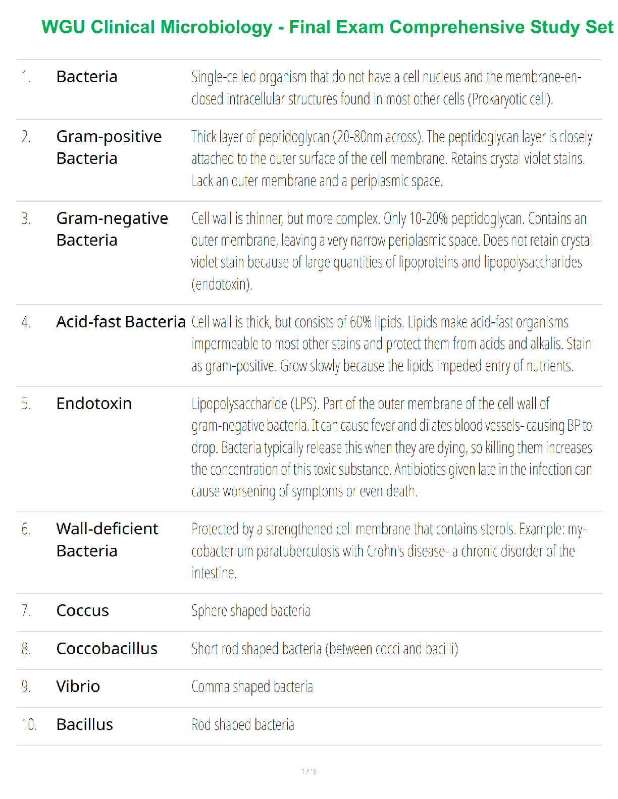
• Describe Gram positive, Gram negative, and Acid Fast cell wall/membrane structures including molecular components (lipids, acids, endotoxins, periplasmic space).
Gram Positive= has a thick layer of peptidoglycan, contains teichoic acids (endotoxin) appear purple after staining.
Gram negative= thin layer of peptidoglycan, contains a bilayer membrane, lipopolysaccharide (LPS lipid A portion is responsible for fevers), appears pink after staining.
Acid Fast- A gram positive bacterium that has a thick layer of peptidoglycan but also up to 60% mycolic acid. Causes TB and leprosy
• Compare/contrast methods of transport into and out of cells
The plasma membrane is responsible for allows molecules in and out of the cell because it is selectively permeable. Molecules are only able to enter in (endocytosis) and out (exocytosis) of the cell by passive, active and bulk transport.
• Describe passive, active, bulk transport
1. Passive transport- is the move of molecules in and out of the cell without energy. Types of passive:
• Diffusion- through the phospholipid bilayer from high concentration to low concentration
• Facilitated diffusion- nonspecific channel protein
• Osmosis-diffusion of water through a water channel protein
2. Active transport- uses ATP as energy to transport molecules in and out of the cell. Pumps force molecules from a high concentration to a low concentration.
3. Bulk transport- requires vesicle and metabolic energy to move molecules in and out of the cell
• Compare/contrast archaea and bacteria in terms of cellular structures and how they were separated by Carl Woese.
• Compare/contrast prokaryotes and eukaryotes in terms of size, shape, reproduction, motility, organelles, metabolic processes
TCO 3 – Given several types of microscopes, evaluate how scientists use the microscopes to identify and study microorganisms.
• Describe the principles of various types of microscopy and their applications. Types, applications, alive vs. dead, stained vs. unstained
1. Light field Microscopy
Simple Light field has one lens Compound- has two lenses REQUIRES STAINING (STAINING KILLS THE ORGANISM)
2. Dark Field Microscopy
Best for observing pale objects. Specimen appears light against a dark background. Organism is alive (INCREASES CONTRAST AND MORE DETAILS ARE SEEN)
3. Phase- Contrast
• Compare the sizes of typical sizes of eukaryotes, prokaryotes, and viruses using scientific units (mm, um, nm)
• Describe various staining methods used in microscopy
• Describe how Gram stain works (& why is each step important)
• Analyze the different methods of classification and identification of microorganisms.
TCO 4 – Given a culture of a microorganism, examine its physical and nutritional requirements.
• Explain how environmental conditions (temperature, pH, oxygen, and osmotic and hydrostatic pressure) affect microbial growth.
• Correctly identify and name organisms that live in extreme conditions (e.g. psychrophiles, barophiles, etc.) or normal conditions (e.g. mesophiles)
• Correctly identify organisms based on oxygen requirements (obligate anaerobe, obligate aerobe, facultative anaerobe, microaerophile, aerotolerant anaerobe)
• Describe the information that can be interpreted from a growth curve; name the four phases and know what makes each unique
TCO 5 – Given a nutritional requirements of a microorganism, analyze the processes of microbial metabolism
• Compare and contrast respiration and fermentation.
Cellular Respiration- is a process that a cell can use to make ATP and process it into 3 main types of ways: glycolysis, the krebs cycle, and the electron transport chain.
Fermentation- Takes place without oxygen (anaerobic), so less ATP is produced from each molecule of glucose .
• Discuss the macromolecular structure as well as function of enzymes in a cell Macromolecules are made up of basic molecular units. They include proteins, nucleic acids, carbohydrates, and lipids. Enzymes function to catabolize large nutrient molecules into smaller molecules that can be absorbed or transported into the cell.
• How do enzymes work? What do they bind, what do they form, what is activation energy?
Enzymes speed up reactions by lowering activation energy. They work with substrates; the substrates will bind to a region on the enzyme which is called the active site. The enzymes will form new molecules. Activation energy is the minimum quantity of energy that the reacting species must possess in order to undergo a specified reaction.
• Under what conditions do most enzymes function optimally?
Enzymes function optimally at a temperature that is in the range of 35-40 Celsius (95-104 Fahrenheit).
• Define apoenzyme, holoenzyme, enzyme-substrate complex
Apoenzyme- An inactive enzyme, activation of the enzyme occurs upon binding of an organic or inorganic cofactor.
Holoenzyme- An apoenzyme together with its cofactor.
Enzyme-substrate complex- Is when a substrate molecule interacting with the active site of an enzyme.
• Describe the three stages of aerobic glucose catabolism.
2. Lysis stage: Fructose 1,6-bisphosphate is cleaved into glyceraldehyde 3-phosphate and dihydroxyacetone phosphate.
3. Energy-conserving stage- Each glyceraldehyde 3-phosphate is oxidized to pyruvic acid, yielding two ATP molecules each, for a total of four ATP; It also oxidizes to pyruvic acid, yielding another two ATP molecules, for a total of four ATP molecules.
• Know major products of each stage and relative # of ATP produced for each stage
• Know where in the cell each stage occurs (prokaryotic and eukaryotic) Prokaryotic- Located in multicellular organisms (has nucleus) Eukaryotic- Located in the nucleoid (no nucleus)
• Compare aerobic and anaerobic metabolism in terms of products and # ATP produced Aerobic metabolism uses oxygen uses 36-38 ATP molecules
Anaerobic metabolism uses without oxygen; the process uses a respiratory electron transport chain but does not use oxygen as the electron acceptors. Uses between 36-2 ATP molecules.
TCO 6 – Given the characteristics of a eukaryotic organism, classify it into its right kingdom based on genetic relatedness.
• Describe the characteristics of protozoa, their nutritional requirements, and their mode of reproduction, and relevant vocab (structural features).
Some characteristics of a protozoa is that they are able to move independently. Nutritional requirements are that they require organic materials. Asexual reproduction; binary fission and multiple fission.
• Know general life stages of blood-born protists (e.g. malaria) with relevant vocab
• Know general life stages of intestinal protists (e.g. Giardia lamblia) with relevant vocab
• Describe the characteristics of fungi that distinguish them from other eukaryotes.
Fungi usually disperse themselves by releasing spores, they’re heterotrophic, they usually grow as tubular filaments called hyphae, the walls of the hyphae are often strengthened with chitin.
• Discuss the role of arthropod vectors in disease transmission; differentiate mechanical and biological vectors
The arthropod will salivate into the wound that they create.
Mechanical vectors- housefly, cockroach, or other animal that passively carries pathogens to new hosts on its feet or other body parts and is not infected by the pathogens it carries. Biological vectors- biting arthropod or other animal that transmits pathogens and serves as host for the multiplication of the pathogen during some stage of the pathogen’s life cycle.
TCO 7 – Given the characteristics of an acellular pathogen, identify the pathogen to design the appropriate treatment strategy.
• Discuss the classification strategies of a virus. 1. RNA vs. DNA (type of nucleic acid)
2. Single-stranded vs. double-stranded nucleic acid
3. Enveloped vs. non-enveloped
4. "sense" of RNA (+/-): + indicates sense is same as mRNA; - indicates sense is complementary to mRNA
5. Icosahedral vs. helical morphology of capsid
• Describe the intracellular and extracellular structures of viruses
Extracellular- virion; in this form the virus will consist of a protein coat called a capsid which surrounds the nucleic acid.
Intracellular- the capsid will be removed
• Discuss the various modes of replication of a bacteria virus and animal virus.
Bacteria virus- usually reproduce by binary fission, replicates the DNA then divides itself into two identical cells.
Animal virus- they can enter in two different ways, when a protein in the viral capsid binds to its receptor on the host cell, the virus may be taken inside the cell by a vesicle during the normal process of receptor-mediated endocytosis.
• Discuss the different ways viruses can be cultured in a lab.
Viruses need living cells to support their replication so they cannot grow in culture media or on agar plates alone. The best way to grow viruses are bacteriophages (reason is the easiest cells to grow in lab are bacteria).
• Compare and contrast viruses, virions, prions, and viroids.
Viruses- infective agent that typically consists of a nucleic acid molecule in a protein coat, is too small to be seen by light microscopy, and able to multiply only within the living cells of a host.
Virions- a virus outside of a cell, consisting of a proteinaceous capsid surrounding a nucleic acid core.
Prions- proteinaceous infectious particle that lacks nucleic acids and replicates by converting similar normal proteins into new prions.
Viroids- extremely small, circular piece of RNA that is infectious and pathogenic in plants.
TCO 8 –Analyze methods of controlling microbial growth in the environment and in the body.
• Describe the appropriate physical and/or chemical methods of controlling microbial growth when given a particular scenario (e.g. sterilizing insulin, saline solution, fresh fruits/veggies, goggles; de-germing local injection site, table tops, etc.)
• Summarize the mode of action of major groups of antimicrobial and antiviral drugs.
• Summarize practices that have led to increased bacterial antibiotic resistance and strategize
ways to limit this increased resistance.
• Describe factors to be taken into account when selecting antimicrobial and antiviral drugs, including drug effects on patients.
o Discuss common outcomes of antibiotics on normal patients
o Discuss superinfections and how to avoid these infections
o Discuss teratogenic antibiotics
TCO 9 – Given an illustration of the three lines of defense that protect the human body from pathogens, analyze how the components of each work to prevent or overcome infection
• Describe nonspecific defense mechanisms, differentiating between the first and second lines of
defense.
• Differentiate skin and mucous membranes
• Describe the events of phagocytosis
• Describe specific immunity, differentiating the types of cells in this system from nonspecific
defense mechanisms
• Differentiate B cells and T cells , including where they mature and their primary functions
• Describe the roles of inflammation and fever in innate immunity
• Define antibody (5 types), antigen (3 types), epitope, BCR, TCR, MHC, APC and give examples of
each.
• Differentiate between humoral and cell-mediated immunity.
• Differentiate between the five different classes of antibodies.
o Structure, function, abundance, when they are made in immune responses (first exposure vs second exposure)
IgM IgG IgA IgE IgD
pentamer monomer Monomer, dimer monomer monomer
First antibody produced; later switches to IgG
They are the most common type of antibody in the blood during the initial phases of an immune Most common and longest- lasting antibody NK cells interacts agglutination Associated with body secretions (ex. Breast milk)
agglutination Involves in response to parasitic infections and allergies
A cause of basophil and unknown
response. eosinophil degranulation
• Differentiate between passive and active acquired immunity.
o In terms of source of antibodies, response time, & memory
o Identify the use of immunizations
TCO 10 –Given data on an outbreak of a microbial disease, propose a plan that outlines the steps taken to identify the cause of the disease and to stop or limit the spread of the infection.
• Discuss symbiotic relationships, including commensal, mutualistic, and parasitic (i.e., biofilms,
normal microbiota, etc.).
• Discuss when normal microbiota can become pathogenic, and how to avoid these situations
• Explain the general mechanisms of invasion and virulence that pathogenic agents must employ
to cause disease.
• Describe categories of disease reservoirs and how the diseases may be eliminated or controlled (i.e., zoonoses).
• Describe modes of disease transmission and ways of preventing transmission, including in clinical settings.
• Describe and differentiate the 5 stages of disease
• Differentiate types of healthcare-associated diseases
• State how diseases are tracked and how their frequency is defined.
TCO 11 – Given a patient with the symptoms of influenza or another infectious disease, identify the causative agent and analyze its pathogenicity.
• Describe the portal of infection for the various organ systems.
• List axenic areas of the body
• Differentiate signs and symptoms, giving examples of each
• Describe the signs and symptoms of major infectious diseases of humans.
• Describe virulence factors and pathogenic mechanisms that enable microorganisms to infect
humans.
• Differentiate between the different types of infectious diseases, including eukaryotic, viral, and prion diseases.
• Summarize the diagnosis, treatment, and prevention of infectious diseases
• Describe the portal of infection for the various organ systems: respiratory, digestive, urinary, and
reproductive.
MICROBES TO KNOW:
(unique features, associated diseases, targets for antimicrobials)
Bacillus and Clostridium Candida
Chlamydia trachomatis Clostridium difficile Cytomegalovirus (CMV) Enterobacteria Escherichia coli
Epstein-Barr virus (HHV- 4)
Dermatophytes (fungal) Giardia
Helicobacter pylori Hepatitis (A, B, C) HIV
HPV
Influenza
Lactobacillus
Listeria monocytogenes
Malaria Methanogens Mycobacterium tuberculosis
Mycobacterium leprae Mycoplasma – various forms
Neisseria gonorrhoeae Neisseria meningitidis. Parasitic worms
Papillomavirus Prions
Pneumocystis pneumonia Saccharomyces cerevisiae Salmonella Staphylococcus – various species
Streptococcus – various species
Treponema pallidum
Vibrio cholerae
-Acne
-Acute bacterial meningitis
-African sleeping sickness Alzheimer’s disease
-Anthrax
-Bacteremia
-Botulism
-Brucellosis Bubonic plague
-Candidiasis Cervical cancer
-Chlamydia
-Chicken pox (Varicella) Varicella zoster
-Common cold
-Cytomegalovirus (CMV)
-Encephalitis
-Foliculitis (Furuncle)
-Gastroenteritis
-Glomerulonephritis
-Gonorrhea
-Hansen’s disease
-Hepatitis (all forms)
-Herpes zoster
DISEASES TO KNOW:
(causes, symptoms, signs, prevention)
-Histoplasmosis
-Impetigo Listeriosis
-Lyme disease Lymphangitis
-Malaria
-Measles Mononucleosis, infectious =Epstein- Barr virus (HHV-4)
-Mumps
-Necrotizing fasciitis
-Peptic ulcers Pertussis Petechiae
-Pneumonia
-Pneumonic plague
-Rabies Rheumatic fever Ringworm Rocky Mountain spotted fever
-Rubella
-Salmonella Scarlet fever
-Shingle
-Smallpox (Herpes zoster)
Spongiform encephalitis
-Staphylococcal scalded skin syndrome
-Streptococcal pharyngitis Swimmer’s itch
-Syphilis (primary, secondary, tertiary) Tuberculosis
-Tetanus
-Traveler’s diarrhea
-Tularemia
-Variant Creutzfeld- Jakob disease walking pneumonia Whooping Cou
Disease Causative
Agent Signs
+Symptoms Diagnos
is Transmitted Treatment
Folliculitis Staph epi & aureus Infection of the hair follicles (furuncles) Isolation of gram positive bacteria (grape like
clusters) Direct contact by fomites Dicloxacillian Vancomycin
Staphylococc al Skin Syndrome Staph aureus Wrinkled red skin with blisters (skin peels in sheets) Diagnosed by skin sloughing
(peeling) Person to person Antimicrobial
Impetigo Staph aureus & Strep pyogenes Red patches forms on the face and limbs The presence
of vesicles for impetigo Person to person Penicillin
Necrotizing Fasciitis Strep pyogenes Nonspecific Early diagnosis is different because symptoms are
nonspecific Person to person Clindamycin and penicillin
Acne Propionibacteriu m acnes Commonly found on skin Visual examination Antimicrobial drugs Accutane treats severe
acne
Anthrax Bacillus anthracis Presence of an eschar black painless ulcer fever chills loss of appetite if ingested Visual of black painless ulcer (if inhaled Chest X-ray) (if ingested difficult to
recognize) Contact Antimicrobial drugs
Smallpox Chickenpox (Herpes zoster) Shingles (VZV) Varicella Zoster Virus Lesions on the back and trunk that spread (involves sensory nerves Visual diagnosis after rash appears Reactivation of
chickenpox Occurs mostly in children (leads to shingles in adults) Vaccine available against chicken pox
Rubella Rubella virus Children: Causes slightly swollen lymph nodes, red rash
Adults: encephalitis
and arthritis Serological testing and visual of rash Person to person MMR vaccine available No cure or medications to shorten
symptoms
Measles (Rubeola) Measles virus Red skin rash (Koplik Spots) , muscle pain, runny nose fever Diagnosed based on symptoms Respiratory droplets Vitamin A and fever reducers help with symptoms no
treatment
Hansens Disease Mycobacterium leprae Discolored patches of skin, painless ulcers painless swelling loss of sensation (Leprosy) Diagnosed based on symptoms Confirmed by acid fast
samples Person to person by nasal secretions or droplets Antimicrobials (can be lifelong treatment) BCG vaccine available
Botulism Clostridium botulinum Flaccid paralysis Foodborne, Wound
botulism and Infant Diagnostic based on
symptoms Contact, water or soil, and
foodborne Washing intestinal tract,
admin. Of
botulism all show symptoms of difficulty swallowing, speaking
and facial weakness botulism immune globulin
Tetanus Clostridium tetani Tightening of the jaw, muscle spasms, irregular heartbeat
sweating Diagnostic based on symptoms Contact, break in skin or mucus membranes Wound cleaning, antimicrobial, vaccine available
Bacterial Meningitis/ Viral Meningitis Viral: Coxsackie A Coxsackie B Echovirus Bacterial: S. agalactiae (during birth and newborns) Listeria momocytogense (food), S. Pneumoniae &
N. Meningitidis
(most prevalent) High fever, meningeal inflammation, increased WBC’s in the CSF, develops infection to the brain (encephalitis) Diagnostic based on symptoms culturing CFS Contact and food contamination Penicillin at birth for newborns, intravenous antimicrobial drugs
Rabies Rabies virus (ssRNA virus) Perpetual muscle contractions Flu like symptoms, if reaches CNS hydrophobia, seizures, hallucinations,
paralysis Diagnosed based on neurological symptoms Transmitted via infected animal Treated with rabies immunoglobulin, vaccine available, cleaning site
Encephalitis Many viral infections cause encephalitis mumps, measles, HSV, VZV
Arboviral: arthropod borne
virus Confusion, headache, fever, vomiting, stiff neck, muscle soreness, extreme weakness Diagnosed based on symptoms
Confirmed by arbovirus antibodies in CSF Mosquitos/insect transmit arbovirus encephalitis Preventative measures using a net to avoid mosquitos
African sleeping sickness Trypanosoma brucei Lesion, fever, lymph node swelling, headache, can cause meningoencephalitis Observation of trypanosome s in the blood, lymph
node biopsy Glossina African Fly (tsetse fly) Bed nets to avoid flies based on disease stage
Creutzfeldt Jakob disease
Spongiform encephalitis Abnormal Prion Protein Insomnia, weight loss, and memory failure, loss of muscle control Diagnosed based on symptoms Can be
confused Medical procedures (vCJD occur in young people) No treatment Prevention: avoid prion contaminated
meat
with other
dementia
Bacteremia Gram negative bacteria Fever, chills, nausea, diarrhea, malaise Hemorrhagic lesion, osteomyelitis if bacteria invades
bones Diagnosed based on symptoms Contact surgery blood Treated with prompt diagnosis
Brucellosis Brurcella melitiensis Fluctuating fever that spikes every afternoon Serological testing and presence of fever Consumption of contaminated dairy products Contact with animal blood,
urine or placenta Usually require to treatment.
Attenuated vaccine available
Tularemia Funcisella tularensis Skin lesions and swollen lymph nodes at infection site Serological testing Contact bites diverse host range includes mammal’s birds
fish and insects Treated with antimicrobials
Plague Yersinia pestis Bubonic plague: large lymph nodes Pneumonic plague: occurs in the lungs and makes it hard to
breathe Diagnosed based on symptoms Contact with infected animal or flea feces Antimicrobial drugs
Lyme Disease Spirochete Borrelia burgdorferi Three phases is untreated patients Bulls-eye rash at infection site Neurological symptoms and severe
arthritis Diagnosed based on signs and symptoms of the disease One of the most vector borne diseases in the U.S. Antimicrobials in the earlier stages and treatment in the later stages is difficult
Mononucleos is Epstein-Barr virus
(EBV and HHV- 4) Severe sore throat fever occurs initially followed by swollen lymph nodes fatigue loss of appetite and
skin rash Diagnosed by presence of large lobed B lymphocyte Via saliva Relieving symptoms
Cytomegalov irus Disease Cytomrgalovirus Asymptomatic in most cases.
Complications in neonates and the immunocompromised Diagnosed by identifying enlarged cells with inclusions Direct contact with bodily fluid or transparently One of the most common infection of
humans Fomivirsen administered for infections No vaccine available
Malaria Plasmodium species P. falciparum causes the most
severe malaria Associated with parasites life in erythrocytes. Fever, chills, diarrhea,
headache, anemia, Diagnosed made by identifying plasmodium
in blood Mosquitoes Antimicrobials
weakness, fatigue
Streptococcal Pharyngitis Group A streptococci (S. pyogenes) Sore throat , difficulty swallowing, fever, malaise, and headache (can spread to lower respiratory infections and may progress to Scarlett fever) Often confused with viral pharyngitis, common cold symptoms fever sore throat headache lymph node
enlarged Respiratory droplets Oral penicillin
Common Cold Enterovirus (rhinovirus) Sneezing, runny nose, congestion, sore throat, malaise, and cough Diagnostic based on symptoms Spread by coughing and sneezing person to person contact Flushing nasal and sinus cavities with saline solution reduces
symptoms
Bacterial Pneumonia Streptococcus pneunoniae Fever chills congestion chest pain rapid breathing rust colored sputum lung inflammation fluid filled alveoli and
bronchioles Diagnosis by identifying diplococci in sputum Inhalation of bacteria Penicillin Vaccine available
Histoplasmos is Histoplasma capsulation Asymptomatic in most cases.
Symptoms infection causing coughing with bloody sputum
or skin lesions Diagnosed by identifying the fungus in clinical
samples Inhalation of airborne spores from soil Resolve with treatment
Peptic Ulcer H. pylori Abdominal pain is the main symptoms X-ray to identify ulcers Fecal or oral transmission Antimicrobial drugs that can withstand
stomach acids
Bacterial Gastroenteriti s Species of shigella Nausea vomiting diarrhea abdominal cramps inflammations of the intestines and stomach Diagnosed based on symptoms Contaminated food or water and poor living conditions Supportive treatment and antimicrobials
Travelers Diarrhea E. Coli Diarrhea Diagnosed based on
symptoms Contaminated food or water Fluid and electrolyte
replacement
Salmonellosis Salmonella
enterica Typhoid fever Diagnosis is
made by Contaminated
food or water Salmonellosis is
self limiting
finding salmonella in stool Salmonellosis is often consumed from eggs Typhoid fever can be treated with
antimicrobials
Mumps Mumps virus Fever headache muscle aches tiredness loss of appetite swollen salivary glands on
both sides Diagnosed based on symptoms No specific treatment of mumps Infected cases have long life immunity.
Vaccine available
Hepatitis HAV HBV HCV HDV HEV (all are viruses) Jaundice abdominal pain fatigue vomiting weight loss symptoms may occur years after infection Liver damage Observation of jaundice enlarge liver or fluid on the abdomen Contact blood sexual contact Alpha interferon and nucleotide analogs helps reduce levels of the virus Vaccines available for
each virus
Streptococcal Glomerulone phritis Streptococci strains Cause inflammation of glomeruli and nephrons produce hypertension and low
urine output Urinalysis Contact
Candidiasis Candida albicans Severe vaginal
itching and burning Symptoms or
diagnostic Contact Azole or
Fluconazole
Gonorrhea Neisseria gonorrhoeae Men: painful urination purulent discharge Women: Pelvic
inflammatory disease and can infect babies
upon delivery Genetic probes are used and asymptomati c infection Sexual encounters Treated with broad spectrum cephalosporin
Syphilis (primary, secondary, Tertiary) Treponema pallidum Discharge and burning during urination Serological testing and culture Sexual encounters (transmission from mother to
child Penicillin
Chlamydial infections Chlamydia trachomatis Women are asymptomatic, and men have painful urination Detection of chlamydial DNA by PCR
diagnostic Sexual contact Antimicrobials
Herpes Herpes virus Blisters may form at sites of initial infection Diagnostics based on characteristic lesions Contact Sexual contact for genital Antiviral agents to lessen symptoms Males have lower risk
of infection
Alzheimer’s disease Amyloid beta protein Memory loss, mood swings, impaired
judgement, Diagnosed based on
symptoms Transmission during surgical
procedures No cure
confusion, repetitive
speech
Cervical cancer HPV Abnormal menstrual bleeding, pelvic pain, fatigue, weight loss,
loss of appetite Pap smear , HPV testing Sexual transmission Vaccine available
Listeriosis Listeria monocytogenes Food poisoning, fever diarrhea, headache
Pregnant women: flu like symptoms, can
cause miscarriage Diagnostic testing for L. monocytoge nes Contaminated foods, infected animals, soil, and water Antimicrobial
Lymphangitis Streptococcus pyogenes Chills swollen lymph glands fever malaise loss if appetite headache aching
muscles Diagnosed based on symptoms Direct contact Antimicrobials
Pertussis (Whooping cough) Bordetella pertussis Coughing, and mid fever, runny nose Babies: experience
apnea no coughing Serological testing Person to person Antimicrobials Vaccine available
Petechiae Meningococcal septicemia Red spot on skin shows signs of a low
platelet count
Rheumatic fever
Scarlet Fever (Streptococci)
Untreated strep throat Does not occur to all
individuals who had strep throat, Culture for strep Person to person Antimicrobials
Ringworm Microsporum spp Red itchy skin, raised patches develops
blisters defined edges Diagnosed based on
symptoms Person to person Antifungals
Rocky mountain spotted fever R. rickettsii Headaches chills muscular pain later develops rash covering the body that can ulcerate
fever malaise, Diagnosed based on symptoms serological testing Ticks Antimicrobials Doxycycline
Swimmers itch Schistosome cercariae Tingling burning small red pimples itching can blister Diagnosed based on symptoms and related
events Contact: Birds, parasites, lakes, and ponds Corticosteroid creams for itching
Tuberculosis Mycobacterium tuberculosis Coughing, chills fatigue fever loss of weight loss of
appetite PPD test, X- ray serological
testing Airborne Vaccine available Antimicrobials
[Show More]