Rasmussen College :NUR 2063 Exam 2 Blueprint Fall 2021,100% CORRECT
Document Content and Description Below
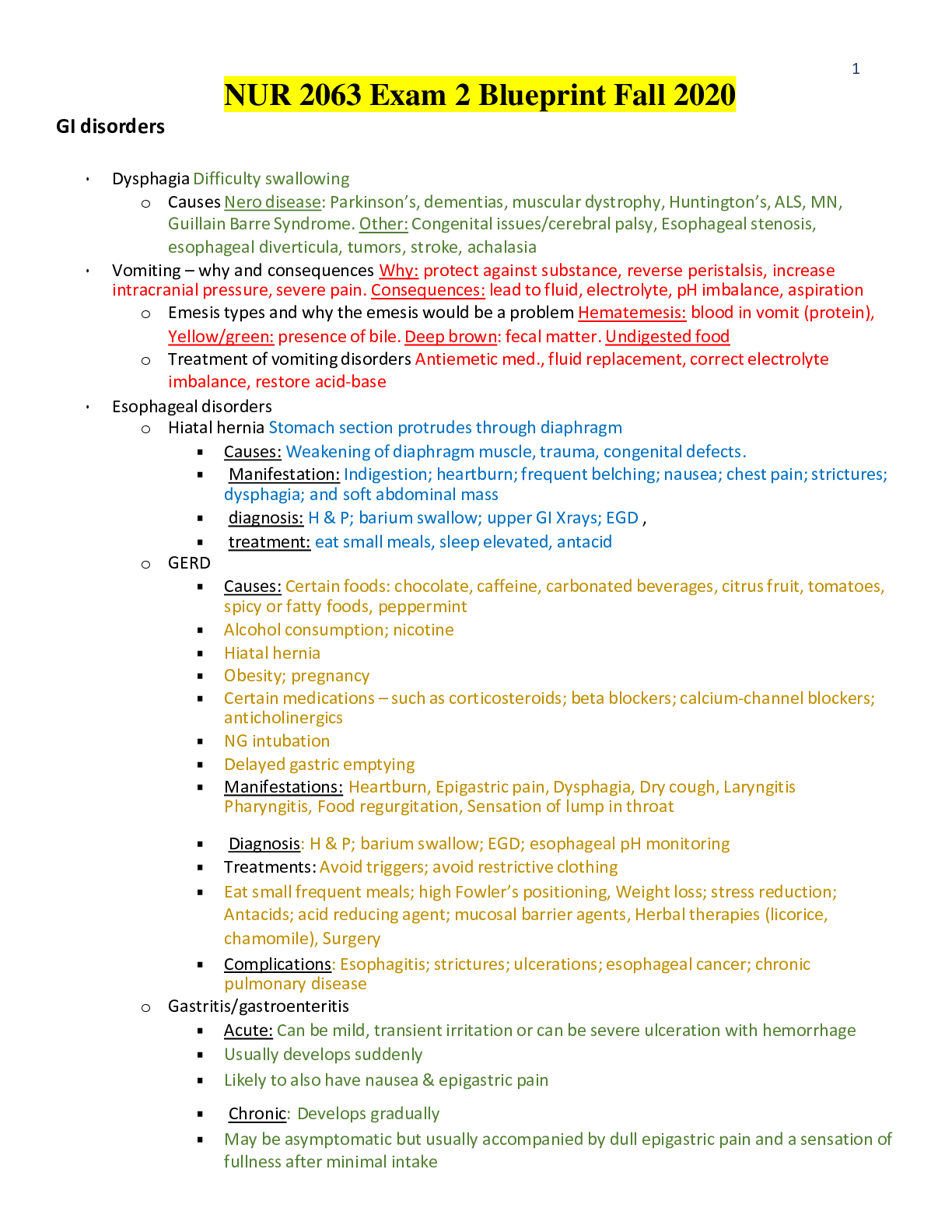
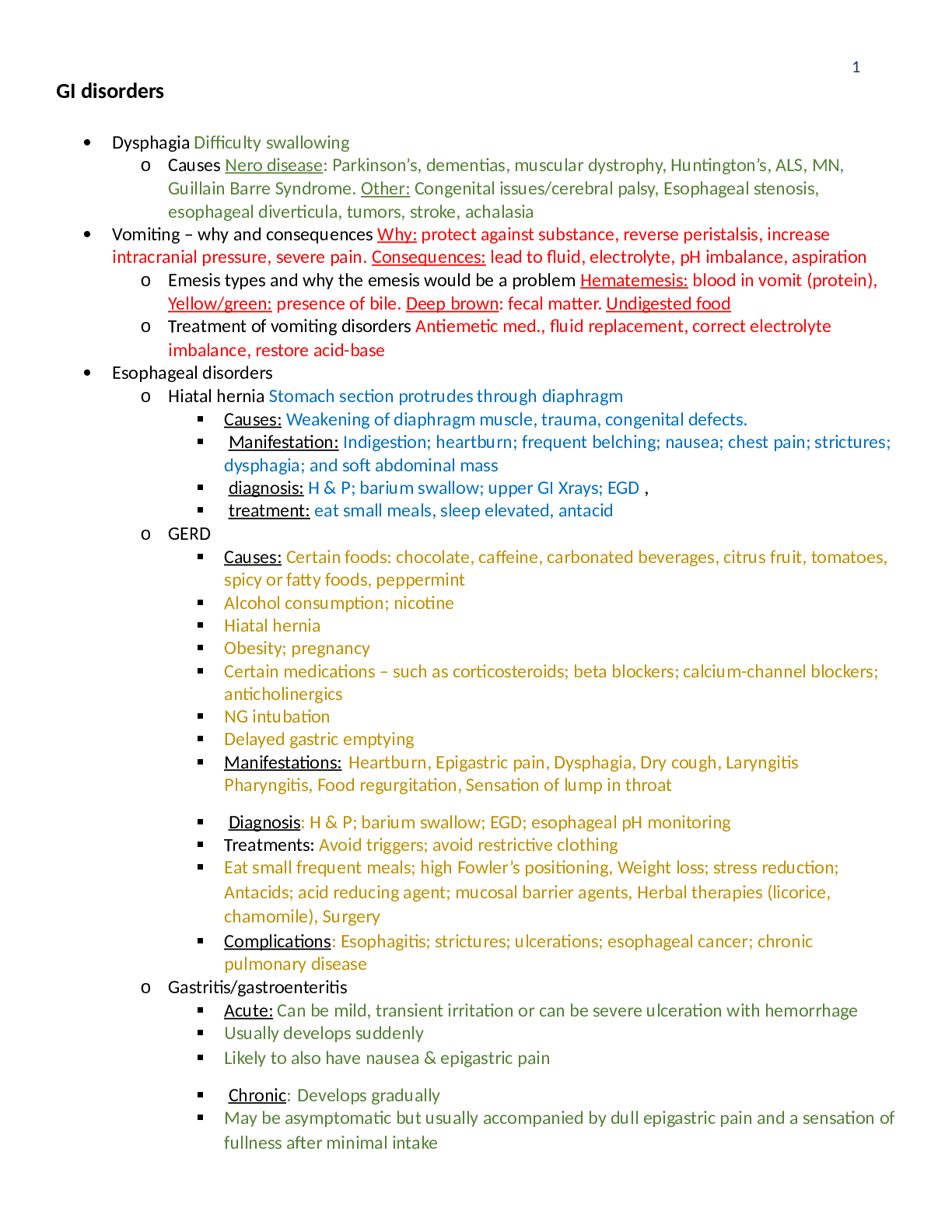
Rasmussen College :NUR 2063 Exam 2 Blueprint Fall 2021 GI disorders • Dysphagia Difficulty swallowing o Causes Nero disease: Parkinson’s, dementias, muscular dystrophy, Huntington’s, ALS,... MN, Guillain Barre Syndrome. Other: Congenital issues/cerebral palsy, Esophageal stenosis, esophageal diverticula, tumors, stroke, achalasia • Vomiting – why and consequences Why: protect against substance, reverse peristalsis, increase intracranial pressure, severe pain. Consequences: lead to fluid, electrolyte, pH imbalance, aspiration o Emesis types and why the emesis would be a problem Hematemesis: blood in vomit (protein), Yellow/green: presence of bile. Deep brown: fecal matter. Undigested food o Treatment of vomiting disorders Antiemetic med., fluid replacement, correct electrolyte imbalance, restore acid-base • Esophageal disorders o Hiatal hernia Stomach section protrudes through diaphragm ▪ Causes: Weakening of diaphragm muscle, trauma, congenital defects. Manifestation: Indigestion; heartburn; frequent belching; nausea; chest pain; strictures; dysphagia; and soft abdominal mass. diagnosis: H & P; barium swallow; upper GI Xrays; EGD, treatment: eat small meals, sleep elevated, antacid o GERD ▪ Causes: Certain foods: chocolate, caffeine, carbonated beverages, citrus fruit, tomatoes, spicy or fatty foods, peppermint , Alcohol consumption; nicotine, Hiatal hernia, Obesity; pregnancy, Certain medications – such as corticosteroids; beta blockers; calcium-channel blockers; anticholinergics, NG intubation, Delayed gastric emptying ▪ Manifestations: Heartburn, Epigastric pain, Dysphagia, Dry cough, Laryngitis Pharyngitis, Food regurgitation, Sensation of lump in throat ▪ Diagnosis: H & P; barium swallow; EGD; esophageal pH monitoring ▪ Treatments: Avoid triggers; avoid restrictive clothing, Eat small frequent meals; high Fowler’s positioning, Weight loss; stress reduction; Antacids; acid reducing agent; mucosal barrier agents, Herbal therapies (licorice, chamomile), Surgery ▪ Complications: Esophagitis; strictures; ulcerations; esophageal cancer; chronic pulmonary disease o Gastritis/gastroenteritis ▪ Acute: Can be mild, transient irritation or can be severe ulceration with hemorrhage, Usually develops suddenly, Likely to also have nausea & epigastric pain ▪ Chronic: Develops gradually ▪ May be asymptomatic but usually accompanied by dull epigastric pain and a sensation of fullness after minimal intake ▪ Complications: peptic ulcer; gastric cancer; hemorrhage ▪ H. pylori: Most common cause of chronic gastritis ▪ Bacteria embeds in mucous layer; activates toxins & enzymes that cause inflammation ▪ Genetic vulnerability & lifestyle behaviors (smoking, stress) may increase susceptible ▪ Other causes: Organisms through food/water contamination, LT NSAID use, Excess alcohol use, Severe stress, Autoimmune conditions ▪ Manifestations of GI bleeding: Indigestion; heart burn, Epigastric pain; abdominal cramping, N/V; anorexia, Fever; malaise, Hematemesis, Dark, tarry stools = ulceration & bleeding • GI tract disorders o Peptic ulcer disease ▪ Duodenal: Most commonly associated with excess acid or H.pylori infections, Typically present with epigastric pain relieved by food ▪ Gastric: Less frequent; more deadly, typically associated with malignancy and NSAIDs, Pain worsens with food ▪ Symptoms: ▪ Curling’s ulcer from what: associated with burns ▪ Cushing’s ulcer from what: associated with head injuries ▪ Complications of ulcers: GI hemorrhage; obstruction; perforation; peritonitis ▪ Manifestations: Epigastric or abdominal pain, Abdominal cramping, Heartburn; indigestion, N/V ▪ Diagnosis: same as gastritis ▪ Treatment: Same as for gastritis, Surgical repair may be necessary for perforated or bleeding ulcers, Prevention is crucial – may need prophylactic medications (ex: acid- reducers) for at-risk clients o Gallbladder disorders ▪ Cholelithiasis: Gallbladder stones ▪ Cholecystitis: Inflammation or infection in the biliary system caused by calculi ▪ Manifestations: Biliary colic; abdominal distension; N/V; jaundice; fever; leukocytosis ▪ Diagnosis: H & P; abdominal Xray; gallbladder US; laparoscopy ▪ Treatments: Low-fat diet, medications to dissolve calculi, Antibiotic therapy, NG tube with intermittent sxn, Lithotripsy, Choledochostomy, Laparoscopic surgery o Liver disorders ▪ Hepatitis – infectious: A, B, C, D, E vs. noninfectious: Giant cell hepatitis, Ischemic hepatitis, Non-alcoholic fatty liver hepatitis, Autoimmune hepatitis, Toxic & drug-induced hepatitis, Alcoholic hepatitis ▪ Transmission of viral hepatitis: If it’s a Vowel, it comes from the Bowel. All others are blood ▪ Define: acute: Proceeds through 4 stages—asymptomatic stage then 3 symptomatic stages chronic: Characterized by continued liver disease > 6 months, Symptom severity and disease progression vary by degree of liver damage, Can quickly deteriorate with declining liver integrity fulminant: Uncommon, rapidly progressing form that can quickly lead to ▪ Liver failure, hepatic encephalopathy, or death within 3 wks • Diagnosis: H & P, Serum hepatitis profile, Liver enzymes, Clotting studies, Liver biopsy, Abdominal US • treatment for viral hepatitis: treat with interferon & antiviral mediations ▪ Cirrhosis • Common causes: Hep C and chronic alcohol abuse most common cause in U.S. Hepatitis and all factors that can lead to hepatitis • What happens to liver: Leads to fibrosis, nodule formation, impaired blood flow, and bile obstruction liver failure • Manifestations: Portal hypertension, Varicosities, Bleeding –slow or severe, Muscle wasting, Bile accumulation, Clay-colored stools, Dark urine, Ulcers/GI bleeding, Encephalopathy, Spontaneous bacterial peritonitis • Diagnosis & treatments: H & P; liver biopsy; abdominal Xray; liver enzymes; EGD; clotting studies; stool exam for occult blood • Hepatic encephalopathy: o Pancreatitis ▪ Causes: Cholelithiasis, Alcohol abuse, Biliary dysfunction, Hepatotoxic drugs, Metabolic disorders, Trauma, Renal failure, Endocrine disorders, Pancreatic tumors, Penetrating peptic ulcer ▪ What happens to the pancreas in the disorder? pancreatic enzymes to leak into the pancreatic tissue and initiate autodigestion - -results in edema, vascular damage, hemorrhage & necrosis ▪ Acute pancreatitis importance & complications: Acute respiratory distress syndrome (ARDS), DM, Infection, Septic or hypovolemic shock, Disseminated intravascular coagulation (DIC), Renal failure, Malnutrition, Pancreatic cancer Pseudocyst – pancreatic fluids & necrotic debris accumulate & eventually rupture, Abscess • Manifestations: Upper abdominal pain that radiates to the back, worsens after eating, somewhat relieved by leaning forward or pulling knees to chest, N/V Mild jaundice, Low-grade fever, BP and pulse changes ▪ Chronic pancreatitis manifestations: Upper abdominal pain, Indigestion, Losing weight without trying, Steatorrhea, Constipation, Flatulence ▪ Pancreatitis diagnosis: H & P, Serum amylase & lipase, Serum calcium level, CBC, Liver enzymes, Serum bilirubin level, ABG, Stool analysis (lipid & trypsin levels), Abdominal Xray, CT/MRI, Abdominal US, ERCP (endoscopic retrograde cholangiopancreatography) ▪ treatment: Rest pancreas by fasting; administer IV nutrition; gradually advance diet from clears as tolerated to low fat, Pancreatic enzyme supplements when diet resumed Maintain hydration with IVF, NG to intermittent suction, Antiemetic agents, Pain management, Antacids/acid-reducing agents, Anticholinergic meds, Antibiotics, Insulin • Bowel disorders o Diarrhea – acute: Often d/t bacterial or viral infections, Certain medications such as antibiotics, antacids, laxatives, usually self-limiting depending on cause o Chronic: Lasts longer than 4 weeks o Causes: Inflammatory bowel diseases, Malabsorption syndromes, Endocrine disorders, Chemo/radiation ▪ Manifestations: If small bowel: Large and loose, provoked by eating, Usually accompanied with pain in right lower quadrant, If large bowel: Stools are small and frequent, Frequently accompanied by pain and cramping in the left lower quadrant, Acute diarrhea is generally infectious, Accompanied by cramping, fever, chills, N/V Blood (may be frank, occult, or melena), pus, or mucus may be present Bowel sounds may be hyperactive, Fluid, electrolyte and pH imbalances, Metabolic alkalosis ▪ Diagnosis/Bristol stool chart: H & P, include usual bowel habits and complete Bristol Stool chart, Stool analysis –include cultures & occult blood, CBC, Blood chemistry ABG, Abdominal US ▪ Treatment: Fasting for acute diarrhea with infection, Antidiarrheal agents maybe or not Antibiotics may be needed, Anticholinergics, Antispasmodic agents, Clear liquid diet until diarrhea subsides, then advance diet as tolerated. Dietary fiber to manage chronic diarrhea, maintain hydration and correct electrolyte and pH imbalance, Meticulous skin care in presence of incontinence o Constipation ▪ Causes: Low-fiber diet, Inadequate physical activity, Insufficient fluid intake, Delaying urge to defecate, Laxative abuse, Stress, Travel, Bowel diseases, Certain medications Mental health problems, Neuro diseases, Colon cancer ▪ Manifestations: Pain while passing stool ▪ Inability to pass stool after straining or pushing for more than 10 minutes ▪ No bowel movements for more than 3 days ▪ Hypoactive bowel sounds ▪ Complications: Anal bleeding, Anal fissure, pH disturbances, Hemorrhoids, Diverticulitis, Impaction, Intestinal obstruction, Fistula ▪ Diagnosis: History (including usual bowel pattern and completion of the Bristol Stool chart) and physical examination, including digital exam, Abdominal Xray, Upper GI series Barium swallow, Colonoscopy, Proctosigmoidoscopy ▪ Treatment: Increase dietary fiber along with hydration, Increase physical activity Defecate when initial urge present, Take stool softeners, Limit use of laxatives & enemas Digitally remove impaction o Intestinal obstructions ▪ Mechanical: Foreign bodies, Tumor, Adhesion, Hernia, Intussusception or volvulus, Strictures, Crohn’s disease, Diverticulitis, Hirschsprung’s disease, Fecal impaction ▪ Functional: (aka paralytic ileus): Neuro impairment, Intra-abdominal surgery complications, Chemical, electrolyte, mineral disturbances, Intra-abdominal infections Abdominal blood supply impairment, Renal and lung disease, Use of certain medications (ex: opiates) ▪ The problem with obstructions: Can develop suddenly or gradually and can be partial or incomplete, Chyme and gas accumulate at the site of the blockage, Saliva, gastric juices, bile, and pancreatic secretions begin to collect as blockage lingers, Intestinal blood flow can become impaired, leading to strangulation and necrosis, Intestinal contents can seep into the abdomen as pressure increases ▪ Manifestations: Abdominal distension, Abdominal cramping, Colicky pain, N/V Constipation/diarrhea, Borborygmi, Intestinal rushes, Decreased or absent bowel sounds, Restlessness, Diaphoresis, Tachycardia, Progressing to: Weakness, Confusion, Shock ▪ Diagnosis: H & P (including bowel patterns), Blood chemistry, ABG, CBC, CT Abdominal, Xray/US, Sigmoidoscopy, Colonoscopy ▪ Treatment: Strategies depend on underlying cause, Correct fluid, electrolyte, pH imbalances, NG tube to intermittent sxn, Fasting & TPN until bowel function restored Ambulation, Avoid laxatives until obstruction resolved, Surgery o Appendicitis ▪ What it is: Inflammation of vermiform appendix, Usually caused by infection Local edema triggered; fluid builds up and obstructs structure, Ischemia/necrosis develop, Pressure increases and forces out bacteria/toxins = perforation ▪ Complications: Abscess, Peritonitis, Gangrene, Death ▪ Manifestations: Range asymptomatic to sudden & severe Sharp pain develops and gradually intensifies over 12-24 hrs and localized to RLQ (McBurney point) When appendix ruptures, pain may temporarily subside, but pain will return and escalate, N/V, abdominal distension, changes in bowel pattern Indications of peritonitis, Abdominal rigidity, Tachycardia, Hypotension ▪ McBurney’s point: RLQ (McBurney point) ▪ Treatment: Surgery – open or laparoscopic – may need irrigation, Drainage tubes, LT antibiotics, Analgesics, Avoid activities that increase intra-abdominal pressure (ex: coughing & straining) o Peritonitis: Serious! Inflammation of peritoneum ▪ Causes: Chemical irritation (ex: ruptured gallbladder or spleen) ▪ Direct organism invasion (ex: appendicitis or peritoneal dialysis) ▪ Manifestations: Usually sudden & severe, Abdominal rigidity (classic), Abdominal tenderness & pain, Large volumes of fluid leak into peritoneal cavity, N/V, Decreased peristalsis, Intestinal obstruction, Indications of infection, Indications of sepsis Tachycardia, hypotension, restlessness, diaphoresis ▪ Diagnosis: H & P, CBC, Abdominal Xray, Abdominal US, Abdominal CT, Paracentesis with peritoneal fluid analysis, Laparoscopy ▪ Treatments: Surgery, LT antibiotics, Correcting fluid & electrolytes, NG tube with low intermittent sxn, TPN o Celiac disease ▪ What is this disorder? Inherited, autoimmune, malabsorption disorder Results from combination of the immune response to an environmental (gliadin) and genetic predisposition, Intestinal enzyme defect that prevents further digestion of gliadin (from gluten digestion) ▪ Complications: Anemia, Arthralgia, Myalgia, Bone disease, Dental enamel defects & discoloration, Intestinal cancers, Depression, Hair loss, Hypoglycemia, Mouth ulcers Increased bleeding tendencies, Neuro disorders, Skin disorders, Vitamin or mineral deficiency, Endocrine disorders ▪ Manifestations: Abdominal pain & distension, Bloating, gas, indigestion, Constipation, diarrhea, Changes in appetite, Lactose intolerance, N/V, Steatorrhea, Unexplained weight loss, Irritability, lethargy, malaise, Behavioral changes ▪ Diagnosis: H & P, duodenal biopsy, celiac blood panel (includes antibody/antigens immunoglobulins) ▪ Treatment: Gluten-free diet, Periodic monitoring for cancer, LT support o Inflammatory bowel diseases ▪ Crohn’s • Characteristics: patchy areas of inflammation involving full thickness of the intestinal wall and ulcerations, Form fissures divided by nodules, giving the intestinal wall a cobblestone appearance, Entire wall becomes thick & rigid; lumen becomes narrowed and potentially obstructed, Granulomas develop on intestinal wall and nearby lymph nodes • Complications: Malnutrition, Anemia (esp. iron deficiency), Fistula/perforation Adhesion, Abscesses, Intestinal obstruction, Anal fissure, Fluid, electrolyte, and pH imbalances • Manifestations: Abdominal cramping & pain (RLQ), Diarrhea/steatorrhea Constipation, Palpable abdominal mass, Melena, Anorexia, weight loss Indications of inflammatory reaction • Diagnosis: H & P, stool analysis, CBC, blood chemistry, Abdominal Xray, abdominal CT/MRI, Barium studies, Sigmoidoscopy, Colonoscopy, Biopsy • Treatment: Low-residue, high-calorie, high-protein diet, Oral supplements & multivitamins, TPN, Antidiarrheal agents, Medications, Surgical intestine resection, Stress management & support ▪ Ulcerative colitis • Characteristics: Included epithelial loss, surface erosion, and ulceration, Becomes inflamed, edematous, frail, Necrotic epithelial tissue can result in abscesses, Granulation tissue formed is fragile and bleeds easily • Complications: Malnutrition, anemia, Hemorrhage, perforation, Strictures, fistulas, Pseudopolyps, Toxic megacolon, Colorectal cancer, Liver disease • Manifestations: Diarrhea –frequent, up to 20/day, Watery stools with blood and mucus, Vomiting, weight loss, Indications of inflammatory process • Diagnosis: Similar to Crohn’s, Differences: barium enema, colonoscopy • Treatment: Similar to Crohn’s, Surgical intervention = ileostomy or colostomy ▪ IBS • Characteristics: exacerbations associated with stress, Includes alterations in bowel pattern & abdominal pain not explained by other causes, Less serious than IBD = no permanent intestinal damage • Complications: Hemorrhoids, nutritional deficits, social issues, sexual discomfort • Manifestations: Stress, mood disorders, some foods, and hormone changes often worsen symptoms, Abdominal distension, fullness, flatus, bloating Intermittent abdominal pain exacerbated by eating and relieved by defecation Chronic & frequent constipation or diarrhea, Nonbloody stool which may have mucus, Bowel urgency, Intolerance to certain foods, Emotional distress, Anorexia • Diagnosis (Rome III) Improvement with defecation, 2. Change in frequescy of stool, 3. Change in form of stool. • Treatment: Antidiarrheal agents, Laxatives, Antispasmodics, Antidepressants Avoid triggers, Maintain adequate fiber intake, Stress management, Support ▪ Diverticular disease • What it is: Conditions related to outpouching (diverticula) of intestinal wall layer that occur when mucosa sections or large intestine submucosa layers herniate through a weakened muscular layer. Thought to be d/t low fiber diet and poor bowel habits leading to chronic constipation • Diverticulosis: asymptomatic disease with multiple diverticula • Diverticulitis: diverticula are inflamed usually d/t retained fecal matter • Manifestations: Abdominal cramping followed by passing large quantity of frank blood, Low-grade fever, Abdominal tenderness (usually LLQ), Abdominal distension, Constipation/obstipation, N/V, Palpable abdominal mass, Leukocytosis • Diagnosis: H & P, Stool analysis (including for occult blood), Abdominal CT/MRI Colonoscopy, Barium enema, Biopsy • Treatment: High-fiber diet, Omitting foods with seeds, Decrease food intake during active bleeding, Adequate hydration, Colon resection, Proper bowel habits, Antibiotics, Analgesics • GI cancers o Oral cancer ▪ Risk factors: Tobacco product use, Alcohol consumption, Viral infections (esp. HPV) Immunodeficiencies, Inadequate nutrition, Poor dental hygiene, Exposure to UV light ▪ Manifestations: Lump, thickening, or soreness in mouth, throat, or tongue ▪ Difficulty chewing or swallowing ▪ Treatment: surgery, radiation, and speech therapy o Esophageal cancer ▪ Complications: Esophageal obstruction, Respiratory compromise, Esophageal bleeding ▪ Manifestations: Dysphagia, Chest pain, Weight loss, Hematemesis ▪ Treatment: Surgery, Chemo/radiation, Speech therapy o Stomach cancer ▪ Risk factors: Low-fiber diet, Constipation, H. pylori infection, Smoking, Pernicious anemia, Gastric polyps ▪ Manifestations: Abdominal pain & fullness, Epigastric discomfort, Palpable abdominal mass, Dark stools/melena, Dysphagia that worsens over time, Excess belching, N/V, hematemesis, anorexia, Unintentional weight loss, Weakness, fatigue ▪ Treatment: Gastrectomy, Chemo/radiation, Nutritional support o Liver cancer ▪ Manifestations: Anorexia, N/V, Jaundice, Abdominal pain (RUQ), Hepato- and splenomegaly, Edema, Third spacing, Ascites, Diaphoresis, Weight loss ▪ Treatment: Varies depending on primary site and progression of cancer Chemotherapy, Surgical removal, Liver transplant, Cryoablation, Radiofrequency ablation, Pure alcohol injected into tumor o Pancreatic cancer ▪ Risk factors: Family history, long-standing diabetes mellitus, Chronic pancreatitis, cirrhosis, Alcohol abuse, tobacco use ▪ Manifestations: Progressive upper abdominal pain that may radiate to the back, Jaundice, Dark urine, Clay-colored stools, Indigestion, anorexia, Weight loss, malnutrition, Hyperglycemia, Increased clotting tendencies ▪ Treatment: Surgical tumor removal, Chemo/radiation, Surgery or endoscopy repair of biliary leakages o Colorectal cancer ▪ Risk factors: Family history, Advanced age, Obesity, Tobacco use, Physical inactivity, Inflammatory bowel disease ▪ Manifestations: Lower abdominal pain & tenderness, Blood in stool (occult or frank), Diarrhea/constipation, Intestinal obstruction, Narrow stools, Unexplained anemia (usually iron deficiency), Unintentional weight loss ▪ Treatment: Removal during colonoscopy, Chemo, Surgical resection, Colostomy, Lifestyle changes Urinary system disorders • What is GFR and why is it important? Rate that blood flows through the glomerulus, Best indicator of renal function, Normal = 125mL/min • Kidney hormone function – what hormones, what do they do? Antidiuretic hormone (ADH): more water reabsorbed into blood • Aldosterone: Increases water an Na+, increases blood volume • Renin-angiotensin-aldosterone: arteries to constrict increasing blood pressure, decrease GFR resulting in water retention, increased thirst • Urinary incontinence o Difference between types listed in PPT: Enuresis: bedwetting after 4-5 years old Transient: temporary condition caused by Delirium, infection, Certain meds such as diuretics, sedatives, Psychologic factors, High urine output, restricted mobility, Fecal impaction Alcohol/caffeine use Stress: coughing, laughing, sneezing, exercise, weak sphincter, Urgent: sudden need to urinate following by involuntary loss of urine caused by UTI, bladder irritants, bowel conditions, smoking, Parkinson disease, stroke, injury, nervous system damage Reflex: Urinary incontinence caused by trauma or damage to nervous system Overflow: Inability to empty bladder or retention. May have dribbling and a weak urine stream Causes: bladder damage, urethral blockage, nerve damage, prostate condition Functional: Occurs in many older adults, especially in LTC with physical or mental impairment Mixed: Occurs when symptoms of more than one type of urinary incontinence are experienced Gross total: Continuous leakage of urine, day or night, Bladder has no storage capacity o Risk factors: Female, Advanced age, Overweight, Smoking, Other diseases & complications: Female, Advanced age, Overweight, Smoking, Other diseases o Diagnosis: H & P, Bladder diary, UA/UC, Cystourethrogram, Cystoscopy, Pelvic ultrasound, Postvoid residual measurement, Urodynamic testing o Treatment—simple to invasive: Bladder training, scheduled toileting, absorbent pads, skin barrier cream, pelvic floor exercise, medication, fluid/diet management, Pessary/urethral inserts, Sling procedure, Bladder neck suspension, Artificial urinary sphincter, Botox A injections into bladder muscle, Sacral nerve stimulator • Neurogenic bladder: Bladder dysfunction d/t interruption of normal bladder innervation o Causes: Brain or spinal cord injury/infections, Nervous system tumors, Dementia/Parkinson disease, Spinal bifida, Diabetes mellitus, Stroke, Medications, Vaginal childbirth, Multiple sclerosis, Chronic alcoholism, SLE, Heavy metal poisoning, Herpes zoster o Manifestations: Symptoms of an over- or underactive bladder, diagnosis: Bladder diary, UA/UC, Cystourethrogram, Cystoscopy, Pelvic ultrasound, Postvoid residual measurement, Urodynamic testing, treatment: dependent on cause and includes those for incontinence • UTIs o Risk factors: Female, BPH, Immobility, Urinary or bowel incontinence, Kidney stones (renal calculi), Urinary catheters, Decreased cognition, Pregnancy, Impaired immune response, Improper personal hygiene o Manifestations: May be asymptomatic, Urgency, Dysuria, Frequency, Hematuria, Bacteriuria, Cloudy/foul-smelling urine, Symptoms of infection/diagnosis: H & P, UA/UC, Cystoscopy, CBC treatments: Antibiotics, Increased hydration, Proper perineal care, Cotton undergarments, Do not delay urination, Adequately empty bladder, Proper urinary catheter care o Cystitis: Inflammation of the bladder, Due to infection & irritants • Pyelonephritis o What it is: Infection that located to one or both kidneys o Complications: Renal failure, Recurrent UTIs, Sepsis • Nephrolithiasis: kidney stones o Risk factors: pH changes, Excessive concentration of insoluble salts in urine, Urinary stasis, Family history, Obesity, Hypertension, Diet o Manifestations: Colicky pain in the flank area that radiates to lower abdomen and groin, Bloody, cloudy, or foul-smelling urine, Dysuria, Frequency, Genital discharge, N/V, Fever, chills o Diagnosis: H & P, urine exam, KUB, CT, US, IV pyelogram, calculi analysis, serum studies & treatment: Strain all urine, Increase fluid intake to 2.5-3.5 L, Lithotripsy, Ureteroscopy, Surgery, Pain management, Diet changes, Increase physical activity • Polycystic kidney disease (remember, this is genetic): Fluid-filled cysts in both kidneys o Manifestations: Hematuria, Nocturia, Drowsiness, In adults: Hypertension, Lumbar pain, Increased abdominal girth, Swollen, tender abdomen, Grossly enlarged, palpable kidneys o Diagnosis: H & P, UA, Blood chemistry, US, CT/MRI, IV pyelogram o treatment: Antibiotics, Analgesics, Antihypertensive agents, Adequate hydration, Low-salt diet, Surgical drainage of cyst abscesses or retroperitoneal bleeding, Dialysis o Kidney transplant • Cancers o Kidney (renal) cancer: Most frequent occurring. ▪ Manifestations: Asymptomatic, Painless hematuria, Abnormal urine color, Dull, achy flank pain, Urinary retention, Palpable mass over kidney, Unexplained weight loss, Anemia, Hypertension, Fever, Paraneoplastic syndromes ▪ Diagnosis: H & P, UA, CT/MRI, Bone scan, CXR, IV pyelogram, Renal arteriogram, Liver function, CBC/blood chemistry ▪ treatment: Surgical removal, Chemotherapy, Immunotherapy o Bladder cancer ▪ Risk factors: Advancing age, Males, Caucasians, working with chemicals, Smoking, Use of analgesics, Recurrent UTIs, LT urinary catheters, Chemo/radiation ▪ Manifestations: Painless hematuria, Abnormal urine color, Frequency/dysuria, UTIs, Back or abdominal pain ▪ Diagnosis: H & P, UA, CT/MRI, Bone scan, CXR, IV pyelogram, Renal arteriogram, Liver function, CBC/blood chemistry ▪ treatment: Surgical removal, Radiation, Chemotherapy, Immunologic agents • Intrarenal disorders o Glomerulonephritis – what it is: Bilateral inflammatory disorder of glomeruli typically after strep infection ▪ Diagnosis: ▪ Treatment o Nephrotic syndrome: Results from antibody-antigen complexes lodging in glomerular membrane, Triggers activation of complement system o vs. nephritic syndrome: Inflammatory injury to glomeruli that may occur d/t antibodies interacting with normally occurring antigens in the glomeruli ▪ Causes: Systemic diseases, Gold therapy, May be idiopathic. Diseases which initiate the inflammatory process ▪ Complications: Risk for infection & atherosclerosis. impaired renal function ▪ Manifestations: Gross hematuria, Urinary casts & leukocytes, Low GFR, Azotemia, Oliguria, ▪ High blood pressure • Renal failure o Acute vs. chronic ▪ Acute • Causes: Prerenal: Extremely low BP or blood volume, Heart dysfunction Intrarenal: Reduced blood supply to kidneys, Hemolytic uremic syndrome, Renal inflammation, Toxic injury Postrenal: Ureter obstruction, Bladder obstruction/dysfunction • Risk factors: Advanced age, Autoimmune disorders, Liver disease • Phases: 1. Asymptomatic phase 2. Oliguric phase: Daily urine output is 400ml or less, Waste products accumulate 3. Diuretic phase: Daily urine increases up to 5 L per day, Nephrons attempting to heal 4. Recovery phase: Glomerular function gradually returns to normal o Manifestations: Oliguric phase: Decreasing urine output, Electrolyte disturbances ( K), Fluid volume excess, Azotemia (BUN: Cr ratios abnormal), Metabolic acidosis Diuretic phase: Increased urine output Electrolyte disturbances, Dehydration, Hypotension, Recovery phase: Symptoms begin to resolve • Diagnosis: H&P, Blood chemistry, ABGs, UA, CBC, Renal ultrasound, Biopsy • Treatment: Correct fluid/electrolyte imbalances, Diet high in calories, Restrict: Protein, Sodium, Potassium, Phosphates, Hypertension management, Infection prevention strategies, ST dialysis, Treat anemia with Epogen ▪ Chronic • Causes (most common causes!): Diabetes mellitus, HTN • Stages and associated GFR: Stage 1: Kidney damage but GFR normal or increased (>90) Stage 2: Mildly decreased GFR (60-89) Stage 3: Moderately decreased GFR (30-59) Stage 4: Severely decreased GFR (15-29 Stage 5: ESRD (<15); need dialysis • Manifestations: neurological weakness, fatigue, confusion. High BP, pitting edema, ammonia odor to breathe, • Diagnosis: H & P, UA, Blood chemistry, CT/MRI, Renal ultrasound, Biopsy, CBC, ABGs • Treatment: Manage and prevent complications Alternative medications: Epogen, Phosphate management, Potential antibiotics, Dialysis vowels: A: acid-base problems, E: electrolyte problems, I: intoxication O: overload of fluids U: uremic symptoms Reproductive disorders • Male alterations o Epispadias: Urethral meatus appears on dorsal surface of penis, Urinary defects also often present: inside out bladder o Diagnosis: Physical exam, Determine any other conditions, IV pyelogram, X-ray, CT/MRI Ultrasound o Treatment: Surgical repair of urethral meatus open, Surgical repair of bladder o Hypospadias: Urethral meatus appears on ventral surface of penis ▪ Diagnosis: H & P, Imaging ▪ Treatment: Surgical repair in stages, Usually done as infant o Testicle disorders ▪ Cryptorchidism • Risk factors: Prematurity, Low birth weight/size, Multiple fetuses, Maternal use of, Estrogen use during first trimester, Alcohol use during pregnancy, Tobacco use or secondhand smoke, Maternal diabetes, Parental exposure to pesticides • Diagnosis: H & P, Self-testicular exams, Abdominal US, MRI, Hormone levels, Genetic studies • Treatment: May not be required, Manual manipulation, Hormones or surgery Testicle implants o Erectile dysfunction ▪ Causes: Circulatory impairment, Diabetes mellitus, MS, Prostate disease, HTN, Neuro dysfunction, Medications, Low testosterone, Alcohol & tobacco use, Cirrhosis ▪ Diagnosis: H & P, Hormone analysis, US, Penile structure and function exams ▪ Treatment: Identify & manage underlying cause, Psychological counseling, Testosterone replacement, Phosphodiesterase inhibitors, Implants, Vascular surgery o Priapism: Prolonged, painful erection ▪ Causes: Sickle cell disease, leukemia, trauma, diabetes, spinal cord injuries, neuro diseases, medications, alcohol/illicit drugs, poisonous venom ▪ Diagnosis: H & P, ultrasound, CBC, toxicology, CT ▪ Treatment: Needle aspiration of blood; injection of medication into penis; cold application; urinary catheterization; if trauma –surgical repair o Phimosis (review in book) & o Paraphimosis: Foreskin retracted and cannot be replaced, Constricts penis & becomes edematous, Medical emergency – if not repaired can lead to gangrene Treatment: Circumcision, foreskin stretching, replace foreskin after hygiene o Testicular torsion ▪ What is it: Abnormal rotation of testes on spermatic cord ▪ Causes: trauma; may be spontaneous ▪ Manifestations: sudden scrotal pain & edema ▪ Diagnosis: H & P; scrotal ultrasound ▪ Treatment: manual manipulation and surgery o Hydrocele: Fluid accumulation – can affect one or both testes o Causes: congenital defect; inflammation; infection; trauma; tumors o Diagnosis: painless scrotal enlargement & scrotal heaviness o Treatment: may not be required; scrotal elevation; heat/cold application; aspiration; surgical removal o Spermatocele: Sperm-containing cyst between testes & epididymis o Manifestations: painless, small, movable cyst o Cause: unknown; may be blockage of duct system; infection, inflammation, or trauma o Diagnosis: similar to hydrocele o Treatment: usually not required but may need surgical removal if large o Varicocele ▪ What it is: Dilated vein in spermatic cord; more common in left testicle d/t anatomic factors ▪ Causes: congenital defects and obstructions ▪ Manifestations: ‘bag of worms’ feeling in scrotum; scrotal heaviness ▪ Treatment: often unnecessary; surgical repair; embolectomy; sclerotherapy o Prostate disorders ▪ Prostatitis • Female alterations • Causes: Conditions that trigger the inflammatory process • Diagnosis: H & P, including digital rectal exam, UA/UC, Cystoscopy, Transrectal US • Manifestations: Dysuria, Frequency/urgency, Nocturia, Pain in: Abdomen, Groin, Lower back, Perineum, Genitals, Painful ejaculation, S/sx of infections • Treatment: LT organism-specific antibiotics, Analgesics/antipyretics, Hydration, Prostatic massage o Menstrual disorders – causes, diagnosis, manifestations, treatment ▪ Amenorrhea: Absence of menstruation ▪ Causes: Genetic, Hypothalamus disorders, Stress, Sudden weight loss, Extreme reduction in body fat, Chemotherapy, Pregnancy (of course) & lactation, Menopause ▪ Dysmenorrhea: Painful menstruation ▪ Causes: Unknown, Reproductive conditions, Endometriosis, repro cancers, After childbirth ▪ Diagnosis: H & P, pelvis US, laparoscopy, hysteroscopy ▪ Treatment: Analgesics, OCPs, heat application ▪ Menorrhagia: Increased menstrual flow (apprx. 80 ml) and duration (8-10 days) metrorrhagia: Premenopausal bleeding between periods ▪ Polymenorrhagia: Short, less than 21 days, menstrual cycle = frequent menstruation ▪ Oligomenorrhea: Long (>42 days) menstrual cycle, resulting in infrequent menstruation o Premenstrual syndrome ▪ Manifestations: mood swings, headache, fatigue, ache, food craving, tender/swallow breast, cramps, constipation, diarrhea ▪ Premenstrual dysphoric syndrome ▪ Diagnosis: H & P – gyn complaints ▪ Treatment: Hormone therapy, diuretics, Antidepressants, comfort measures, Analgesics, Decrease intake: Caffeine, Soda, Chocolate, Fat, Processed sugars, Alcohol o Pelvic support disorders: Supportive structures weakened o Causes: Weakened pelvic support from excess straining o Childbirth, chronic constipation, heavy lifting o Manifestations: May be asymptomatic, Visualization of bladder from vaginal opening, Stress incontinence, Urinary retention, Frequency, urgency, Pain or urine leakage during sexual intercourse o Treatment: Pessary devices, surgical repair, estrogen therapy (if postmenopausal), incontinence interventions, Kegel exercises, avoidance of straining ▪ Cystocele: Bladder protrudes into anterior wall of vagina ▪ Rectocele: Rectum protrudes through posterior wall of vagina ▪ Uterine prolapse: Descent of uterus or cervix into the vagina o Uterine disorders ▪ Endometriosis: Endometrial tissue grows in areas outside the uterus Most common areas: Fallopian tubes, Ovaries, Perineum • Complications: Pain, cysts, scarring, adhesions, infertility • Manifestations: Dysmenorrhea, menorrhagia • Pelvic pain, pain during/after intercourse • Treatment: Analgesics, Hormone therapy, Surgical repair ▪ Leiomyoma: ‘Fibroids’ –firm, rubbery growth of the myometrium -size varies • Cause: Unknown, but seems to grow during menstrual years in presence of estrogen and shrink after menopause • Manifestations: asymptomatic; menorrhagia; pain in pelvis, back or legs; urinary frequency and retention; UTIs; constipation; abdominal distention; pain during intercourse; anemia • diagnosis: H & P; abdominal transvaginal ultrasound; hysteroscopy; biopsy; laparoscopy; CT/MRI; CBC • treatment: may not need treatment; monitoring; hormone therapy; analgesics; surgery; myolysis; endometrial ablation; anemia treatment o Ovarian cysts: Benign, fluid-filled sacs on ovary. Often form during ovulation process. May rupture, causing discomfort ▪ Manifestations: Asymptomatic; abdominal pain or discomfort; abnormal menstrual bleeding; abdominal distention ▪ Diagnosis: H & P; CT/MRI; abdominal US; laparoscopy; biopsy; hormone ▪ Treatment: May not be required; hormone therapy; analgesics; manage metabolic & other disorders; and surgery o Polycystic ovary syndrome ▪ Manifestations: Infertility (anovulation), Amenorrhea, Hirsutism, Acne o Breast disorders ▪ Fibrocystic breast condition • What it is: Numerous benign nodules in the breast • What causes this to happen: More frequent during childbearing years, Contributing factors, Family history, High-fat diet, Excessive caffeine intake • Manifestations: Dense, irregular, bumpy breast tissue, Dull, heavy breast pain & tenderness, Feeling of breast fullness, Occasional non-bloody nipple discharge • Diagnosis: H & P, Mammogram, Breast US, Biopsy • Treatment: Usually not necessary, Needle aspiration of fluid, Surgical removal of cysts, Analgesics, Limit dietary fat, salt, caffeine, Avoid OCPs ▪ Mastitis • What it is – why is this happening? • Manifestations: Breast tenderness, Swelling, redness or warmth, Pain or a burning sensation continuously or while breastfeeding, Fever, malaise • Treatment: Antibiotic therapy, hydration, rest, analgesics, supportive bra, cold application, adequate milk expression, needle aspiration o Candidiasis (yeast infection) ▪ Cause: Antibiotic therapy, Bubble baths, Feminine products, Decreased immune response, Increase glucose in vaginal secretions ▪ Manifestations: Thick white, vaginal discharge that resembles cottage cheese, Vulvular erythema and edema, Vaginal and labial itching and burning, White patches on vaginal walls, Dysuria, Painful intercourse ▪ Treatment: antifungal agents, Perineum care – avoid douching Resist urge to scratch, Eat yogurt or take Lactobacillus acidophilus tablets, Avoid feminine sprays, fragrances or powders, Control blood glucose if diabetic, Use feminine pads instead of tampons during menstruation o Pelvic inflammatory disease – either acute or chronic ▪ Causes: STIs; bacteria introduced during childbirth, Endometrial procedures; abortions, Bacterial invasion from bloodstream ▪ Complications: Reproductive structure obstructions; peritonitis, Abscesses; septicemia, Adhesions; strictures, Chronic pelvic pain; ectopic pregnancies, Infertility; problems with surrounding structures ▪ Manifestations: Indications of infection, Pain or tenderness in pelvis, lower abdomen, or lower back, Abnormal vagina or cervical discharge, Bleeding after intercourse, Painful intercourse, Urinary frequency, Dysuria, Dysmenorrhea; metrorrhagia, Anorexia; N/V ▪ Diagnosis: H & P; culture discharge; CBC; pelvic US; CT; laparoscopy ▪ Treatment: Antibiotic therapy, Screen and treat sex partner(s), Practice safe sex Avoid douching, Treat abscesses, Follow up exam, Infertility evaluation Sexually transmitted diseases • Bacterial o Which are reportable to CDC? o Chlamydia ▪ Transmission ▪ Complications ▪ Manifestations, diagnosis, treatment o Gonorrhea ▪ Transmission ▪ Complications ▪ Manifestations, diagnosis, treatment o Syphilis ▪ Transmission ▪ Stages ▪ Complications ▪ Manifestations, diagnosis, treatment • Viral o Herpes simplex 1 & 2 (how do these present—meaning what triggers this infection?) ▪ Transmission ▪ Complications ▪ Manifestations, diagnosis, treatment o Human papilloma virus (HPV) ▪ Complications of HPV ▪ Transmission ▪ Manifestations, diagnosis, treatment • Trichomonas vaginalis (good to know info) Reproductive system cancers • Those with high rates of treatment vs. those with high mortality rates • Prostate cancer o Diagnosis o Manifestations o Complications o Treatment • Testicular cancer o Diagnosis o Manifestations o Complications o Treatment • Breast cancer o Diagnosis (genetics?) o Manifestations o Complications o Treatment • Cervical cancer o Causes o Diagnosis o Manifestations o Complications o Treatment • Endometrial (uterine) cancer o Diagnosis o Manifestations o Complications o Treatment • Ovarian cancer o Diagnosis o Manifestations o Complications o Treatment Endocrine disorders • Pituitary disorders o Hypopituitarism: Condition which pituitary does not produce some or all of hormones. May be insufficient quantities ▪ Dwarfism: Short stature d/t deficient levels of growth hormone, somatotropin or somatotropin-releasing hormone ▪ Diabetes insipidus: Excess fluid excretion (water!) in kidneys caused by insufficient ADH levels o Causes: Cerebral or pituitary trauma, Autoimmune conditions, Pituitary tumors, Sarcoidosis o Manifestations: Fatigue, headache, Cessation of menstruation, infertility (women), Decreased libido, Low stress tolerance, Weight loss or gain, Short stature; delayed growth and development, Thirst; excess urination o Treatment: Lifelong hormone replacement o Hyperpituitarism: Secretion of excess amounts of one or all pituitary hormones ▪ Gigantism ▪ Acromegaly ▪ SIADH: Syndrome of Inappropriate Antidiuretic Hormone ▪ Cushing’s syndrome ▪ Hyperthyroidism o Causes: Most commonly caused by tumors that secrete hormone or hormone-like substances o Manifestations: Headache; vision changes; excess sweating; sleep apnea; joint pain and stiffness; muscle weakness; paresthesia o Treatment: Depends on underlying cause, Tumors usually require surgery, radiation/chemo • Thyroid disorders o Hypothyroidism (Hashimoto’s vs. iatrogenic) ▪ Diagnosis: H & P, Serum thyroid hormone levels, Serum TSH (thyroid-stimulating hormone), Cholesterol panel ▪ Manifestations: Fatigue, sluggishness, Sensitivity to cold, unexplained weight gain, high cholesterol, Facial edema, hoarseness, hair thinning or loss, brittle nails, Myalgia, arthralgia, muscle weakness, pale/dry skin, Bradycardia, hypotension, Depression, goiter ▪ Complications: Myxedema: Rare, life-threatening advanced hypothyroidism Manifestations: Marked hypotension, Respiratory depression, Hypothermia, Lethargy, Coma ▪ Treatment: Thyroid hormone replacement (ex: levothyroxine), Weight management, Constipation measures, Avoid cold temperatures o Hyperthyroidism: Excess levels of thyroid hormones = hypermetabolic state ▪ Diagnosis: H & P, Serum thyroid levels; serum TSH, Radioactive iodine uptake test, Thyroid scan ▪ Manifestations: Sudden weight loss/increased appetite, Tachycardia/hypertension, Nervousness, anxiety or anxiety attacks, Irritability/difficulty sleeping, Diaphoresis, Sensitivity to heat, Goiter, Exophthalmos ▪ Complications: ▪ Treatment: Radioactive iodine/antithyroid agents, Beta blockers, Surgery, Strategies for exophthalmos, Increased caloric and calcium intake • Adrenocortical disorders o Cushing’s syndrome (excessive cortisol) ▪ Diagnosis ▪ Manifestations ▪ Complications ▪ Treatment o Addison’s disease (deficiency of cortisol) ▪ Diagnosis ▪ Manifestations ▪ Complications ▪ Treatment • Adrenal medulla disorders o Pheochromocytoma ▪ Release of catecholamines (epi/norepi): rare tumor ▪ Manifestations: Hypertension, tachycardia, forceful heartbeat, Profound diaphoresis Sudden onset of severe headaches, Anxiety, feeling of extreme fright, pallor, weight loss Abdominal pain ▪ Can be life-threatening/complications: Hypertensive crisis; stroke; renal failure; psychosis; seizures ▪ Diagnosis: H & P; serum levels of epinephrine & norepinephrine ▪ Abdominal CT/MRI; biopsy ▪ Treatment: Surgery and antihypertensive medications • Parathyroid disorders o Calcium regulation • Antidiuretic hormone disorders o Diabetes insipidus o SIADH Diabetes • Type I diabetes o Cause o Complications o Manifestations o Diagnosis o Treatment • Type II diabetes o Cause o Complications o Manifestations o Diagnosis o Treatment • Gestational diabetes o Risk factors o Treatment • Metabolic syndrome o What is it? o Diagnosis o Treatment Medications: (Therapeutic Effect) Medication Levothyroxine Medication Insulin Medication • Glyburide • Metformin Medication: • PPIs (Omeprazole) • H2 blockers (ranitidine) Laboratory Values Essentials of Pathophysiology (NUR 2063) – Exam 2 blueprint 18 [Show More]
Last updated: 2 years ago
Preview 1 out of 35 pages

Buy this document to get the full access instantly
Instant Download Access after purchase
Buy NowInstant download
We Accept:

Reviews( 0 )
$16.00
Can't find what you want? Try our AI powered Search
Document information
Connected school, study & course
About the document
Uploaded On
Feb 17, 2022
Number of pages
35
Written in
Additional information
This document has been written for:
Uploaded
Feb 17, 2022
Downloads
0
Views
30


