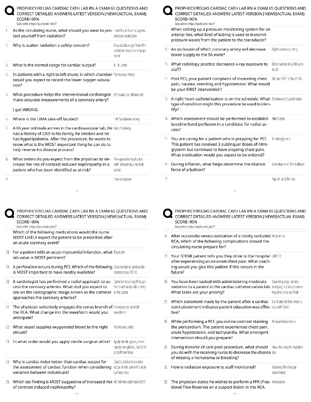
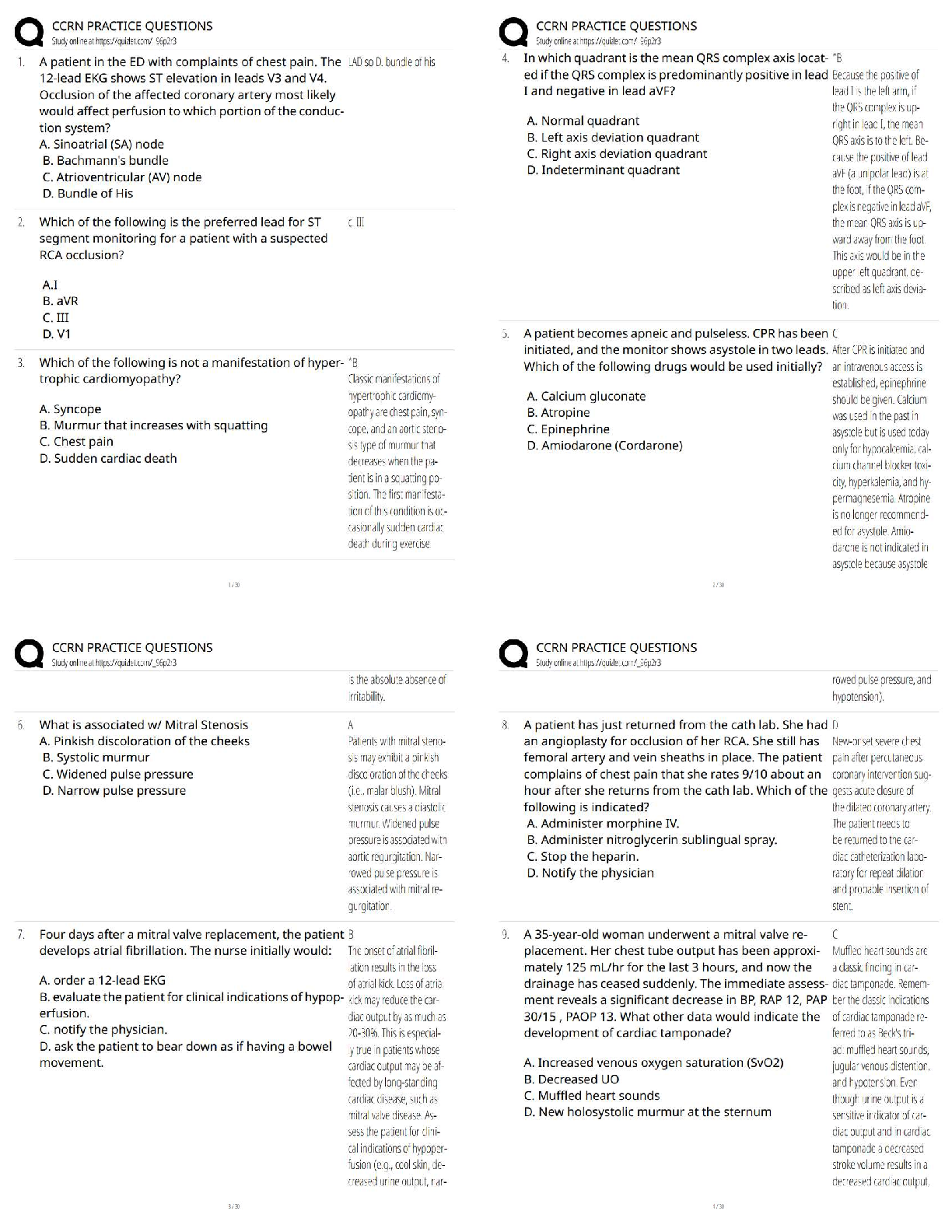
Cardiology > QUESTIONS & ANSWERS > Cardiology EKG Rhythms and 60 Interventions. (All)
Cardiology EKG Rhythms and 60 Interventions.
Document Content and Description Below
Last updated: 3 years ago
Preview 1 out of 12 pages
Instant download

Buy this Document to get the Full Access Instantly
Provided by Students Who Aced it
We Verify Document Content to Gurantee Accuracy
Reviews( 0 )
Document information
Connected school, study & course
About the document
Uploaded On
May 07, 2022
Number of pages
12
Written in
All

Additional information
This document has been written for:
Uploaded
May 07, 2022
Downloads
0
Views
237
Document Keyword Tags
Recommended For You
Get more on QUESTIONS & ANSWERS »
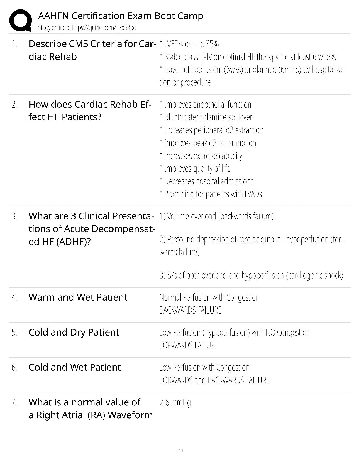
AAHFN Certification Exam Boot Camp / Heart Failure Nursing / 2...
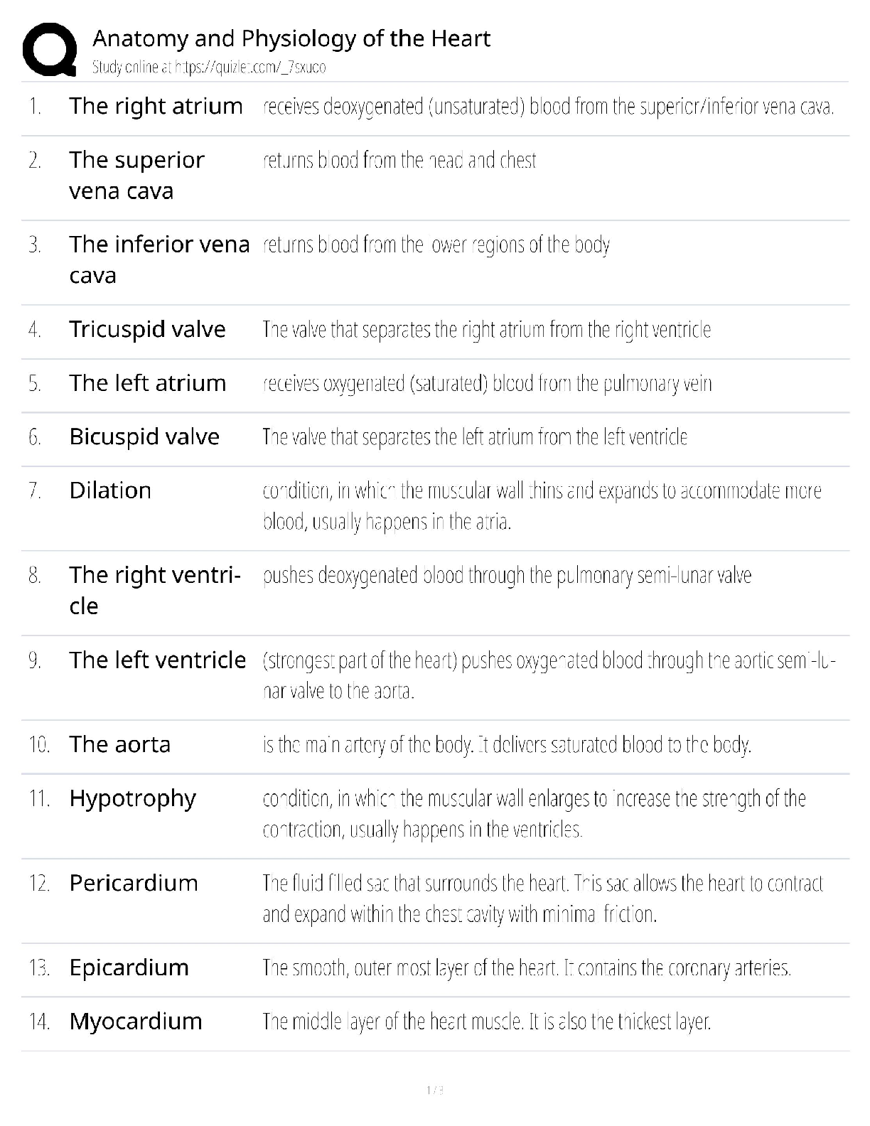
Heart Anatomy & Physiology / Cardiology Exam / 2025 Practice T...
.png)

Advanced Cardiovascular Life Support (ACLS) Exam Version A (5...
.png)
.png)
CCI Echocardiography Practice Questions with COMPLETE SOLUTION...
.png)
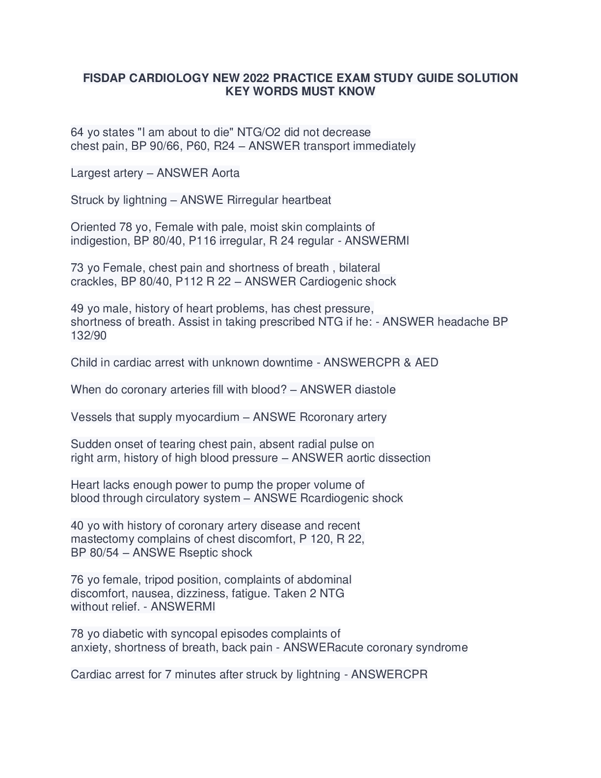
FISDAP CARDIOLOGY NEW 2022 PRACTICE EXAM STUDY GUIDE SOLUTION...
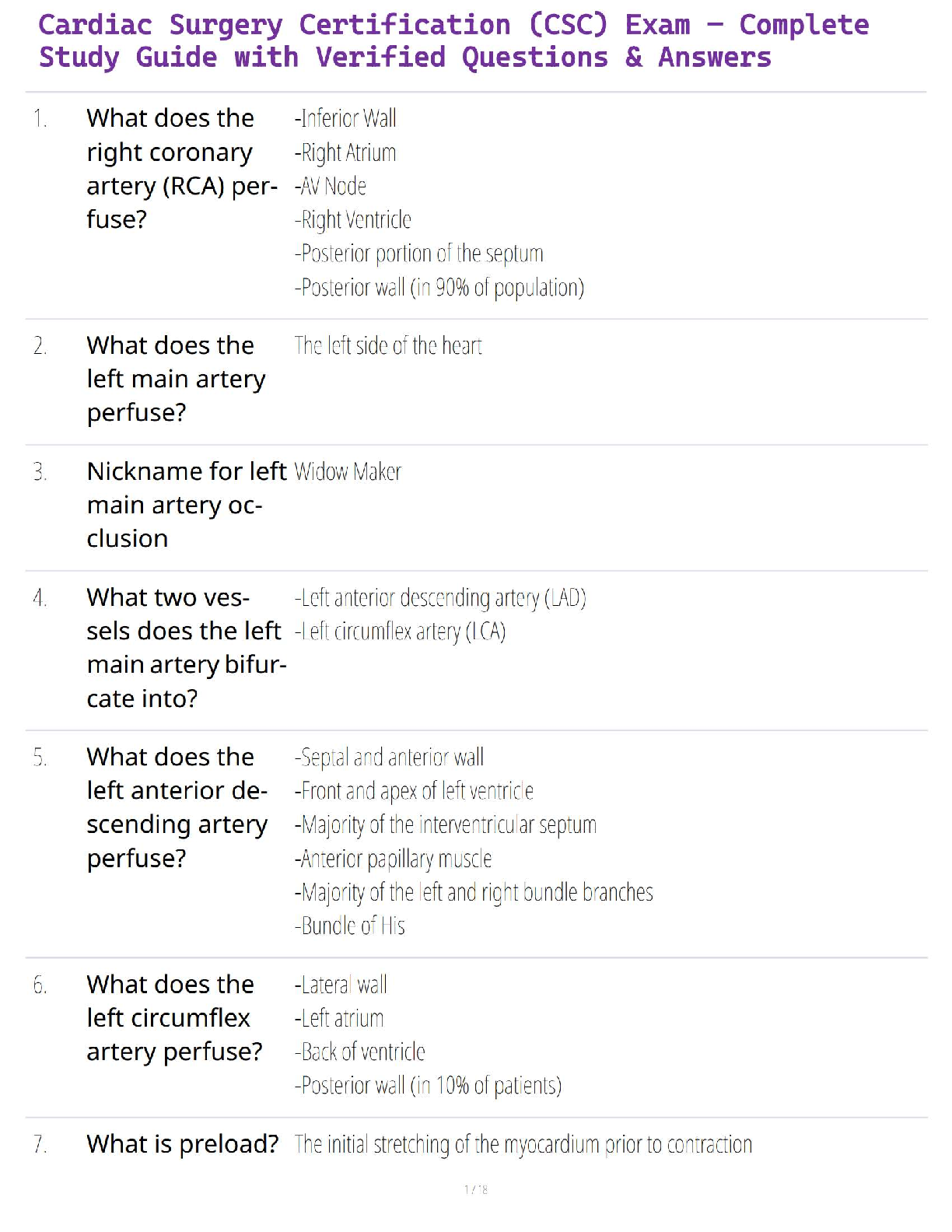
Cardiac Surgery Certification (CSC) Exam – Complete Study Guid...
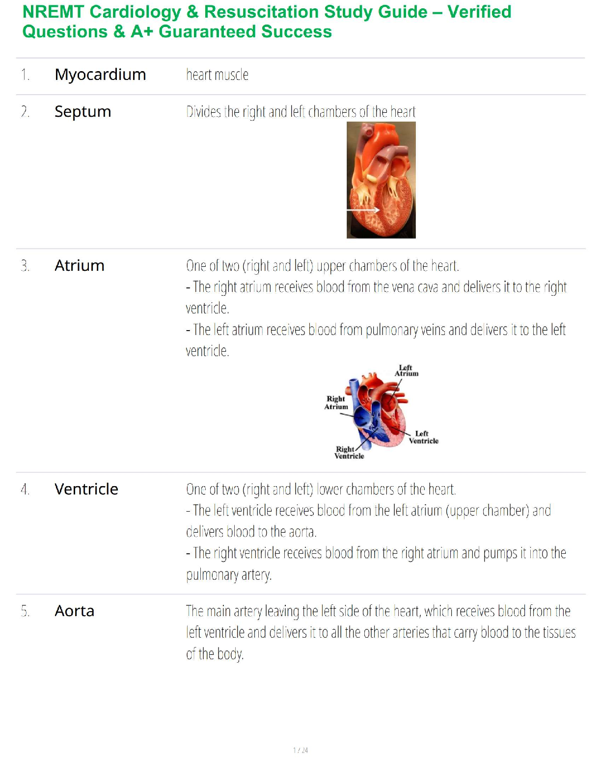
NREMT Cardiology & Resuscitation Study Guide – Verified Questi...