BIO 152 A&P 2 Module 1 Problem Set
Anatomy and Physiology of the Nervous System: Introduction
1. What is the difference between anatomy and physiology?
2. The function of the nervous system is to integrat
...
BIO 152 A&P 2 Module 1 Problem Set
Anatomy and Physiology of the Nervous System: Introduction
1. What is the difference between anatomy and physiology?
2. The function of the nervous system is to integrate and control the other body systems. Explain how the nervous system does this.
3. List the 2 parts of the nervous system.
4. How are the parts of the central nervous system protected?
5. Collections of cell bodies inside the central nervous system are
called , and the collection of nerve axons in the central
nervous system are called .
6. What is included in the peripheral nervous system?
7. Collections of cell bodies inside the peripheral nervous system are called , and the collection of nerve axons in the peripheral nervous system are called .
8. What are the 2 divisions of the peripheral nervous system?
9. Describe the movement of nerve impulses in the peripheral nervous system.
10. What are the 2 divisions of the efferent division of the peripheral nervous system?
11. What is controlled by the somatic and autonomic nervous systems?
12. What are the 2 divisions of the autonomic nervous system?
13. What is the function of the sympathetic nervous system?
14. What is the function of the parasympathetic nervous system?
Your Answer:
1. Anatomy focuses on the structure of the body whereas physiology looks at how the body parts function together
2. The nervous system receives and processes information and sends out signals to the muscles and glands to elicit an appropriate response. The nervous system integrates and controls the other systems of the body
3. Brain and spinal cord
4. Skull protects the brain and the vertebrae protect the spinal cord
5. Nuclei and tracts
6. All the nerves not in the brain or spinal cord
7. Ganglia and nerves
8. Sensory (afferent) and motor (efferent)
9. The PNS receives impulses from the sensory organs via the afferent division and then relays signals or impulses from the CNS to muscles and glands via the motor or efferent division
10. Somatic and autonomic
11. The somatic system nerves control skeletal muscles, skin, and joints. The autonomic system nerves control the glands and smooth
muscles of the internal organs and are not generally under conscious control
12. Sympathetic and parasympathetic
13. Activates and prepares the body for vigorous muscular activity, stress, and emergencies
14. Lowers activity, operates during normal situations, permits digestion, and conserves energy
1. Anatomy focuses on the structure of the
body. Physiology looks at how the body parts function together. The physiology of the body depends on the anatomy of the body.
2. The nervous system receives and processes information and sends out signals to the muscles and glands to elicit an appropriate response. In this way, the nervous system integrates and controls the other systems of the body.
3. CNS and PNS (central nervous system and peripheral nervous system)
4. The skull protects the brain and the vertebrae protect the spinal cord.
5. nuclei, tracts.
6. The PNS includes the cranial nerves and the spinal nerves.
7. ganglia, nerves.
8. Afferent, efferent
9. The peripheral nervous system receives impulses from the sensory organs via the afferent division and then relays signals or impulses from the central nervous system to muscles and glands via the motor or efferent division.
10. Somatic system and the autonomic system.
11. The somatic system controls the movements of skeletal muscles, skin, and joints. The autonomic system nerves control the glands and smooth muscles of the internal organs.
12. sympathetic and parasympathetic nervous systems.
13. The sympathetic nervous system activates and prepares the body for vigorous muscular activity, stress, and emergencies.
14. The parasympathetic nervous system generally operates during normal situations, permits digestion, and conserves energy.
Question 2
0 / 0 pts
Neurons
15. Label the components of a neuron.
16. List 3 unusual characteristics of neurons.
17. List the 3 parts that all neurons contain.
18. Describe the structure and function of the neuron cell body.
19. Describe the structure and function of the dendrite.
20. Describe the axon, including the number in each neuron, function, structure and organelles.
21. Describe the function and site of synthesis and storage of neurotransmitters.
22. What is a synapse?
23. A post-synaptic neuron is a neuron that is found the synapse.
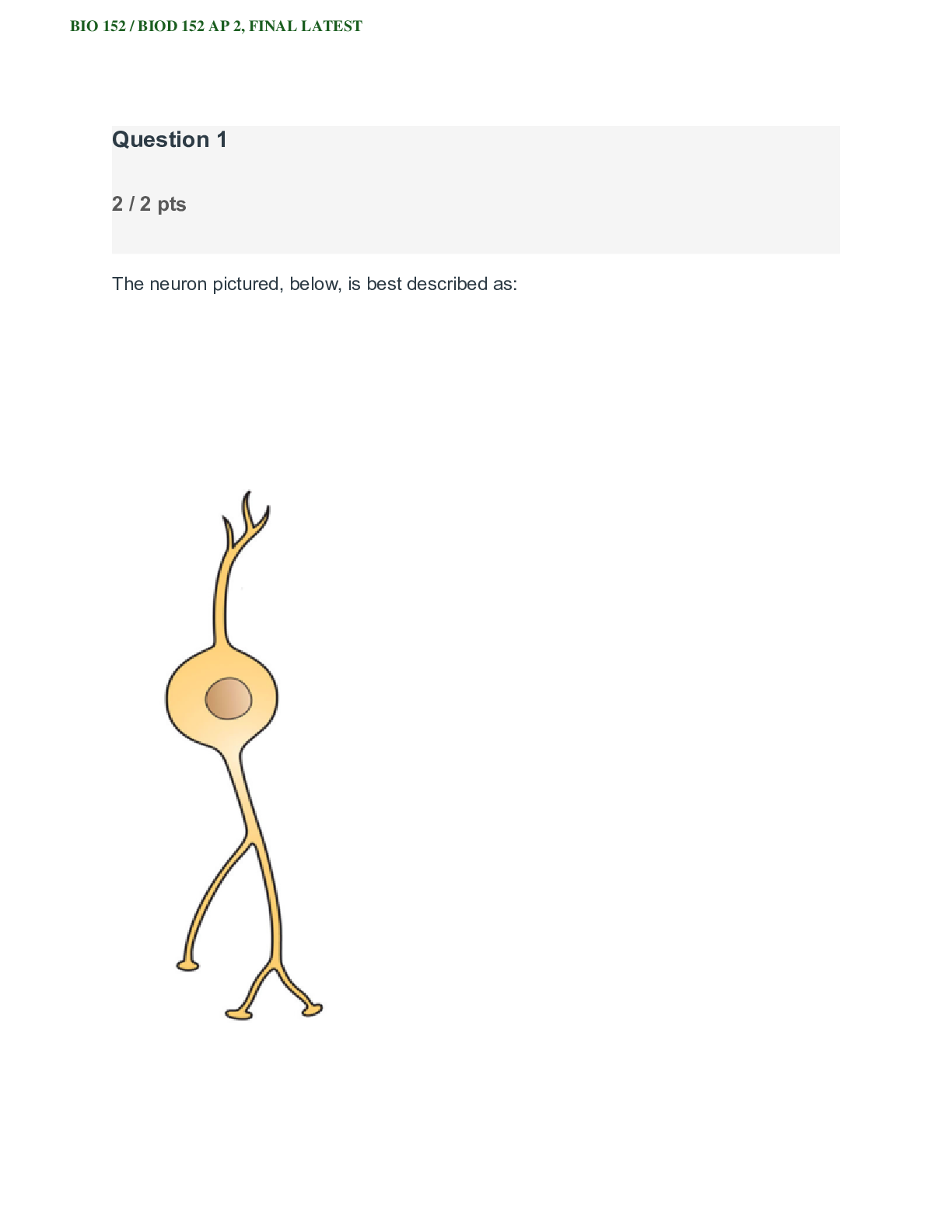
24. neurons have three or more extensions from the cell body and have one axon and many dendrites.
25. neurons have a central cell body with two extensions.
26. neurons have one extension off the cell body which branches into two: one central process running to the CNS and another peripheral process running to the sensory receptor.
27. neurons are unipolar and function to carry information from the peripheral to the central nervous system.
28. These types of neurons are also called association neurons.
29. neurons send messages from the central nervous system to the peripheral.
Your Answer:
15. dendrite, cell body, nucleus, axon, myelin sheath, schwann cell, node of ranvier, axon terminal
16. Neurons do not undergo mitosis, require enormous amounts of fuel being able to survive just minutes without oxygen and can last an entire human lifetime
17. Dendrites, cell body, axon
18. Synthesizes all nerve cell products, consists of a large nucleus with surrounding cytoplasm containing the normal organelles
19. Numerous short extensions that emanate from the cell body which receive info from other neurons conducting those nerve impulses toward the cell body
20. Conducts nerve impulses away from the cell body to its axon terminals where it is emitted across a synapse to the dendrite of another neuron. Can vary in length being as long as three feet. Composed of cells like the cell body but lack rough endoplasmic reticulum
21. One of the main functions of the cell body is to manufacture neurotransmitters, which are chemicals stored in secretory vesicles at
the end of axon terminals. When neurotransmitters are released by the axon terminal vesicles, they participate in the transmission of the nerve impulse from one neuron to another
22. a junction between two nerve cells, consisting of a minute gap across which impulses pass by diffusion of a neurotransmitter.
23. after
24. Multipolar
25. Bipolar
26. Unipolar
27. Sensory neurons
28. Interneurons
29. Motor neurons
15. See figure in module
16. Neurons do not undergo mitosis (cell division), require enormous amounts of fuel being able to survive just minutes without oxygen and can last an entire human lifetime.
17. The dendrites, the cell body, and the axon.
18. Synthesizes all nerve cell products, consists of a large nucleus with surrounding cytoplasm containing the normal organelles
19. Numerous short extensions that emanate from the cell body which receive information from other neurons conducting those nerve impulses toward the cell body.
20. Conducts nerve impulses away from the cell body to its axon terminals where it is emitted across a synapse to the dendrite of another neuron. Axons are composed of cells like the cell body but lack rough endoplasmic reticulum.
21. One of the main functions of the cell body is to manufacture neurotransmitters, which are chemicals stored in secretory vesicles at the end of axon terminals.When neurotransmitters are released by the axon terminal vesicles, they carry the transmission of the nerve impulse from one neuron to another.
22. A synapse is a gap between two neurons.
23. After
24. Multipolar
25. Bipolar
26. Unipolar (pseudounipolar)
27. Sensory
28. Interneurons
29. Motor
Neuroglial Cells
30. What is the function of neuroglial cells?
31. What are the peripheral nervous system neuroglial cell types?
32. True or False: Axons cannot regenerate in the peripheral nervous system.
33. True or False: Myelin sheath is continuous and has no gaps.
34. List the four types of support neuroglial cells in the central system and a function of each.
Your Answer:
30. Do not transmit impulses, function as support cells for the neurons. They never lose their ability to divide
31. Schwann cells and satellite cells
32. True
33. False
34. Schwann cells, myelin sheath, nodes of ranvier, and satellite cells.
30. Neuroglial cells are support cells, helping to support neurons to enable them to thrive in their needed environment.
31. Schwann cells; satellite cells
32. False
33. False
34. Ependymal cells circulate cerebrospinal fluid and allow fluid exchange between brain, spinal cord and cerebrospinal fluid (CSF). Oligodendrocytes act as the insulation for central nervous system axons. Astrocytes control chemical environment of neurons by wrapping around the blood capillaries, forming the blood brain barrier.
Microglial cells protect the CNS by scavenging dead cells and infectious microorganisms.
Action Potentials
35. What is the technical term used to describe a nerve impulse and what causes the impulse?
36. An axon's membrane is polarized with a resting potential of -70 mV. Explain what this means and what maintains this resting potential.
37. What are the four steps of an action potential in order?
38. Describe what happens to the charges on the axon cell membrane during depolarization and what causes this to happen.
39. Describe what happens to the charges on the axon cell membrane during repolarization and what causes this to happen.
40. Describe what happens during afterpolarization.
41. What causes the difference in intensity of a sensation?
42. True or False: An impulse from a neuron moves in both directions.
43. What is meant by neuron signals being electrochemical in nature?
44. What is the chemical portion of neuron signal transmission?
45. How is an impulse passed from one nerve cell to another?
46. What prevents continuous stimulation of a nerve synapse and how is this accomplished?
47. What neurotransmitter helps regulate emotional responses and muscle tone?
48. What neurotransmitter is found at the neuromuscular junctions?
49. Once ACh is released in the NMJ, what happens to cause muscle contraction to occur?
Your Answer:
35. Action potential caused by the movement of unequally distributed ions on either side of an axon's plasma membrane
36. The axon plasma membrane is polarized, meaning that one side has a different charge than the other side. This difference called a resting potential means that the charge on the inside of the axon's cell membrane is 70 millivolts less than the outside of the membrane. A sodium-potassium pump using active transport carries ions across the plasma membrane and because three Na+ ions are pumped out as two K+ ions are pumped in a relative positive charge develops and is maintained on the outside of the membrane
37. 1. Resting state, 2. depolarization, 3. repolarization, 4. afterpolarization
38. During the resting phase both sodium and potassium gates that control the relative charges on sides of the membrane are closed. During depolarization the sodium gates open and sodium rushes into the axon and the inside becomes more positive than the outside causing the membrane potential to become more positive
39. During repolarization the sodium gates close and potassium gates open allowing potassium to rush out of the axon. This returns a negative charge to the inside of the axon reestablishing the negative potential
40. The potassium gates that open during repolarization are slow to close and there is generally an afterpolarization undershoot of the potential
41. Number of neurons stimulated and the frequency with which they are activated
42. False
43. nature as chemicals called neurotransmitters allow the signal to jump the synaptic gap
44. Neurotransmitters
45. There is a minute fluid-filled space, called a synapse, between the axon terminal of the sending neuron and the dendrite of the receiving neuron. When a nerve impulse reaches the end of an axon, neurotransmitters are released into the synapse. These bind with a receptor on the next neuron, opening Na+ gates in the receiving dendrite which causes depolarization and the impulse is carried
46. The short existence of neurotransmitters in the synapse prevents continuous stimulation. Some synapses contain the enzymes that rapidly inactivate neurotransmitters and other synapses rapidly absorb the neurotransmitter
47. Dopamine
48. Acetylcholine
49. The muscle tightens
35. A nerve impulse is called an action potential and is caused by the movement of unequally distributed ions on either side of an axon’s plasma membrane.
36. The axon plasma membrane is polarized, meaning that one side has a different charge than the other side. This difference called a resting potential means that the charge on the inside of the axon's cell membrane is 70 millivolts less than the outside of the membrane. A sodium- potassium pump using active transport carries ions across the plasma membrane and because three Na+ ions are pumped out as two K+ ions are pumped in a relative positive charge develops and is maintained on the outside of the membrane.
37. Resting Potential, Depolarization, Repolarization, Afterpolarization (hyperpolarization)
38. Sodium gates open and sodium rushes into the axon and the inside becomes more positive than the outside causing the membrane potential to become more positive.
39. The sodium gates close and potassium gates open allowing potassium to rush out of the axon. This returns a negative charge to the inside of the axon re-establishing the negative potential.
40. The potassium gates that open during repolarization are slow to close and there is an afterpolarization undershoot of the potential.
41. Due to the number of neurons stimulated and the frequency with which they are stimulated.
42. False
43. The signal moves from electrical (through the neuron) to chemical (in the synapse) to electrical again once the signal reaches the next neuron.
44. Neurotransmitters.
45. There is a minute fluid-filled space, called a synapse, between the axon terminal of the sending neuron and the dendrite of the receiving neuron. When a nerve impulse reaches the end of an axon, neurotransmitters are released into the synapse. These bind with a receptor on the next neuron, opening Na+ gates in the receiving dendrite which causes depolarization and the impulse is carried.
46. The short existence of neurotransmitters in the synapse prevents continuous stimulation. Some synapses contain enzymes that rapidly inactivate neurotransmitters and other synapses rapidly absorb the neurotransmitter.
47. Dopamine
48. Acetylcholine
49. Acetylcholine binds to receptors on the muscle fiber that cause sodium channels to open. Sodium rushes out of the muscle cell, triggering an action potential which reaches the sarcoplasmic reticulum. Calcium ions are released from the sarcoplasmic reticulum of the muscle cell causing the muscle to contract.
Reflexes
50. Define the term reflex. Give an internal and an external example.
51. Sensory information travels into the spinal cord via the of a nerve
52. What is the gray and white matter of the spinal cord?
53. What does the DRG contain?
54. Sensory neurons synapse on cells in the of the spinal cord.
55. Motor neuron cell bodies are in the of the spinal cord.
56. Motor neurons (axons) leave the spinal cord via the .
57. When does a ventral root transition to a spinal nerve?
58. Label the components of the cross section of a spinal cord.
59. Why is a spinal reflex faster than a conscious decision to move by the brain?
60. List the 5 components of a reflex arc.
61. The stretch reflex utilizes what type of specialized receptor to detect over-stretch?
62. What is the purpose of the stretch reflex? Your Answer:
50. Reflexes are nearly instantaneous, automatic, involuntary motor responses to stimuli occurring inside or outside of the body. 51. Dorsal root
52. The gray matter is where neurons synapse with other neurons. The white matter contains the axons
53. cell bodies of sensory neurons
54. Posterior horn
55. anterior horn
56. ventral root
57. When axons leave the spinal cord
59. because they involve fewer neurons, but also because the electrical signal does not have to travel to the brain and back
60.
1. The receptor at the end of a sensory neuron reacts to a stimulus.
2. The sensory (afferent) neuron conducts nerve impulses along an afferent pathway towards the central nervous system (CNS).
3. The integration center consists of one or more synapses in the CNS.
4. A motor (efferent) neuron conducts a nerve impulse along an efferent pathway from the integration center to an effector.
5. An effector responds to the efferent impulses by contracting (a muscle) or secreting a product (a gland).
61. Muscle spindles
62. protect the muscle against increases in length that may tear or damage muscle fibers.
50. Reflexes are nearly instantaneous, automatic, involuntary motor responses to stimuli occurring inside or outside of the body. A subconscious reflex is the regulation of blood sugar by the hormones. An external example is touching a very hot object and immediately withdrawing your hand.
51. Dorsal root
52. Gray matter contains the cell bodies of neurons. The white matter of the spinal cord contains the axons of neurons.
53. Contains the cell bodies of sensory neurons.
54. Posterior horn
55. Anterior horn
56. Ventral root
57. A spinal nerve contains both sensory and motor neurons from the ventral and dorsal roots.
58. See figures in module.
59. Spinal reflexes are faster not only because they involve fewer neurons, but also because the electrical signal does not have to travel to the brain and back. Spinal reflexes only travel to the spinal cord and back which is a much shorter distance.
60. The receptor, the afferent neuron, the integration center, the efferent neuron and the effector.
61. Muscle spindles
62. Stretch reflexes are a special type of muscle reflex which protect the muscle against increases in length which may tear or damage muscle fibers.
[Show More]


.png)