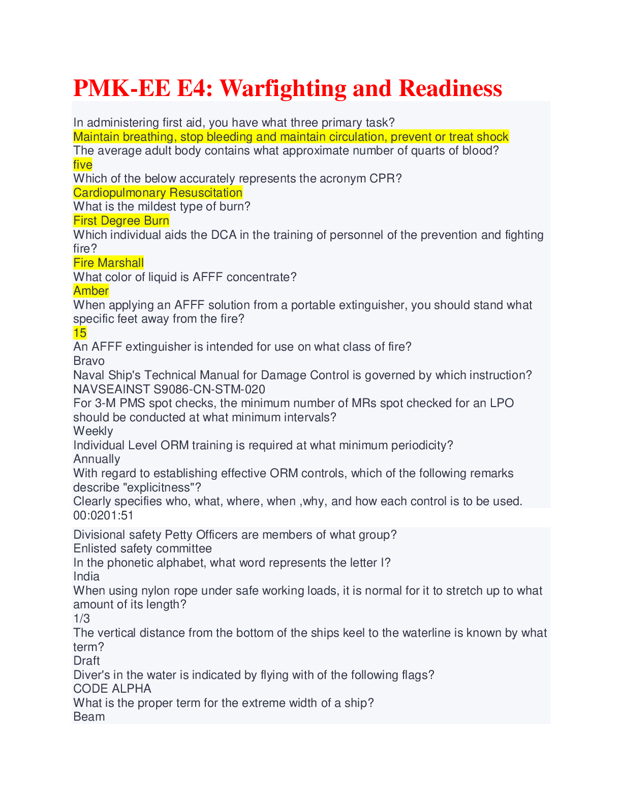
ATLS Written Review Latest 2021 Graded A
What is the primary goal of treating TBI? How is this done? Correct Answer- preventing secondary brain
injury. This is done by maintaining blood pressure and providing adequate
...
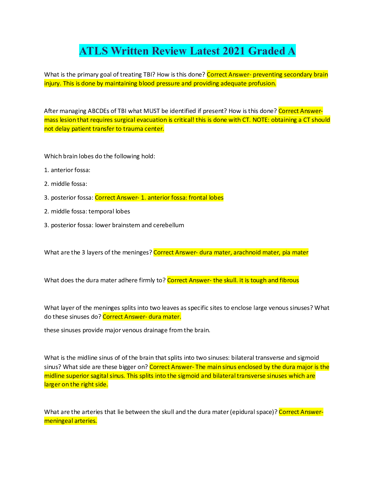
ATLS Written Review Latest 2021 Graded A
What is the primary goal of treating TBI? How is this done? Correct Answer- preventing secondary brain
injury. This is done by maintaining blood pressure and providing adequate profusion.
After managing ABCDEs of TBI what MUST be identified if present? How is this done? Correct Answermass lesion that requires surgical evacuation is critical! this is done with CT. NOTE: obtaining a CT should
not delay patient transfer to trauma center.
Which brain lobes do the following hold:
1. anterior fossa:
2. middle fossa:
3. posterior fossa: Correct Answer- 1. anterior fossa: frontal lobes
2. middle fossa: temporal lobes
3. posterior fossa: lower brainstem and cerebellum
What are the 3 layers of the meninges? Correct Answer- dura mater, arachnoid mater, pia mater
What does the dura mater adhere firmly to? Correct Answer- the skull. it is tough and fibrous
What layer of the meninges splits into two leaves as specific sites to enclose large venous sinuses? What
do these sinuses do? Correct Answer- dura mater.
these sinuses provide major venous drainage from the brain.
What is the midline sinus of of the brain that splits into two sinuses: bilateral transverse and sigmoid
sinus? What side are these bigger on? Correct Answer- The main sinus enclosed by the dura major is the
midline superior sagital sinus. This splits into the sigmoid and bilateral transverse sinuses which are
larger on the right side.
What are the arteries that lie between the skull and the dura mater (epidural space)? Correct Answermeningeal arteries.What is the most commonly injured meningeal artery and where is it located? Correct Answer- middle
meningeal artery.
Located over the temporal fossa
T/F: the arachnoid mater is fused to the dura mater? Correct Answer- FALSE: not attached. This
produces a potential space for a subdural hematoma
In a subdural hematoma, what is the cause? Correct Answer- injury to bridging veins that extend from
brain surface to the sinuses within the dura.
_______ fills the space between the arachnoid and pia mater? Correct Answer- CSF. this cushions the
brain and spinal cord.
What location of brain hemorrhage is frequently seen in brain contusion or injury to major blood vessels
at base of brain? Correct Answer- subarachnoid.
The ____ and _____ contain the reticular activating system which is responsible for ____. Correct
Answer- midbrain and upper pons
state of alertness
What important function resides in the medulla? Correct Answer- cardiorespiratory centers.
What important functions are in the following brain segments:
1. left hemisphere:
2. frontal lobe:
3. parietal lobe:
4. temporal: Correct Answer- 1. left hemisphere: language center
2. frontal lobe: executive function, emotions, motor
3. parietal lobe: sensory function/spatial orientation
4. temporal: memory functionsWhat divides the brain into supratentorial and infratentorial compartments? Correct Answer- tentorium
cerebelli. (tent over cerebellum)
What is the physiology behind a blown pupil? Correct Answer- blown pupil: dilation of pupil
-CN III runs along the tentorium cerebelli. parasympathetic fibers that constrict the pupil run along CN III
(oculomotor). When temporal lobe is herniated, it can compress these fibers. Unapposed sympathetic
activity causes pupillary dilation.
What is the tentorial notch/hiatus Correct Answer- this is where the midbrain passes through into the
infratentorial compartment.
what part of the brain most commonly herniates through the tentorial notch? Correct Answer- Uncus
(medial part of temporal lobe)
does weakness occur on the same or opposite side of the uncal herniation? Correct Answer- OPPOSITE.
the corticospinal tract of the midbrain is compressed and then crosses at the foramen magnum.
state: Ipsilateral/contralateral
____ pupillary dilation associated with _____ hemiparesis is the classic sign of uncial herniation. Correct
Answer- ipsi
contra
average ICP is _____ mmHg. Correct Answer- 10
The monro-kellie doctrine states that the total volume of intracranial contents must remain constant,
because the cranium is ___ Correct Answer- a rigid, non expandable container.
The monro-kellie doctrine states that _____ and _____ may be compressed out of the skull providing a
degree of buffering. Correct Answer- CSF and venous blood.Once the CSF and venous blood reach a certain level of displacement the ICP rapidly increases.
What is the equation for CPP (cerebral perfusion pressure)? Correct Answer- CPP=MAP-ICP
in TBI, Every effort should be made to reduce ______, while normalizing ____, ___, and _____. Correct
Answer- ICP
MAP, oxygenation, intravascular volume
What GCS ranges for the following classes:
1. Minor
2. Moderate
3. Severe Correct Answer- 1. 13-15
2. 9-12
3. 3-8
What nerve palsy may occur with basilar skull fracture? Correct Answer- seventh nerve.
A GCS of ___ is accepted definition of coma? Correct Answer- 8 or less
How do you assess a GCS of someone with asymmetric responses? Correct Answer- Use the best
possible because this will be the best predictor of outcome
Basilar fractures of the skull usually require what type of imaging? Correct Answer- this requires CT with
bone-window setting.
What are the typical clinical signs of basilar skull fractures? Correct Answer- 1.periorbital ecchymosis
(raccoon eyes)
2. retroauriculor ecchymosis (battle sign)
3. CSF leak from nose or ears
4. 7th or 8th CN dysfunction (facial paralysis and hearing loss)What should be a primary consideration for any patient with a skull fracture, especially a linear skull
fracture? Correct Answer- hematoma. linear skull fracture increases likelihood of intracranial hematoma
by about 400x
What mechanism is common with diffuse axonal injury and what is the likely outcome? Correct Answerthese injury often occur with high velocity or deceleration injures. They appear as diffuse cerebral
hemorrhage often between grey and white matter. These are associated with variable but often poor
outcomes.
Epidural hematomas often occur in the _____ area of the skull and result from a tear of the _______
arteries. Correct Answer- temporal
middle meningeal artery
What is the classic presentation of a epidural hematoma? Correct Answer- a lucid interval between time
of injury and neurologic a deterioration.
What are more common brain injury: epidural or subdural? Correct Answer- subdural 30%
epidural 0.5%
Subdural hematoma occur from tear of _________. Correct Answer- bridging vessels of the cerebral
cortex
Contusion occur in ___% of TBI. They often occur in _____ or ______ lobes of brain. They may coalesce
to form ______ in as many as 20$%. Correct Answer- 20-30%
frontal or temporal
intracerebral hematoma.
What is the imaging protocol for a patient with cerebral contusion? Correct Answer- get CT at
presentation. then get another within 24 hours to assess for coalesced hematoma.
What factors would require a CT in minor brain injury? Correct Answer- 1. suspected open skull frac2. basilar frac
3. >2 episode vomitting
4. pt older than 65
5. LOC >5 min
6. amnesia before impact of >30 min
How long after discharge should patient with mild brain injury be observed by friend? Correct Answer-
24 hours
What type of brain injury requires serial GCS? Correct Answer- ALL. minor. moderate. major
What imaging is done in all patient with moderate brain injury? Correct Answer- CT
What factor of ABCDE must be monitored closely in moderate brain injury? Correct Answer- Airway and
breathing. rapid deterioration may occur. hypoventilation and hypercapnia may ensue requiring
intubation. close monitoring in ICU is required.
What should immediately follow the secondary survey in major/severe brain injury? Correct Answer- CT.
REMEMBER: CT should never delay patient transfer
When assessing ABCDE of severe brain injury, when does DPL or FAST come before neuro exam? Correct
Answer- if the systolic blood pressure cannot be brought above 100, DPL or FAST is done first as to
assess source of hypotension
[Show More]


.png)