NURS 3056 Final Info Test 2.
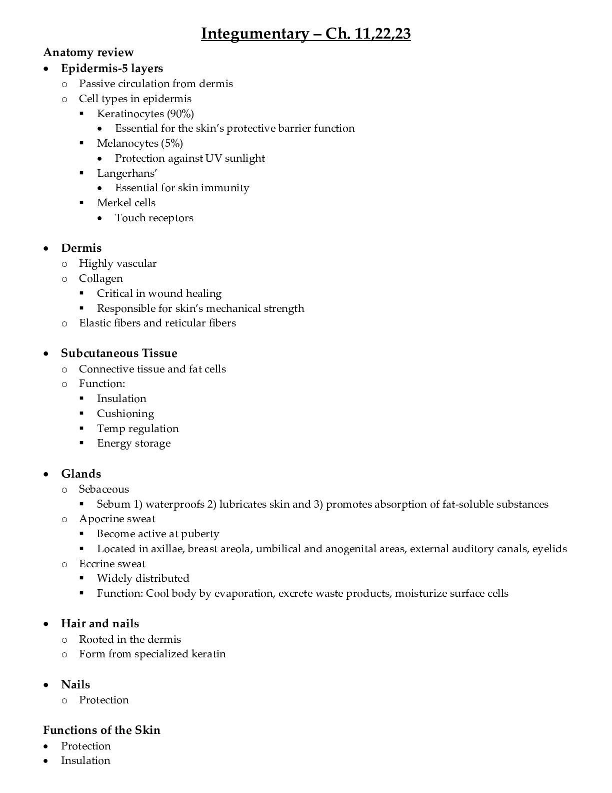
Integumentary – Ch. 11,22,23
Anatomy review
• Epidermis-5 layers
o Passive circulation from dermis
o Cell types in epidermis
Keratinocytes (90%)
• Essential for the skin’s protective
...
NURS 3056 Final Info Test 2.
Integumentary – Ch. 11,22,23
Anatomy review
• Epidermis-5 layers
o Passive circulation from dermis
o Cell types in epidermis
Keratinocytes (90%)
• Essential for the skin’s protective barrier function
Melanocytes (5%)
• Protection against UV sunlight
Langerhans’
• Essential for skin immunity
Merkel cells
• Touch receptors
• Dermis
o Highly vascular
o Collagen
Critical in wound healing
Responsible for skin’s mechanical strength
o Elastic fibers and reticular fibers
• Subcutaneous Tissue
o Connective tissue and fat cells
o Function:
Insulation
Cushioning
Temp regulation
Energy storage
• Glands
o Sebaceous
Sebum 1) waterproofs 2) lubricates skin and 3) promotes absorption of fat-soluble substances
o Apocrine sweat
Become active at puberty
Located in axillae, breast areola, umbilical and anogenital areas, external auditory canals, eyelids
o Eccrine sweat
Widely distributed
Function: Cool body by evaporation, excrete waste products, moisturize surface cells
• Hair and nails
o Rooted in the dermis
o Form from specialized keratin
• Nails
o Protection
Functions of the Skin
• Protection
• Insulation
• Sensation
• Fluid balance
• Temperature regulation
• Vitamin production
• Immune response function
• Can act as a delivery system for drugs
Primary vs. Secondary Lesions
• Primary lesions
o Develop on previously unaltered skin
o Macule
Circumscribed, flat area with a change in skin color, <0.5cm diameter. If lesion is >0.5cm, it’s a patch
Ex. Freckles, petechiae, measles, flat mole (nevus), café-au-lait spots, vitiligo (complete depigmentation)
o Papule
Elevated, solid lesion, <0.5cm diameter. If >0.5cm diameter it’s a nodule
Ex. Wart (verruca), elevated moles, lipoma, basal cell carcinoma
o Vesicle
Circumscribed, superficial collection of serous fluid. <0.5cm diameter
Ex. Varicella (chickenpox), herpes zoster (shingles), second-degree burn
o Plaque
Circumscribed, elevated, superficial, solid lesion. >0.5cm diameter
Ex. Psoriasis, seborrheic and actinic keratoses
o Wheal
Firm, edematous, irregularly shaped area, diameter variable
Ex. Insect bite, urticaria
o Pustule
Elevated, superficial lesion filled with purulent fluid
Ex. Acne, impetigo
• Secondary skin lesions
o Change with time
o Occur because of secondary factors (scratching, infection, etc.)
o Fissure
Linear crack or break from epidermis to dermis, dry or moist
Ex. Athletes foot, cracks at corner of the mouth
o Scale
Excess, dead epidermal cells produced by abnormal keratinization and shedding
Ex. Flaking skin after drug reaction or sunburn
o Scar
Abnormal formation of connective tissue that replaces normal skin
Ex. Surgical incision, healed wound
o Ulcer
Loss of epidermis, extending into dermis, crater-like irregular shape
Ex. Pressure ulcer, chancre
o Atrophy
Depression in skin resulting from thinning of the epidermis or dermis
Ex. Aged skin, striae
o Excoriation
Area in which epidermis is missing, exposing the dermis
Ex. Abrasion, scratch
Lesion distribution
Term Description
Annular Circular, begins in center and spreads to periphery (tinea corporis - ringworm)
Asymmetric Unilateral distribution
Confluent Merging together (urticaria - hives)
Discrete Distinct individual lesions that remain separate (acne)
Gyrate Twisted, coiled spiral, snakelike
Grouped Clusters of lesions (multiple vesicles of contact dermatitis)
Localized Limited areas of involvement that are clearly defined (confined to one area)
Polycyclic Annular (circular) lesions grow together (psoriasis)
Solitary Single lesion
Symmetric Bilateral distribution
Zosteriform Linear arrangement along a dermatome area (herpes zoster)
Assessment
• Subjective: health history, medications, surgery or other treatments, functional health patterns (ex: nutrition)
• Objective – Physical Assessment - inspection and palpation
o Nails
Color
Texture
Consistency
Thickness
Shape/angle of nail
o Hair
Color
Texture
Distribution
Quantity
o Skin
Color
Texture
Turgor
Temperature
Moisture
Lesions?
Vascular changes?
Skin changes can point to other disease processes that we will study throughout this year
Effects of Drugs on Integumentary System
• Drugs that cause photosensitivity (Table 23-2 for full list)
o Examples of common offenders:
Antidepressants: amitriptyline, venlafaxine (Effexor)
Antidysrhythmics: amiodarone
Antihistamines: diphenhydramine (Benadryl), cetirizine (Zyrtec)
Antimicrobials: tetracycline, azithromycin (Zithromax), ciprofloxacin (Cipro)
Antifungals: griseofulvin, ketoconazole
Antipsychotics: haloperidol
Diuretics: furosemide (Lasix), hydrochlorothiazide
Hypoglycemics: glipizide, glyburide
NSAIDS: diclofenac (Voltaren), sulindac
Categories Examples
Antidepressants amitriptyline, doxepin, venlafaxine
Antidysrhythmics quinidine, amiodarone (Cordarone)
Antihistamines diphenhydramine, chlorpheniramine, clemastine, cetirizine (Zyrtec)
Antimicrobials tetracycline, sulfamethoxazole, azithromycin (Zithromax), ciprofloxacin (Cipro)
Antifungals griseofulvin, ketoconazole
Antipsychotics chlorpromazine, haloperidol
Chemotherapy methotrexate, dacarbazine (DTIC), 5-fluorouracil (5-FU)
Diuretics furosemide (Lasix), hydrochlorothiazide
Hypoglycemics tolbutamide, glipizide (Glucotrol), glyburide
Nonsteroidal antiinflammatory drugs diclofenac (Voltaren), piroxicam (Feldene), sulindac
Diagnostic tools
• Inspection and history
• Dermatoscopy
• Biopsy
• Potassium hydroxide (KOH)
• Tzanck test
• Stains and cultures
• Curretage
• Mineral oil slides
• Wood’s lamp
• Patch testing
Study Description and Purpose Nursing Responsibility
Biopsy
Punch Special punch biopsy instrument of appropriate size used. Instrument rotated to appropriate level to include dermis and some fat. Suturing may or may not be done. Provides full-thickness skin for diagnostic purposes Before: Verify that consent form is signed (if needed).
During: Assist with site preparation, anesthesia, procedure, and hemostasis. Properly identify specimen.
After: Apply dressing, give postprocedure instructions to patient.
Excisional Used when good cosmetic results and/or entire lesion removal desired. Skin closed with subcutaneous and skin sutures Same as above
Incisional Wedge-shaped incision made in lesion too large for excisional biopsy. Useful when larger specimen than shave or punch biopsy is needed Same as above
Shave Single-edged razor blade used to shave off superficial lesions or small sample of a large lesion. Provides thin specimen for diagnostic purposes Same as above
Microscopic Tests
Potassium hydroxide (KOH) Hair, scales, or nails examined for superficial fungal infection. Specimen put on glass slide and 10%-20% concentration of KOH added Before: Instruct patient regarding purpose of test.
During: Prepare slide.
Tzanck test (Wright's and Giemsa's stain) Fluid and cells from vesicles examined. Used to diagnose herpes infections. Specimen put on slide, stained, and examined microscopically Before: Inform patient of purpose of test.
During: Use sterile technique for collection of fluid.
Culture Test identifies fungal, bacterial, and viral organisms. For fungi, scraping or swab of skin performed. For bacteria, material obtained from intact pustules, bullae, or abscesses. For viruses, vesicle or bulla and exudate taken from base of lesion Before: Instruct patient regarding purpose and procedure.
During: Properly identify specimen. Follow instructions for storing specimen if not immediately sent to laboratory.
Mineral oil slides To check for infestations, scrapings are placed on slide with mineral oil and viewed microscopically. Before: Instruct patient about purpose of test.
During: Prepare slide.
Immunofluorescent studies Some skin diseases have specific, abnormal antibody proteins that can be identified by fluorescent studies. Both skin tissue and serum can be examined. Before: Inform patient about purpose of test.
During: Assist in obtaining specimen. For punch biopsy, place specimen in special fixative (e.g., Michel's) and not formalin.
Miscellaneous
Wood's lamp (black light) Examination of skin with long-wave ultraviolet light causes specific substances to fluoresce (e.g., Pseudomonas fungal, infections, vitiligo). Before: Explain purpose of examination. Inform patient it is not painful.
During: Darken room.
Patch test
(Fig. 22-10) Used to assess for allergic dermatitis and photoallergic reactions. Application of allergens to the patient's skin (usually on the back) for 48 hr. Test sites examined 48 hr later for a reaction, characterized by the presence of erythema, papules, vesicles, or all of these. Additional readings beyond 48 hr may be done. Before: Explain purpose and procedure to patient.
After: Instruct patient that patches are left in place for 48 hr, during which time it is important not to wash the area or play vigorous sport because if the adhesive tapes peel off the process will have to be repeated. The patches should not be exposed to sunlight or other sources of ultraviolet (UV) light.
Skin care
• Environmental Hazards
o Sun exposure
Ultraviolet (UV) rays
• UVA—tanning
• UVB—sunburn
Sun protection
• Protective clothing
• Sunscreen
• Risk factors
o Fair skin
o Blonde or red hair, blue eyes
o Outdoor sunbathing
o Living near the equator or high altitudes
o History of skin cancer
Nonmelanoma skin cancers
• Actinic keratosis
o Premalignant skin lesions
• Basal cell carcinoma
o Most common type of skin cancer
o Least deadly
Inflammatory response
• Clinical Manifestations
o Local response to inflammation
Redness
Heat
Pain
Swelling
Loss of function
o Systemic response to inflammation
Increased WBC count with a shift to the left
Malaise
Nausea and anorexia
Increased pulse and respiratory rate
Fever
Beneficial aspects of fever include:
• Increased killing of microorganisms
• Increased phagocytosis
• Increased proliferation of T cells
• Types of inflammation
o Acute
Healing occurs in 2 to 3 weeks, usually leaving no residual damage
Neutrophils are predominant cell type at site of inflammation
o Subacute
Has same features as acute inflammation but persists longer
o Chronic
May last for years
Injurious agent persists or repeats injury to site
Predominant cell types involved are lymphocytes and macrophages
May result from changes in immune system (e.g., autoimmune disease)
Nursing and interprofessional management
• Nursing Implementation
o Health Promotion
Prevention of injury
Adequate nutrition
Early recognition of inflammation
Immediate treatment
• Acute Intervention
o Observation
Immunosuppressed—classic manifestations masked; just “don’t feel well”
o Vital signs
o Fever management
Determine cause
Administer antipyretic or antibiotics
Monitor for seizures or delirium (>104° F)
• Drug therapy
o Aspirin
o Acetaminophen
o NSAIDs
o Corticosteroids
• RICE
o Rest—O2 and nutrients for repair; wound healing
o Ice (first 24 hours)—reduced swelling and pain
o Compression and immobilization—reduce edema and stabilize
o Elevation—reduce edema and pain
Inflammation healing process
• The final phase of the inflammation process is healing
• Healing includes two major components:
o Regeneration
Replace cells and tissues with same type
o Repair
Connective tissue replaces lost cells
Primary, secondary, or tertiary intention
Types of wound repair
• Primary intension
o Wound margins are neatly approximated
Ex: surgical incision or paper cut
Incision with blood clot edges approximated with sutures fine scar
• Secondary intention
o Wounds occurring from trauma, ulceration, infection
o Typically have large amounts of exudate
o Wide, irregular wound margins with extensive tissue loss
o Often has a greater defect and more gaping wound edges than wounds healing by primary intention
o Irregular, large wound with blood clot granulation tissue fills in wound large scar
• Tertiary Intention
o Also known as delayed primary intention
o This type of healing is used when:
A contaminated wound is left open and is sutured together after infection is controlled
Infected wound is opened and allowed to granulate before restoring
o Typically results in larger/deeper scar than healing by primary or secondary intention
o Contaminated wound granulation tissue delayed closure with sutures
Effect of Nutrition on Wound Healing
• Vitamin A: maintenance of normal cell structure, wound healing
• Vitamin B complex: needed for metabolic function
• Vitamin C ascorbic acid: connective tissue formation and wound healing
• Vitamin K: essential for synthesis of clotting factors
• Protein: cell growth and wound healing
Nursing Management of Dermatologic Concerns
• Wet compresses
• Baths
• Topical medications
• Control of pruritus
o Break the itch/scratch cycle
o Cool environment
o Hydration, wet compresses, moisturizers
o Topical drugs
• Prevention of spread
• Prevention of secondary infections
• Specific skin care
Chronic Dermatologic Problems
• Psychologic effects
o Reinforce prescribed regimen
o Support groups
o Camouflage
Common Bacterial Infections
• Impetigo
o Etiology and Patho
Caused by Group A beta-hemolytic strep, staphylococci, or both
Associated with poor hygiene
Contagious
o Clinical manifestations
Most commonly on the face
Thick, honey-colored crusts surrounded by erythema
Pruritic (itchy)
o Treatment
Topical: warm saline soaks followed by soap and water removal of crusts, antibiotic cream/ointment
Systemic: antibacterial: oral penicillin or clindamycin for widespread or systemic manifestations
• Folliculitis
o Etiology and Patho
Usually staphylococci
Increased incidence in those with diabetes mellitus
o Clinical manifestations
Present in areas of increased friction, moisture, rubbing, or oil
Tender, small pustule at opening of hair follicle with minimal erythema
Crusting
Common on scalp, beard, extremities in men
o Treatment
Antistaphylococcal soap and water cleansing
Topical antibiotics (ex: mupirocin)
Warm compresses
• Furuncle
o Etiology and Patho
Deep infection with staph around hair follicle
Associated with severe acne or seborrheic dermatitis
o Clinical manifestations
Tender, erythematous area around hair follicle with draining pus
Common on face, back of neck, axilla, breasts, buttocks, perineum, thighs
o Treatment
Incision and drainage
Antibiotics
Warm, moist compresses
• Carbuncle
o Etiology and Patho
Multiple, connected furuncles
o Clinical manifestations
Multiple pustules with localized erythema
o Treatment
Same as for furuncles
Often recur
Heal slowly
Scars may form
• Cellulitis
o Etiology and Patho
Deep inflammation of subcutaneous tissues
Causative agents: Staphylococcus aureus and strep
Often follows a break in the skin
o Clinical manifestations
Hot, tender, erythematous, edematous area with diffuse borders
Chills, malaise, fever
o Treatment
Topical: moist heat, immobilization, elevation
Systemic: antibiotics, hospitalization for IV antibiotics if severe
• Erysipelas
o Etiology and Patho
Superficial cellulitis involving the dermis
Caused by group A beta-hemolytic strep
o Clinical manifestations
Red, hot, painful
Sharply demarcated plaque with induration
Bacteremia may be possible. Toxic signs: fever, increased WBC count, headache, malaise
o Treatment
Systemic antibiotics (typically penicillin)
Hospitalization likely required
Etiology & Pathophysiology Manifestations Treatment and Prognosis
Impetigo
• Group A β-hemolytic streptococci, staphylococci, or combination of both
• Associated with poor hygiene
• Primary or secondary infection. Contagious • Vesiculopustular lesions that develop thick, honey-colored crust surrounded by erythema
• Pruritic
• Most common on face as primary infection Topical:
• Wound care with warm saline or aluminum acetate soaks followed by soap-and-water removal of crusts and application of topical antibiotic cream or ointment (mupirocin [Bactroban], retapamulin [Altabax])
• Meticulous hygiene essential
Systemic:
• Systemic antibiotics (e.g., cephalosporins, amoxicillin, clindamycin) for widespread infections or systemic manifestations
Folliculitis
• Usually staphylococci
• Present in areas subjected to friction, moisture, rubbing, or oil
• Increased incidence in patients with diabetes mellitus • Small pustule at hair follicle opening with minimal erythema
• Development of crusting
• Most common on scalp, beard, extremities in men
• Tender to touch • Antistaphylococcal soap (e.g., Hibiclens, Lever 2000, Dial) and water cleansing
• Topical antibiotics (e.g., mupirocin)
• Warm compresses of water or aluminum acetate solution
• Usually heals without scarring
• If lesions extensive and deep, possible scarring, loss of involved hair follicles, and treatment with systemic antibiotics
Furuncle
• Deep infection with staphylococci around hair follicle
• Often associated with severe acne or seborrheic dermatitis • Tender erythematous area around hair follicle that is painful
• Draining pus and core of necrotic debris on rupture
• Most common on face, back of neck, axillae, breasts, buttocks, perineum, thighs • Incision and drainage (possibly with packing), antibiotics, meticulous care of involved skin, frequent application of warm, moist compresses
Furunculosis
• Increased incidence in patients who are obese, diabetic, chronically ill, or regularly exposed to moisture, pressure • Lesions as above
• Malaise, regional adenopathy, elevated body temperature Topical:
• Incision and drainage of painful nodules
• Warm, moist compresses to erythematous plaques
• Measures to reduce surface staphylococci include antimicrobial cream to nares, armpits, and groin and antiseptic to entire skin.
• Meticulous personal hygiene
Systemic:
• Systemic antibiotic effective against MRSA pending culture and sensitivity results
• Often recurrent with scarring
Carbuncle
• Multiple, interconnecting furuncles • Many pustules appearing in erythematous area
• Most common at nape of neck • Treatment same as for furuncles
• Often recurrent despite production of antibodies
• Heal slowly with scar formation
Cellulitis
• Inflammation of subcutaneous tissues
• Possibly secondary complication or primary infection
• Often following break in skin
• Staphylococcus aureus and streptococci usual causative agents
• Deep inflammation of subcutaneous tissue from enzymes produced by bacteria • Hot, tender, erythematous, and edematous area with diffuse borders
• Chills, malaise, and fever Topical:
• Moist heat, immobilization, and elevation
Systemic:
• Systemic antibiotic therapy
• Hospitalization if severe for IV antibiotic therapy (dalbavancin [Dalvance], vancomycin, linezolid [Zyvox], ceftaroline fosamil [Teflaro], daptomycin [Cubicin])
• Progression to gangrene possible if untreated
Erysipelas
• Superficial cellulitis primarily involving the dermis
• Group A β-hemolytic streptococci • Red, hot, sharply demarcated plaque that is indurated and painful
• Bacteremia possible
• Most common on face and extremities
• Toxic signs: fever, ↑ WBC count, headache, malaise • Systemic antibiotics, usually penicillin
• Hospitalization often required
Common Viral Infections
• HSV1 and HSV2
o Etiology and Patho
Recurrent, lifelong viral infections
Oral: Herpes Simplex Virus-1
Genital: Herpes Simplex Virus-2
Contagious (transmitted via respiratory droplet or fluid containing virus)
Exacerbated by sun, trauma, menses, stress, systemic infection
o Clinical manifestations
Symptoms occur 2 days-2 weeks after contact
Painful single or grouped vesicles on erythematous base with/without systemic symptoms (fever, malaise)
o Treatment
Symptomatic: soothing moist compress, white petrolatum to lesions
Antiviral drugs: acyclovir, famciclovir, valacyclovir
• Herpes Zoster (Shingles)
o Etiology and Patho
Incidence increases with age
Activation of varicella-zoster virus
Can be contagious to anyone who has not had chickenpox or is immunosuppressed
o Clinical manifestations
Linear distribution of grouped vesicles and pustules on an erythematous base along a dermatome
Usually on trunk, face, lumbosacral area
Burning, pain, neuralgia prior to outbreak
Mild/severe pain during outbreak
o Treatment is symptomatic
Topical: wet compresses, Silvadene for ruptured vesicles
Systemic: antivirals within 72 hrs to prevent postherpetic neuralgia, analgesia, gabapentin for postherpetic neuralgia
• Verruca Vulgaris (common warts)
o Etiology and Patho
Caused by HPV
Mildly contagious in autoinoculation phase
Prevalence greater in young and immunosuppressed
May spontaneously disappear in 1-2 years
o Clinical manifestations
Circumscribed, hypertrophic, flesh-colored papule
Painful if laterally compressed
o Treatment
Surgery
Liquid nitrogen
Blistering agent (cantharidin)
Salicylic acid
CO2 laser destruction
Common Fungal Infections
• Candidiasis
o Etiology and Patho
Caused by Candida albicans
Appears in warm, moist areas (groin, oral mucosa, submammary folds)
Immunosuppression allows yeast to become pathogenic
o Clinical manifestations
Mouth: white, cheesy plaque (think milk curds!)
Vagina: Red, edematous, painful vaginal wall with white patches. Vaginal discharge and itching. Pain with urination and intercourse
Skin: Diffuse papular erythematous rash with pinpoint satellite lesions around edges of affected area
o Treatment and prognosis
Azole antifungals
Sexual abstinence or use of a condom
Skin hygiene (goal to keep skin clean/dry)
Powder for non-mucosal surfaces to prevent recurrence
• Tinea
o Tinea corporis (ringworm)
Ring-like in shape, scaly, well-defined, erythematous
o Tinea cruris (jock itch)
Well-defined, scaly plaque in groin
o Tinea pedis (athlete’s foot)
Scaling and maceration in interdigital space
Plantar surfaces may have erythema and blistering
Can be pruritic and painful
o Tinea unguium (onychomycosis)
Incidence increases with age
Toenails more commonly affected than fingernails
Scaliness under distal nail plate
Brittle/thickened, broken, yellowed nails
Common Infestations
• Pediculosis (lice)
o Etiology and Patho
Can be on the head, body, or pubic area
Parasites suck blood, excrete and leave eggs on skin and hair
Transmission of pubic lice is by sexual contact
o Clinical manifestations
Small, red, noninflammatory
Starts flush with skin. May progress to more of a papular/wheal-like shape
Pruritic. May see excoriation from pruritis
Nits/eggs are firmly attached to hair shift on head/body
o Treatment
γ-Benzene hexachloride or pyrethrins to treat body
Spinosad (Natroba) topical suspension to treat scalp/hair
Close contacts should be screened/treated
• Scabies
o Etiology and Patho
Caused by Sarcoptes scabei
Allergic reaction to the eggs, feces, and mite parts
Spread by direct contact
o Clinical manifestations
Erythematous papules/vesicles with possible crusting
Severe itching, especially at night
Burrows in interdigital webs, flexor surface of the wrists, genitalia, and anterior axillary folds
o Treatment
5% permethrin topical lotion, one overnight application with second application 1 wk later. May yield 95% eradication.
Treat all family members with plastic covering for 5 days, launder all clothes and linen with bleach.
Antibiotics if secondary infection present
• Ticks
o Etiology and Patho
Borrelia burgdorferi causes Lyme disease
Ticks are endemic in the NE, Mid-Atlantic, parts of Midwest and West
o Clinical manifestations
Spreading, ringlike, warm, itchy, painful rash commonly seen in groin, buttocks, axilla, trunk, upper arms, and legs (appearing 3-4 weeks post tick bite)
Flu-like symptoms
Cardiac, arthritic, and neurologic symptoms possible
o Treatment
Oral antibiotics: doxycycline, tetracycline
IV antibiotics if arthritic, neurologic or cardiac symptoms present
Rest/healthy diet
Common Reactions
• Allergic Contact Dermatitis
o Etiology and Patho
Type IV delayed hypersensitivity response
• Notes: The allergic agent is absorbed through the skin and acts as an antigen. Sensitization occurs after one or more exposures to the irritant
Lesions appear 2-7 days after allergic contact
o Clinical manifestations
Sharply circumscribed red papules/plaques
May see occasional vesicles
Pruritic
Dermatitis typically takes shape of causative agent (ex: dermatitis around finger from ring)
o Treatment
Topical or oral corticosteroids (systemic If severe)
Antihistamines
Skin lubrication
Elimination of allergic agent
• Atopic Dermatitis
o Etiology and Patho
Type 1 hypersensitivity
Genetic influence
Typically, chronic and relapsing in nature
Associated with allergic rhinitis/asthma
o Clinical manifestations
Pruritic
Can be erythematous or scaly
May see lichenification from itching
o Treatment
Lubrication
Topical immunomodulators (Elidel and Protopic)
Stress reduction
Corticosteroids or phototherapy if severe
Antibiotics if secondary infection from chronic scratching
Acne
• Etiology and Patho
o Inflammatory disorder of sebaceous glands
o More common in teens but can persist into adulthood
o Flares can occur: 1) before menses, 2) with use of corticosteroids or 3) androgen dominant BCP
• Clinical manifestations
o Noninflammatory: Open comedones (blackheads) and closed comedones (whiteheads)
o Inflammatory: papules and pustules
• Treatment
o Topical: mechanical removal via extraction, topical benzoyl peroxide, retinoids, antimicrobials
o Systemic: systemic antibiotics, Accutane (severe nodulocystic acne)
Psoriasis
• Etiology and Patho
o Autoimmune chronic dermatitis involving excessive turnover of epidermal cells
o Family predisposition
o Typically develops before age 40
• Clinical manifestations
o Well-demarcated silvery, scaly plaques on reddened skin
o Commonly located on scalp, elbows, knees, palms, soles, fingernails
o Itching, burning, pain
• Treatment
o Topical: corticosteroids, tar, salicylic acid, calcipotriene, anthralin, intralesional steroid injection
o Systemic: Natural/artificial UVB light, PUVA, methotrexate, Otezla, biologic therapy (moderate to severe disease)
Seborrheic Keratosis
• Etiology and Patho
o Benign
o Familial
o Usually occur after age 40
• Clinical manifestations
o Irregularly round/oval verrucous papules or plaques
o Stuck-on appearance
o Pigment increase with time
o May be itchy
• Treatment
o Removed via curettage or cryosurgery to eliminate itching or for cosmetic reasons
o Biopsy may be necessary to distinguish between SK and melanoma
Lentigo (age spot)
• Etiology and Patho
o Increased number of normal melanocytes in the basal layer of the epidermis r/t sun exposure and aging
• Clinical manifestations
o Hyperpigmented brown/black macule or patch on a sun-exposed area
• Treatment
o Treatment is only for cosmetic reasons: liquid nitrogen, laser resurfacing
o Biopsy if trying to differentiate between lentigo and melanoma
Atypical or Dysplastic Nevi
• Etiology and Patho
o Can be a precursor for malignant melanoma
• Clinical manifestations
o Typically, greater than 5mm
o Irregular border
o Variegated color
o At least one flat portion, often at edge of mole
• Treatment
o Closely monitor those with family history of melanoma or dysplastic nevi
o Biopsy for suspicious looking lesions
Premalignant Skin Lesion: Actinic Keratosis
• Etiology and Patho
o Caused by sun damage
o Premalignant and common in older whites
o Most common type of skin cancer; least deadly
• Clinical manifestations
o Dry, keratotic, scaly papule
o Can appear as a rough, adherent scale on an erythematous base, which recurs when it is removed
• Treatment
o Cryosurgery
o Topical application of 5-FU, Aldara, or diclofenac
o Chemical peels
o Laser resurfacing
o Photodynamic therapy
Malignant Neoplasms of Skin:
• Basal Cell Carcinoma
o Etiology and Patho
Continuous division of basal cells, causing formation of a mass
Related to sun exposure, genetic skin type, radiation, scars, some types of nevi
o Clinical manifestations
Superficial BCC: erythematous, pearly, well-defined, slightly elevated plaque
Nodular/ulcerative BCC: Small, slowly growing papule that is pearly in appearance. Can develop into ulceration or erosion. May see overlying telangiectasia
o Treatment
5-FU and imiquimod for superficial lesions
Surgical excision, electrodessication and curettage, cryosurgery, radiation therapy, topical or systemic chemo, photodynamic therapy
• Squamous Cell Carcinoma
o Etiology and Patho
Often occurs on previously damaged skin (sun, radiation, area of scarring)
Malignant tumor of squamous cells in epidermis
o Clinical manifestations
Commonly seen on sun exposed areas (think hands and face!)
Superficial SCC: thin, scaly, erythematous plaque
Early SCC: firm nodule with unclear border, scaling, ulceration
Late SCC: Lesion is covered with a scale or horn, may see ulceration
o Treatment
Surgical excision
Cryosurgery
Radiation/chemotherapy
Electrosurgery
• Malignant Melanoma
o Etiology and Patho
Neoplastic growth of melanocytes
Classified according to histological spread
Can metastasize
o Risk factors: red or blonde hair, light-colored eyes, fair skin that freckles, chronic sun exposure, family history
o Clinical manifestations
Irregular color, surface, border
Can be flat or elevated
May be ulcerated or eroded
Typically, less than 1 cm in size
o Diagnosis
Dermoscopy
Incisional biopsy
Tumor thickness
Breslow measurement
Clark level
o Treatment
Treatment determined by
Site of original tumor
Stage of the cancer
Surgical excision with potential sentinel lymph node evaluation
• Table 23.4 – Premalignant and Malignant Conditions of the Skin
Etiology and Pathophysiology Clinical Manifestations Treatment and Prognosis
Actinic Keratosis
• Actinic (sun) damage
• Premalignant skin lesions
• Common in older whites • Flat or elevated, dry, hyperkeratotic scaly papule. Often multiple
• Possibly flat, rough, or verrucous (wartlike)
• Rough adherent scale on red base, which returns when removed
• Often on erythematous sun-exposed area
• Increase in number with age • Cryosurgery. Topical application of 5-FU, imiquimod (Aldara) or diclofenac (Solaraze), chemical peels, laser resurfacing, photodynamic therapy (PDT, with 5-aminolevulinic acid [5-ALA] or methyl aminolevulinate [MAL] followed by light irradiation)
• Recurrence possible even with adequate treatment
Atypical or Dysplastic Nevi
• Morphologically between common acquired nevi and melanoma
• May be precursor of malignant melanoma • Often >5 mm. Irregular border, possibly notched. Frequently multiple
• Variegated color of tan, brown, black, red, or pink within single mole
• Presence of at least one flat portion, often at edge of mole
• Most common site on back, but possible in uncommon mole sites such as scalp or buttocks (Fig. 23-5) • Increased risk for melanoma
• Careful monitoring of persons suspected of familial tendency to melanoma or dysplastic nevi
• Excisional biopsy for suspicious lesions
Basal Cell Carcinoma
• Change in basal cells. No maturation or normal keratinization
• Continuing division of basal cells and formation of enlarging mass
• Related to excessive sun exposure, genetic skin type, x-ray radiation, scars, and some types of nevi Nodular and ulcerative:
• Small, slowly enlarging papule
• Borders semi-translucent or “pearly” with overlying telangiectasia
• Erosion, ulceration, and depression of center
• Normal skin markings lost
Superficial:
• Erythematous, pearly, sharply defined, barely elevated plaques • Surgical excision, electrodessication and curettage, cryosurgery, radiation therapy, topical or systemic chemotherapy, and photodynamic therapy
• 90% cure rate with primary lesions
• Slow-growing tumor that invades local tissue. Metastasis rare
• 5-FU and imiquimod for superficial lesions, photodynamic therapy for small lesions, vismodegib (Erivedge) or sonidegib (Odomzo) for metastatic or recurrent locally invasive lesions
Squamous Cell Carcinoma
• Frequent occurrence on previously damaged skin (e.g., from sun, radiation, scar)
• Malignant tumor of squamous cell of epidermis. Invasion of dermis, surrounding skin • Most common on sun-exposed areas such as face and hands
• Superficial: Thin, scaly erythematous plaque without invasion into the dermis
• Early: Firm nodules with indistinct borders, scaling, and ulceration
• Late: Covering of lesion with scale or horn from keratinization, ulceration • Surgical excision, cryosurgery, radiation therapy, chemotherapy, electrosurgery
• Untreated lesion may metastasize to regional lymph nodes and distant organs.
• 13-cis-retinoic acid and interferon alfa-2A for metastatic lesions
• High cure rate with early detection and treatment
Malignant Melanoma
• Neoplastic growth of melanocytes anywhere on skin, eyes, or mucous membranes
• Classification according to major histologic mode of spread
• Potential invasion and widespread metastases • Irregular color, surface, and border Variegated color, including red, white, blue, black, gray, brown
• Flat or elevated. Eroded or ulcerated
• Often <1 cm in size
• Most common sites in males are back, then chest. In females are legs, then back (Fig. 23-2) • Surgical excision and possible sentinel lymph node evaluation
• Possible use of adjuvant therapy after surgery if lesion >1.5 mm in depth
• Correlation between survival rate and depth of invasion
• Poor prognosis unless diagnosed and treated early
• Spreading by local extension, regional lymphatic vessels, and bloodstream
Cutaneous T-Cell Lymphoma
• Origins in skin. Localized chronic, slowly progressing disease
• Possibly related to environmental toxins and chemical exposure
• Mycosis fungoides (MF) is most common form.
• Sézary syndrome is an advanced form of MF.
• Prevalence twice as high in men as in women • Classic presentation involves 3 stages: patch (early), plaque, and tumor (advanced).
• History of persistent macular eruption followed by gradual appearance of indurated erythematous plaques on trunk that appears similar to psoriasis
• Pruritus, lymphadenopathy • Treatment usually controls symptoms but is not curative. UVB, PUVA, corticosteroids, topical nitrogen mustard, radiation therapy in patch and plaque stage of disease
• Interferon, systemic chemotherapy, extracorporeal photopheresis, romidepsin (Istodax) for progressive disease
• Bexarotene (Targretin), denileukin diftitox (Ontak), and vorinostat (Zolinza) for advanced disease
• Disease course is unpredictable. 10% will have progressive disease.
• Cultural and Ethnic Integumentary Problems
o African Americans and Hispanics have a lower incidence of skin cancer than whites.
o In the United States, rates of BCC and SCC are increasing among Hispanic and Asian women.
o Melanoma can occur in dark-skinned persons but often goes unrecognized until the advanced stages.
o When darker skin heals after injury or inflammation, it tends to be hypopigmented or hyperpigmented.
o African Americans and Asians are more likely to develop a keloid at the site of an injury.
Patient Education
• Self-monitoring for Skin Cancer
o Patients should self-examine monthly
o Teach them ABCDE
A: Asymmetry
B: Border
C: Color change/variation
D: Diameter greater than 6mm
E: Evolving lesion
• Sun Damage
o Sun damage to skin is cumulative
o Greatest risk for sun damage between 10am-2pm
o Sunscreens are not completely water resistant; reapply sunscreen after swimming
o Ears, toes, lips need sunscreen as well
o Reapply every 2 hours
o Certain medications can potentiate the sun’s effects (ex: Cipro can cause photosensitivity)
o Apply sunscreen with at least SPF 15 if normal risk; SPF 30 if history of skin cancer or problems with sun sensitivity. Look for sunscreen that is broad-spectrum
o Teach patients to wear sunglasses, a large-brimmed hat, darker-colored, long-sleeve shirt with tightly woven fabric or carry an umbrella
o There is still a risk of sun damage on cloudy days, up to 80% of sun’s UV rays can penetrate clouds
o Table 23.12 – Drug Therapy
Agent Therapeutic Considerations
Powder Promotion of dryness. Lubricates skinfold areas to prevent irritation. Base for antifungal preparations. Protect patient from inhaling.
Lotion Emulsions of water, alcohol, and/or oil. Cooling and drying. Some leave residual powder film after evaporation of water. Useful in subacute pruritic eruptions
Cream Emulsions of oil and water. Most common base for topical medications. Lubrication and protection
Ointment Oil with differing amounts of water added in suspension. Lubrication and prevention of dehydration. Petrolatum most common
Paste Mixture of powder and ointment, used when drying effect necessary because moisture is absorbed
Gel Non-greasy combination of propylene glycol and water. May contain alcohol
Cutaneous Drug Reactions
• Steven Johnson syndrome (SJS)
• Toxic epidermal necrolysis (TEN)
o Stop offending drugs
o Supportive care
Wounds
Pathophysiology review
• Inflammatory response=reaction to cell injury
o Purpose:
Neutralize/dilute inflammatory agent
Remove necrotic material
Create an appropriate environment for healing
o Made up of:
Vascular response to injury
• Cell injuredBrief vasoconstrictionrelease of chemical mediatorslocal vasodilation and increased blood flow increased capillary permeability and localized edema redness, heat, swelling at injury site
Cellular response to injury
• Neutrophils and monocytes move from circulation to site of injury
Exudate is formed
Healing
Assessment: wound classification
• Cause
• Depth of affected tissue
• Color
• Location
• Size
Nursing Assessment
• Location
• Size (longest length and widest width)
• Depth
• Undermining and tunneling
• Wound margin (normal, macerated, erythema)
• Wound base (eschar, slough, exudate)
• Document
o Consistency, color, odor of drainage
o COCA = Color, odor, consistency, amount
**************************CONTINUES****************************
[Show More]















.png)