NURSING > EXAM REVIEW > NR 340 Critical Care Exam 2 Revised Study Guide. (All)
NR 340 Critical Care Exam 2 Revised Study Guide.
Document Content and Description Below
Last updated: 3 years ago
Preview 1 out of 9 pages
Instant download

Buy this Document to get the Full Access Instantly
Provided by Students Who Aced it
We Verify Document Content to Gurantee Accuracy
Reviews( 0 )
Document information
Connected school, study & course
About the document
Uploaded On
Jan 31, 2021
Number of pages
9
Written in
All

Additional information
This document has been written for:
Uploaded
Jan 31, 2021
Downloads
0
Views
190
Document Keyword Tags
Recommended For You
Get more on EXAM REVIEW »
NR 340 CRITICAL CARE NURSING HESI FINAL EXAM 2021 FIRST CLAS...
 Latest 2022.png)
NR 340 Critical Care Nursing Exam 1(ch 1, 2, 9, 14, 5, 7, 14)...
NR 340 Final Exam Questions and Answers (Verified Answers)

NR 340 HESI Final Exam 2019/2020 | NR340 HESI Final Exam _ Gra...
.png)
NR 340 Final Exam Questions|WITH ALL LATEST GRADE A SOLUTIONS...
.png)


.png)
NR 340 Week 6 Assignment: Clinical Simulation Prep Packet
x.png)
NURS6521 / NURS 6521 Exam Study Guide (Latest): Advanced Pharm...

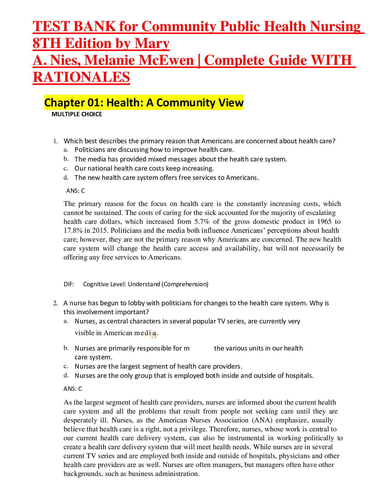
Complete Test Bank For Community Public Health Nursing Promoti...

ATI TEAS 7 | 4 Versions for Reading, Math, Science, English, A...

NR 341 / NR341 CMS Review Updated: Complex Adult Health - Cham...


.png)

.png)
