healthcare > INSTRUCTOR MANUALS > Instructor Manual for Advanced Nutrition and Human Metabolism 7th Edition By Sareen Gropper, Jack Sm (All)
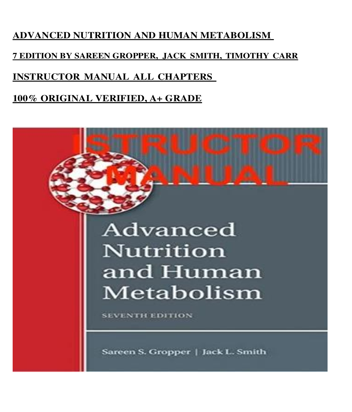
Instructor Manual for Advanced Nutrition and Human Metabolism 7th Edition By Sareen Gropper, Jack Smith, Timothy Carr (All Chapters, 100% Verified) A+ Grade
Document Content and Description Below
Instructor Manual for Advanced Nutrition and Human Metabolism 7th Edition By Sareen Gropper, Jack Smith, Timothy Carr (All Chapters, 100% Verified) A+ Grade-Table of Contents • Chapter Outline •... Resources • Perspectives – Classroom Discussion • Assignment – Group Project • Answer Keys o Case Study o Responding to Research o Labeling It • Worksheet 1: Responding to Research – Mitochondria and Aging • Worksheet 2: Labeling It – A Eukaryotic Cell Chapter Outline I. Introduction 1. This chapter provides a brief review of the basics of a cell, including cellular components, biological energy, and an overview of a cell’s natural life span. 2. Key Terms a. Cells – basic living, structural, and functional units of the human body b. Eukaryotic cells – multicellular organisms c. Prokaryotic cells – primitive cells d. Plasma membrane – sheet-like structure that encapsulates and surrounds the cell, allowing it to exist as a distinct unit 3. Figures and Tables a. Figure 1.1 – three-dimensional depiction of a typical mammalian liver cell II. Components of Cells A. Plasma Membrane 1. Sheet-like structure that encapsulates and surrounds the cell. It is asymmetrical and considered to be a fluid structure 2. Key Terms a. Hydrophobic – molecule or part of molecule that repels water but has strong affinity for nonpolar substances b. Receptors – macromolecules that bind a signal molecule with a high degree of specificity that triggers intracellular events c. Enzymes – protein catalysts that increase the rate of a chemical reaction in the body 3. Figures and tables a. Figure 1.2 – lipid bilayer structure of biological membranes b. Figure 1.3 – fluid model of cell membrane. Lipids and proteins are mobile and can move laterally in the membrane B. Cytoplasmic Matrix 1. Consists of filaments or fibers and provides the cell with structural support, framework, network to direct movement, means of independent location, pathway for intercellular communication, and possible transfer of RNA and DNA 2. Key Terms a. Microtubules – hollow, cylindrical cytoskeletal structures composed of the protein tubulin that act to support the cell structure b. Intermediate filaments – strong, ropelike cytoskeletal fibers that are made of protein and that function to provide mechanical stability to cells c. Microfilaments – solid cytoskeletal structures made of a double-helix polymer of the protein actin that play a role in cell motility 3. Microtubules, intermediate filaments, and microfilaments – make up the cytoskeleton 4. Structural arrangement a. Hexose monophosphate shunt – pentose phosphate pathway 5. Figures and tables a. Figure 1.4 – the cytoskeleton provides a structure for cell organelles, microvilli, and large molecules C. Mitochondrion 1. Cellular organelle that is the site of energy production by oxidative phosphorylation and the site of tricarboxylic acid cycle 2. Key terms a. Mitochondria – primary sites of oxygen use and ATP production in cells b. Oxidative phosphorylation – pathway in the mitochondria that makes ATP from ADP and Pi c. Electron transport chain – sequential transfer of electrons from reduced coenzymes to oxygen that is coupled with ATP formation and occurs within the mitochondria 3. Mitochondrial membrane – consists of a matrix or interior space surrounded by a double membrane 4. Mitochondrial matrix – metabolic enzyme systems that function by catalyzing reactions of the tricarboxylic acid and fatty acid oxidation 5. Figures and tables a. Figure 1.5 – the mitochondrion b. Figure 1.6 – overview of a cross section of a mitochondrion D. Nucleus 1. Largest organelle within the cell, regulating most cellular activities 2. Key terms a. Nuclear envelope – composed of an inner and an outer membrane; surrounds the cell nucleus b. Nucleolus – region of the nucleus containing condensed chromatin and sites for synthesizing ribosomal RNA c. Genes – section of chromosomal DNA that codes for a single protein d. Genome – sum of all the chromosomal genes of a cell e. Nucleotides – phosphate esters of the 5ʹ-phosphate of a purine or pyrimidine in N-glyosidic linkage with ribose or deoxyribose; occurs in nucleic acids f. Complementary base pairing – pairing of nucleotide bases in two strands of nucleic acids; A pairs with T or U, while G pairs with C g. Replication – synthesis of a daughter duplex DNA molecule identical to the parental duplex DNA h. Transcription factors – auxiliary proteins that bind to specific sites in the DNA and alter the transcription of nearby genes i. Sense strand – the strand of DNA that serves as a template for mRNA j. Introns – noncoding regions of a gene k. Exons – coding regions of a gene l. Anticodons – three-base sequences of nucleotides within transfer RNA molecules m. Elongation – (1) extension of the polypeptide chain of the protein product during protein synthesis, (2) the addition of carbons to a fatty acid chain n. MicroRNAs – small noncoding RNAs that silence gene expression by binding to mRNA to inhibit its translation and/or promote its degradation 3. Nucleic acids – macromolecules of nucleotides; consist of a nitrogenous core, a pentose sugar, and a phosphate 4. Cell replication – synthesis of daughter DNA identical to the parental DNA 5. Transcription – taking genetic information in a single strand of DNA and making a specific sequence of bases in a messenger RNA chain 6. Translation – process by which genetic information in an mRNA molecule is turned into the sequence of amino acids in the protein 7. Control of gene expression – controlled through transcription, processing- level control mechanisms determine the path by which mRNA is translated into polypeptide and translation-level control mechanisms determine which mRNA is translated 8. Figures and tables a. Figure 1.7 – steps of protein synthesis b. Figure 1.8 – DNA replication E. Endoplasmic Reticulum and Golgi Apparatus 1. The organelles function together to create a mechanism for communication from the innermost part of the cell to its exterior 2. Key terms a. Endoplasmic reticulum – network of membranous channels pervading the cytosol and providing continuity between the nuclear envelope, the Golgi apparatus, and the plasma membrane b. Sarcoplasmic reticulum – smooth endoplasmic reticulum that is found in muscle cells and is the site of the calcium pump c. Cytochromes – heme-containing proteins that serve as electron carriers d. Oxidation – enzymatic reaction in which oxygen is added to, or hydrogen and its electrons are removed from, the reactant e. Lipophilic – state of being attracted to lipids and thus repelled by water f. Hydrophilic – refers to a molecule or part of a molecule having a strong affinity for water and other polar substances g. Golgi apparatus – the part of the cell responsible for modifying macromolecules synthesized in the endoplasmic reticulum and packaging them to be transported to the cell surface or cytosol F. Lysosomes and Peroxisomes 1. Aid in cell’s digestion and oxidative catabolic reactions 2. Key terms a. Lysosomes – cell organelles that contain digestive enzymes b. Peroxisomes – cell organelles containing enzymes that perform oxidative catabolic reactions c. Catabolism – process by which organic molecules are broken down III. Selected Cellular Proteins A. Receptors 1. Highly specific proteins located in the plasma membrane and act as recognition markers 2. Key terms a. Ligands – small molecules or minerals that bind to a larger molecule 3. Receptors that generate internal chemical signals – an internal chemical signal is generated following interaction between some receptors and ligands 4. Receptors that function as ion channels – in some cases, the binding of the ligand to its receptor causes a voltage change, which then becomes the signal for a cellular response 5. Receptors that internalize stimuli – a stimulus is internalized through a stimulus 6. Receptor’s role in homeostasis – receptors that respond to changes in the external conditions 7. Figures and tables a. Figure 1.9 – example of an internal chemical signal by a second messenger b. Figure 1.10 – internalization of a stimulus into a cell via its receptor B. Catalytic Proteins (Enzymes) 1. These are enzymes that are catalysts and take part in reactions but are not part of the final product of that reaction 2. Key terms a. Oxidoreductases – enzymes that catalyze all reactions in which one compound is oxidized and another is reduced b. Transferases – enzymes that catalyze reactions not involving oxidation and reduction in which a functional group is transferred from one substrate to another c. Hydrolases – enzymes that catalyze cleavage of bonds between carbon atoms and some other kind of atom by the addition of water d. Lyases – enzymes that catalyze cleavage of carbon–carbon, carbon– sulfur, and certain carbon–nitrogen bonds without hydrolysis or oxidation–reduction e. Isomerases – enzymes that catalyze the interconversion of optical or geometric isomers f. Ligases – enzymes that catalyze the formation of bonds between carbon and other atoms g. Ischemia – deficiency of blood in a tissue h. Oncogenes – genes capable of causing a normal cell to convert to a cancerous cell 3. Reversibility – the same enzyme catalyzes a reaction in both directions 4. Regulation – anabolic and catabolic reactions are kept balanced a. Covalent modification – enzyme is inactive until a posttranslational modification is made b. Allosteric enzyme modulation – a secondary regulatory mechanism that is used by certain enzymes called allosteric enzymes. These enzymes possess another site besides the catalytic site c. Induction – creates changes in the concentration of certain inducible enzymes by increasing enzyme synthesis 5. Examples of enzyme types – enzyme participation depends on where the enzyme is located through the cell 6. Clinical applications of cellular enzymes – enzymes are synthesized intracellularly, and most function within the cell they are formed in. Enzymes must have high degree of organ or tissue specificity, steep concentration gradient of enzyme activity between the interior and exterior of cells, must function in the cytosol of the cell, and must be stable for a reasonable time period in the vascular compartment [Show More]
Last updated: 10 months ago
Preview 5 out of 160 pages

Loading document previews ...
Buy this document to get the full access instantly
Instant Download Access after purchase
Buy NowInstant download
We Accept:

Reviews( 0 )
$18.50
Can't find what you want? Try our AI powered Search
Document information
Connected school, study & course
About the document
Uploaded On
Sep 30, 2024
Number of pages
160
Written in
Additional information
This document has been written for:
Uploaded
Sep 30, 2024
Downloads
0
Views
34

