NUR 300 med surg final exam – Galen College
Document Content and Description Below
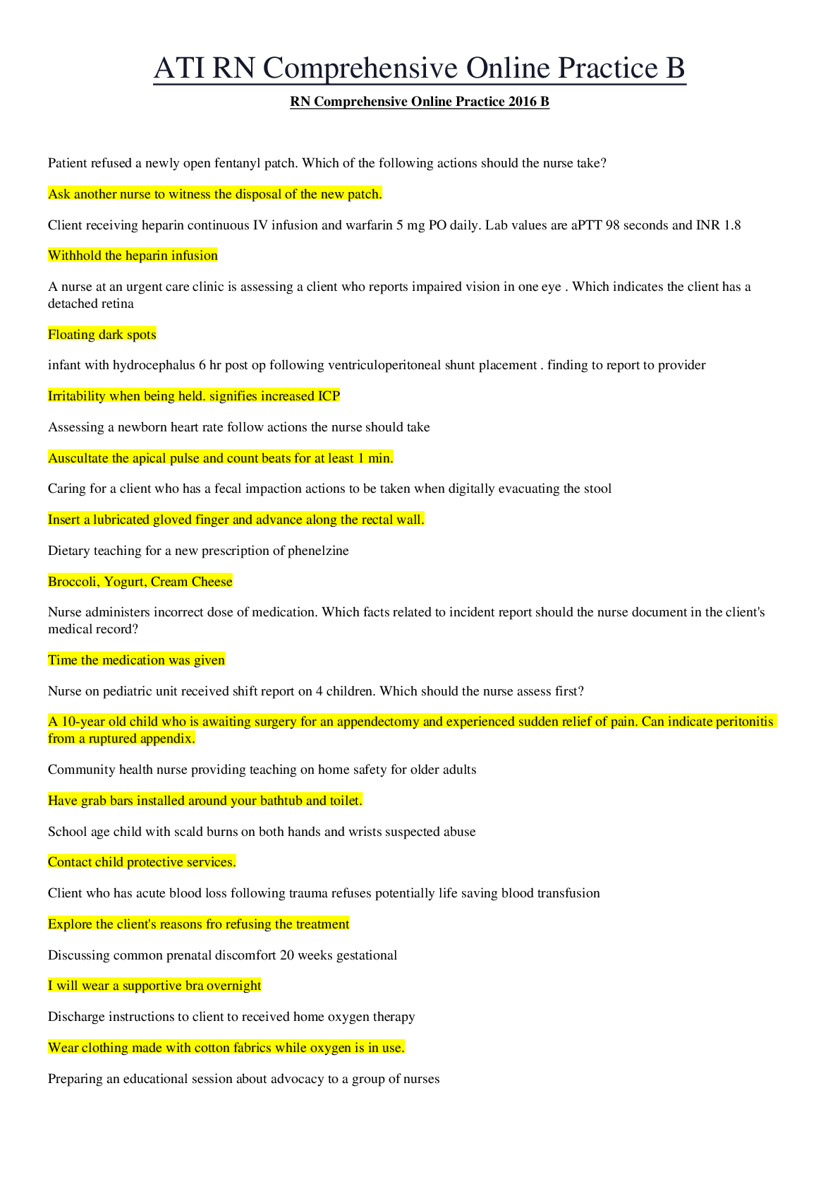
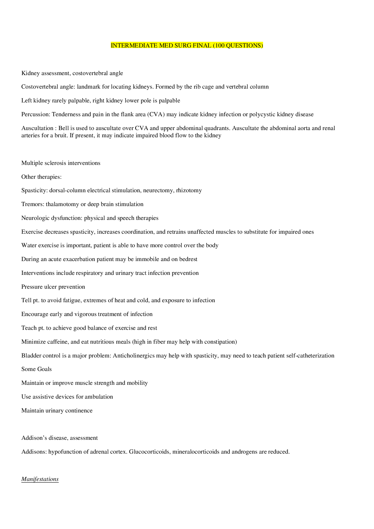
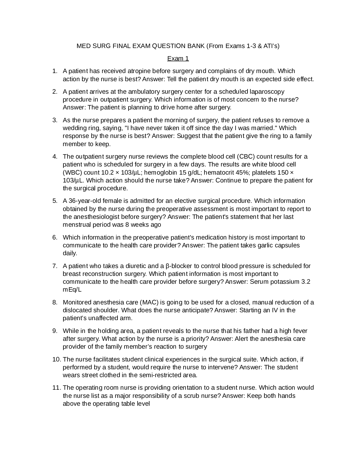
NUR 300 med surg final exam – Galen College COMPREHENSIVE REVIEW 1. Preoperative Care a. Surgical Risk Factors, Current Medications, Data to Obtain in Health History, & Physical/Psychosocial... Assessment i. Health History: Past surgical history and reactions, medications the patient is taking. RED FLAG medications include: Anticoagulants: Coumadin, Warfarin, Plavix, Aspirin, Heparin, Herbal Medications that can thin your blood, and corticosteroids because they delay wound healing. ii. Psychosocial Assessment: You want your patient to feel as good as possible. It is a fact supported by evidence that patients that enter surgery with poorer attitudes experience poorer outcomes. iii. Cultural/Spiritual Assessment: “Is there anything about your cultural or spiritual background that I should know that could affect your surgery and care?” iv. Physical Assessment: This will give us a baseline of information with which to compare during and after surgery. v. Labs: Hgb & Hct (H & H), platelets, PTT, aPTT, PT/INR, Blood Type & Cross, BUN, and CR. vi. Diagnostic Tests b. Pre-operative Care i. What medications are commonly used pre-op and why? 1. To reduce anxiety, pain, and the amount of anesthesia needed, i.e. sedatives: atarax/vistaril (hydroxyzine), hypnotics: Ativan (lorazepam), anxiolytics: versed(midazolam), analgesics: morphine, Demerol (merperidine), fentanyl, dilaudid (hydromorphone). 2. To decrease oral and gastric secretions, anticholinergics: atropine (urecholine) and scopolamine. 3. To reduce nausea & vomiting: Phenergan (promethazine), Zofran (Ondansetron), Reglan (Metoclopramide). ii. What sort of precautions take precedence after giving pre-op medications? 1. Patient safety 2. Monitor status, i.e. vitals, affect c. Preoperative Teaching i. When should you do client teaching about the surgery and why? 1. BEFORE the operation. After surgery, patients are in too much pain to care or be able to retain and understand. ii. Why is client teaching important? 1. It helps the patient understand what to expect after surgery. a. Be sure to explain equipment, tubes, drains, vascular access, and the frequent monitoring for vital signs in great detail. 2. It may help to comfort and prepare patients. 3. It reduces anxiety. d. Informed Consent i. When can the patient withdraw consent for surgery? 1. AT ANY TIME ii. When should the informed consent form be signed? Who informs the patient about the procedure, including its risks and benefits? 1. Before the surgery. The surgeon. iii. What are the RN duties regarding informed consent? 1. Clarify as needed - -- - - - - - - - 5. Electrolyte Imbalances *indicates the symptom you better not miss! (Review Concept Map) a. Potassium i. What is the normal range for potassium? 1. 3.5-5.0 ii. What causes hypokalemia? 1. Loss of GI Content a. NG Suction b. Vomiting c. Diarrhea iii. What are the s/s of hypokalemia? 1. Leg Cramps/Muscle Cramps 2. Irregular Heart Rhythm* a. EKG Result: iv. What is the medical treatment for hypokalemia? 1. PO or IV Potassium a. NEVER EVER EVER PUSH POTASSIUM i. It should be delivered by piggyback, 10 meq/hr ii. Asking the physician to have the order add lidocaine to the potassium in the bag will make it more tolerable for the patient. (Because it burns like FIYAAA) v. NI: Hypokalemia 1. Teach foods high in potassium: a. Dried Fruits & Raw Fruits, like bananas, cantaloupe, kiwi, and oranges b. Vegetables like avocado, greens, and potatoes with skin vi. What causes hyperkalemia? 1. Potassium Sparing Diuretics (Spironolactone) 2. Kidney Failure vii. What are the s/s of hyperkalemia? 1. Muscle Weakness 2. Irregular Heart Rhythm* a. EKG Result viii. What is the medical treatment for hyperkalemia? 1. Kayexulate a. This will cause explosive diarrhea, so think about skin breakdown and patient safety. b. IV insulin and glucose, this will remove excess potassium c. Dialysis-How do we know when? ix. NI: Hyperkalemia 1. Protect skin integrity 2. Protect patient safety when making frequent trips to toilet b. Sodium i. What is the normal range for sodium? 1. 135-145 ii. What causes hyponatremia? 1. Sodium loss through sweat or excessive water on board. iii. What are the s/s of hyponatremia? 1. Cerebral Change, like confusion 2. Muscle Weakness* iv. What is the medical treatment for hyponatremia? 1. PO Sodium tablets or IV Sodium with NS v. NI: Hyponatremia 1. Increase dietary sodium (Remember processed foods are always high in sodium) 2. Risk for Falls vi. What causes hypernatremia? 1. Too much sodium intake 2. Not drinking enough water 3. Diarrhea vii. What are the s/s of hypernatremia? 1. Thirst 2. Dry Mucus Membranes 3. Decreased Urine Output* viii. What is the medical treatment for hypernatremia? 1. Hypotonic IV Solution ix. NI: Hypernatremia 1. Sodium Restriction 2. Teach patient to read labels 3. Avoid processed foods! c. Magnesium i. What is the normal range for magnesium? 1. 1.5-2.5 ii. What causes hypomagnesaemia? 1. Alcoholism iii. What are the s/s of hypomagnesaemia? 1. Cramps in large muscles 2. Irregular Heart Rhythm* iv. What is the medical treatment for hypomagnesaemia? 1. PO or IV Magnesium v. NI: hypomagnesaemia 1. DO NOT PUSH 2. Safe Dose is 1 gram per hour 3. Use seizure precautions when administering vi. What causes hypermagnesaemia? 1. Kidney Failure vii. What are the s/s of hypermagnesaemia? 1. Loss of Deep Tendon Reflexes 2. Cardiac Arrest* viii. What is the medical treatment for hypermagnesaemia? 1. Limit PO intake 2. Dialysis ix. NI: hypermagnesaemia 1. Decrease Dietary Magnesium a. Food Sources: Green Leafys, Nuts, Seeds, and Legumes d. Calcium i. What is the normal range for calcium? 1. 9-10.5 ii. What causes hypocalcemia? 1. Vitamin D Deficiency 2. Hypoparathyroidism iii. What are the s/s of hypocalcemia? 1. Tinging in Extremities (Paresthesia) 2. Positive Chvostek’s Sign 3. Positive Trousseau’s Sign iv. What is the medical treatment for hypocalcemia? 1. PO or IV Calcium or Vitamin D v. NI: Hypocalcemia 1. Increase Dietary Calcium a. Foods: Dairy Products, Leafy Greens, Legumes, and some fruits 2. Could administer two types of IV Calcium a. First choice: Calcium Glucanate b. Only if severe: Calcium Chloride vi. What causes hypercalcemia? 1. Too much vitamin D 2. Hyperparathyroidism vii. What are the s/s of hypercalcemia? 1. General Muscle Weakness 2. Kidney Stones 3. Personality Changes* viii. What is the medical treatment for hypercalcemia? 1. Lasix 2. Calcitonin 3. Dialysis ix. NI: Hypercalcemia 1. Increase PO fluids 2. Decrease dietary calcium a. Be careful making this recommendation to a postmenopausal woman e. How can you evaluate whether or not your treatment of these imbalances is working? i. MONITOR THE LABS, BRO 6. Acid/Base Imbalance *Review worksheet a. pH 7.35-7.45 b. CO2 35-45 c. HCO3 22-26 d. ABG interpretation i. Is the pH acidic or alkalotic? ii. Are both the CO2 and HCO3 abnormal? 1. If no: the one that is abnormal is the problem 2. If yes: which value matches the pH? The matching value is the problem and the other is compensation e. What is the number one indicator to have ABGs drawn? i. Respiratory Distress f. Will compensation save the patient? i. NO. All compensation is temporary, it will eventually fail and WILL KILL YOU. g. What is the pathophysiology of metabolic acidosis? i. Decreased pH ii. Decreased HCO3 h. What can cause metabolic acidosis? i. Shock ii. Diarrhea iii. Cardiac Arrest iv. DKA v. Starvation vi. ASA Overdose vii. Wound Drainage i. What are the S/S of metabolic acidosis? i. Kussmaul’s Respiration (Deep, Regular, and Rapid) ii. Cardiac Dysrhythmias iii. Coma iv. Death j. How do we treat metabolic acidosis? i. IV bicarbonate k. What pathophysiology occurs with metabolic alkalosis? i. Increased pH ii. Increased HCO3 l. What could cause metabolic alkalosis? i. Diuretics ii. Oral bicarbonate drugs iii. Vomiting iv. Gastric Suction v. Hypokalemia m. What are the S/S of metabolic alkalosis? i. Anorexia ii. Nausea and Vomiting iii. Confusion iv. Tetany v. Hypertonic Reflexes n. How do we treat metabolic alkalosis? i. Eliminate the cause ii. Potassium supplements or sodium chloride o. What pathophysiology occurs with respiratory acidosis? i. Decreased pH ii. Increased CO2 p. What could cause respiratory acidosis? i. Head Injuries ii. Pneumothorax iii. Hemothorax iv. Pulmonary Edema v. Asthma vi. Atelectasis vii. Pneumonia q. What are the S/S of respiratory acidosis? i. Respiratory Distress (Hypoventilation) r. How do we treat respiratory acidosis? i. Mechanical Ventilation s. What pathophysiology occurs with respiratory alkalosis? i. Increased pH ii. Decreased CO2 t. What could cause respiratory alkalosis? i. Increased respirations ii. Increased temp iii. Overactive thyroid iv. ASA poison v. Hypoxemia vi. Mechanical Ventilation u. What are the S/S of respiratory alkalosis? i. Increased respiratory rate (Hyperventilation) v. How do we treat respiratory alkalosis? i. Have client breath into paper bag 21. Musculoskeletal a. Normal Serum Calcium Lab Value: i. 9-10.5 b. Arthroscopy i. Go in with a scope to visualize the joint and clean it out. ii. Nursing Considerations: 1. Patient must be able to flex knee; exercises prescribed for ROM. 2. Evaluate the neurovascular status of affected limb frequently. a. This will always be most important to assess distally, not at the actual site of the procedure. We have to be certain that what occurred at the site has not messed with circulation to the distal part of the limb. 3. Analgesics are prescribed for pain relief. 4. Monitor for complications: a. Severe Pain b. Thrombophlebitis c. Infection c. Which test is the best to diagnose damage that has occurred to the musculoskeletal system and the soft tissue? i. MRI. CT shows the MS pretty well, but the MRI is really needed to see the soft tissue damage. d. Osteoarthritis i. NONINFLAMMATORY, LOCALIZED arthritis. This is NOT systemic. NOT autoimmune. ii. The most common type of arthritis. iii. Progressive loss of cartilage, causing joint pain and progressive deterioration in function. iv. Risk Factors: Over 60 years old, obesity. v. Clinical Manifestations: 1. Joint Pain and Stiffness 2. Crepitus 3. Fluid in the joint 4. Atrophy of surrounding skeletal muscle 5. Typically experience pain in the evening. 6. Severe pain may cause depression/anxiety. vi. Diagnostics: MRI will give definitive diagnosis. vii. Goal of Treatment: Avoid surgery for as long as possible. 1. Nonsurgical Management: a. Drug Therapy (Mobic) b. Rest, Immobilization c. Positioning (Elevate when swelling) d. Thermal (Hot/Cold) e. Weight Control (HUGELY IMPORTANT) viii. Surgical Management 1. Total Joint Arthroplasty a. Prevent Complications: i. Dislocation 1. Refer all to PT/OT. a. Shoulder needs OT most. 2. Hip Precautions: a. Don’t cross legs or ankles. b. When standing, stick injured leg out before rising. ii. VTE iii. Infection iv. Atelectasis v. Hypovolemia vi. Neurovascular Compromise 1. Check Pulses, cap refill, etc. distal to surgery. vii. Continuous Passive Motion Machine (CPM) 1. Helps increase mobility in operated joint. Each time they get in, the angle is increased until they can achieve full range of motion. e. Rheumatoid arthritis i. Chronic, progressive, systemic, inflammatory, autoimmune disease. ii. This can occur at any age. The body begins to attack its own joints causing the bones to lose density causing a weakening in the joints and bones. iii. Clinical Manifestations: 1. There will be bilateral joint involvement. 2. It usually affects the upper extremities first. 3. Usually affects the smaller joints. 4. Pain is worst in the morning. 5. Early S/S: joint stiffness, swelling, pain, fatigue, generalized weakness. 6. Late S/S: Joints become progressively inflamed and quite painful. 7. Systemic Complications: a. Weight loss, fever, fatigue b. Exacerbations c. Subcutaneous nodules d. Respiratory, Cardiac complications e. Parenthesis f. Sjogren’s Syndrome i. Dry eyes, mouth, vagina (p. 335) 8. Laboratory Results a. Rheumatoid Factor b. Antinuclear Antibody Titer (ANA) i. These will be present when RA is occurring. 9. RA Drug Therapy a. DMARDs i. Methotrexate 1. Low dose chemo. Acts to suppress body’s immune system. b. NSAIDs i. Naproxen is best. c. BRMs (Biological Response Modifiers) i. Enbrel ii. Humira d. Glucocorticosteroids 10. RA Nonpharmacologic Interventions a. Adequate rest b. Ice and Heat application c. Promotion of self-management d. Management of fatigue e. Enhance body image OA vs RA at a Glance OA RA Noninflammatory Inflammatory Not Autoimmune Autoimmune Localized System Usually affects those over 60 Occurs at any age Usually unilateral involvement Usually bilateral involvement Affects larger joints Affects smaller joints Pain worst at night Pain worst in morning f. Gout i. Type of arthritis ii. Uric acid crystals deposit in joints causing inflammation. iii. S/S: Joint inflammation and pain. iv. Medications: 1. Acute: a. Colchicine (Colsalide) *drug of choice* b. NSAIDs 2. Chronic: a. Allopurinal (Zyloprim) g. Osteoporosis i. A decrease in bone density. ii. Most common in thin, white women. iii. Risk Factors: 1. Old Age 2. Family History 3. Low body weight/thin build 4. Low calcium intake 5. Smoking, Excess alcohol consumption 6. Lack of Exercise iv. Diagnostic Testing: 1. DEXA Bone Scan is the definitive test. v. Clinical Manifestation: 1. Dowager’s Hump 2. Back Pain vi. Primary Problems 1. Decreased Strength 2. Risk for Fracture 3. Injury Prevention 4. Nutritional Status vii. Interventions 1. Diet high in calcium 2. Weight Bearing Exercise (NO SWIMMING) 3. Safety Precautions (Prevent fall!) 4. Medications a. Calcium and Vitamin D b. Biophosphonates i. Taken in the morning with a full glass of water. 5. Surgical management 6. Patient Teaching 7. Community Resources h. Osteomyelitis i. Infection of Bone Tissue ii. Different Types 1. Exogenous a. Organisms enter from outside the body. 2. Endogenous a. Organisms are carried from another infection in the body. 3. Acute hematogenous a. Caused by bacteria, underlying disease or trauma. 4. Contigous a. Occurs in facial bones. 5. Chronic iii. Clinical Manifestations 1. Bone pain that is worse with movement. 2. Fever 3. Swelling/erythema of surrounding tissue. 4. Elevated WBCs. iv. Interventions 1. Non-surgical a. Medications i. IV antibiotics. Heavy duty and for up to 3 months. ii. Severe Pain Management b. Wound Irrigation/Dressing changes (if result of trauma) 2. Surgical a. This is a last resort and usually occurs only in the case that the infection came from a traumatic event. They will create a muscle flap that provides wound coverage and enhances healing after debridement. v. Keep in Mind 1. Pain, fatigue, and increased WBC will be seen in anyone with acute osteomyelitis. However, while a younger adult would also have a high fever, an older adult may only exhibit a low grade fever. i. Fractures i. A break or disruption in continuity of bone. ii. Types: 1. Closed: Does not breakthrough skin. 2. Open/Compound: Breaks through skin. 3. Non-displaced: Bones remain in alignment. 4. Displaced: Bones are knocked out of alignment. 5. Fragmented: Bone shattered in many pieces. 6. Spiral: Twisting type. 7. Oblique: Occurs along an angle. 8. Impacting: Cue to forceful impact. 9. Greenstick: Seen in pediatrics. 10. Pathologic: Spontaneous fractures, seen with cancer and osteoporosis. iii. Stages of Bone Healing 1. Injured site fills with blood and creates a hematoma. 2. New vessels are formed. 3. Bony callus of spongy bone forms. 4. Solid bone forms again. iv. Interventions 1. Non-Surgical a. Immobilization b. Cast care i. Main Priority: 1. Assess circulation distal to cast. Neurovascular checks Q1h for first 24 hrs. Teach patient to assess circulation daily. (Color, temp, etc.) ii. Use ice to reduce swelling. iii. Should be able to fit a finger between cast and skin. iv. CRITICAL COMPLICATION TO AVOID 1. Compartment Syndrome 2. Can begin 6-8 hrs after injury, but may take up to 2 days. 3. S/S: “The 6 P’s” a. Pain more intense than the injury itself b. Pressure c. Paralysis d. Paresthesia (usually occurs 1st) e. Pallor f. Pulselessness 4. Early recognition is critical to prevent loss of limb or function. Teach patient signs to look for as well. 2. Surgical a. ORIF b. Screws fixed on the inside of body. v. External fixation 1. Screws fixed through equipment on the outside of body. a. Risk for infection is high. It is vital to clean areas around the screws well frequently. vi. Complications of Fractures 1. Acute Compartment Syndrome 2. Hypovolemic Shock 3. Fat embolism syndrome 4. VTE 5. Infection 6. Amputations j. Carpal Tunnel Syndrome i. Compression of the median nerve in the wrist. ii. Clinical Manifestations: 1. Pain and numbness, mostly in the wrist, that is worse at night. 2. Paresthesia iii. Interventions 1. Nonsurgical a. NSAIDs b. Splint or Hand Brace 2. Surgical a. “Carpal Tunnel Release” Releases pressure on median nerve. b. Post Op S/S to report: i. Absent Pulse ii. Cold iii. Paresthesia 22. Infection a. Psoriasis i. Thick, reddened papules or plaques covered by silvery white scales. ii. Treatment: 1. Corticosteroids 2. Tar Preparations 3. Other topical therapies 4. UV light therapy 5. Systemic Therapy (Humira) 6. Emotional Support b. Cellulitis i. Staph or step infection in the skin. ii. Usually preceded by minor skin injury. iii. Redness, warmth, edema, pain, and sometimes blisters. iv. How do we manage? 1. Skin Care 2. Transmission Precautions a. Contact 3. Medications a. Antibiotics c. Skin Cancer i. Prevention is KEY. 1. SUNSCREEN HOMIES. d. Herpes Zoster i. Reactivation of the dormant varicella zoster virus in patients who have had chickenpox. ii. Vesicular lesions, look a lot like blisters, which follow along a dermatomal pattern like along the scapula or jaw. Very painful. Postherpetic neuralgia (painfulness in the region after the lesion has gone). iii. Vaccine (Zostavac) is recommended to anyone over 60. iv. Transmission precautions 1. Treat is with both airborne and contact precautions. e. Inflammation does not always =/= infection. f. Five Cardinal Signs of Inflammation: i. Warmth ii. Redness iii. Swelling iv. Pain v. Decreased Function g. Modes of Transmission i. Contact-direct or indirect ii. Droplet-influenza iii. Airborne-Tuberculosis iv. Vector Bourne-Insects/Animals Lyme Disease v. Contaminated Food/Water-Listeria, Salmonella vi. Portal of Exit h. PPE i. Standard Precautions 1. Gloves when there’s a chance of coming into contact with any body fluids except perspiration. 2. Respiratory/Cough Etiquette 3. Hand Hygiene ii. Airborne 1. Negative Pressure Room 2. N-95 Particulate Mask 3. Tuberculosis, Measles, Chickenpox iii. Droplet 1. Can travel three ft. or more, but are not suspended for long periods of time. 2. Gown 3. Gloves 4. Simple Mask (when you’ll be within 3 ft) 5. Influenza, Mumps, Pertussis, Meningitis iv. Contact 1. Gown 2. Gloves 3. MRSA, VRE, Lice, Scabies, RSV, C. diff v. Reverse Isolation (Transport) 1. Mask 2. Dedicated equipment for their safety. 3. Used when transporting sick patients in isolation or for patients with neutropenia, like: a. Chemo b. Radiation c. AIDs I. Staging of Pressure Ulcers: a. Stage One: Non-blanchable, reddened area. b. Stage Two: Shallow, reddened wound in dermis. c. Stage Three: Full thickness, see Sub-Q, fat may be visible. No bone, tendon, etc. Tunneling may be present. d. Stage Four: Now, we can see muscle, tendon, bone, etc. Full thickness. Necrotic tissue may be present. e. Unstageable: Unmeasurable. Slough is in the way and we can’t really see its depth. J. Why is debridement necessary? What kind of dressing changes may be needed? a. Dead tissue can delay or stop wound healing. b. Wet to Dry with Deacons or Normal Saline. [Show More]
Last updated: 2 years ago
Preview 1 out of 104 pages

Buy this document to get the full access instantly
Instant Download Access after purchase
Buy NowInstant download
We Accept:

Reviews( 0 )
$18.00
Can't find what you want? Try our AI powered Search
Document information
Connected school, study & course
About the document
Uploaded On
May 24, 2020
Number of pages
104
Written in
Additional information
This document has been written for:
Uploaded
May 24, 2020
Downloads
0
Views
131


 – University of the People.png)
