
CLG 0010 DoD Governmentwide Commercial Card Exam Spring 2022(100% scored)
*NURSING > PATIENT ASSESSMENTS > EKG Interpretation Packet. Contains the measurements, Calculations, Plus Questions and Answers. A 20 (All)
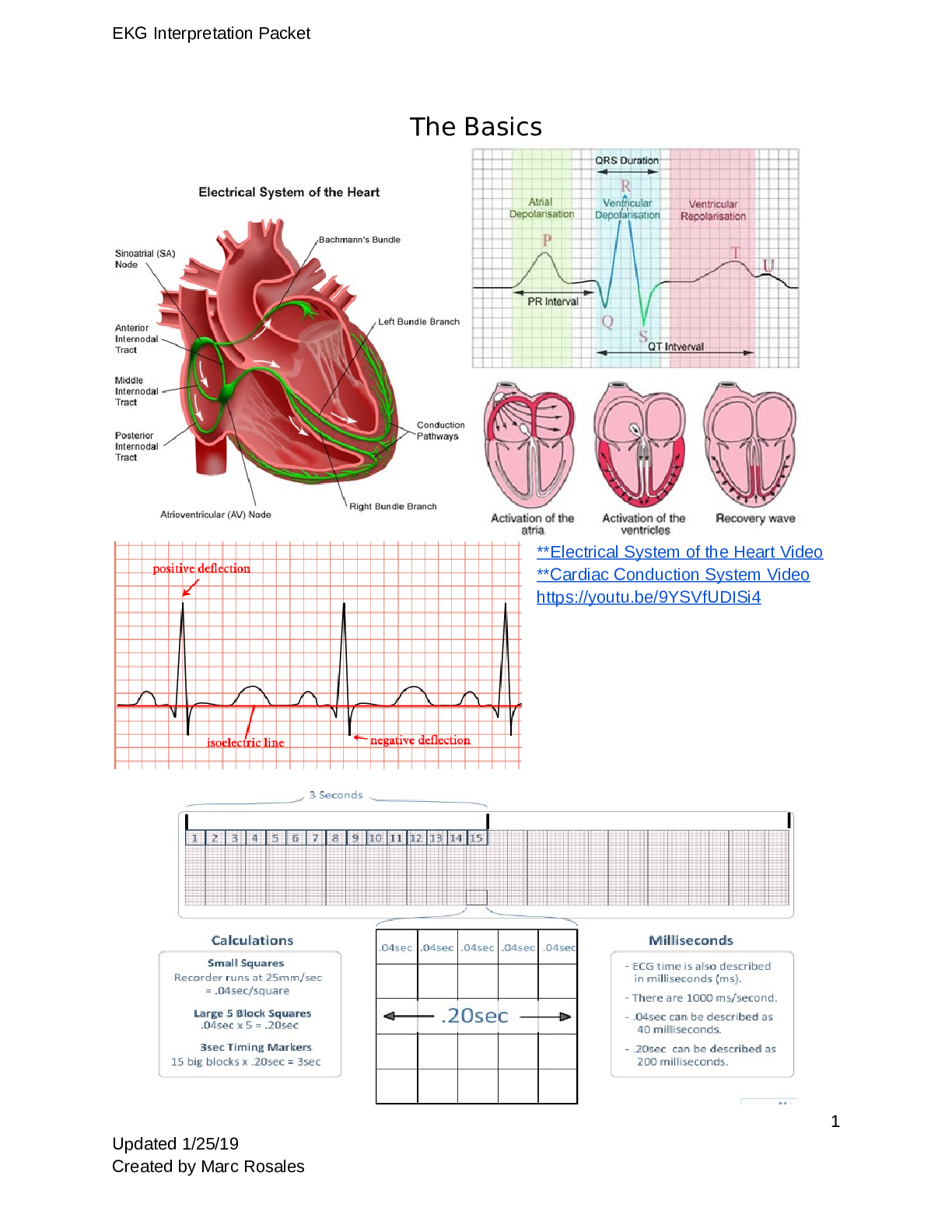
EKG Interpretation Packet. Contains the measurements, Calculations, Plus Questions and Answers. A 2019 Update. SAMPLE CONTENT EKG Interpretation Packet The Basics **Electrical System of the He ... art Video **Cardiac Conduction System Video https://youtu.be/9YSVfUDISi4 1 Updated 1/25/19 Created by Marc RosalesEKG Interpretation Packet Measuring Rate 2 Updated 1/25/19 Created by Marc RosalesEKG Interpretation Packet ST Segment Preview for CHII ● Is the ST segment even with the PR segment? ● Is the ST segment elevated? ● Is the ST segment depressed? Description Source of Possible Variation ST segment: Measured from the S wave of the QRS complex to the beginning of the T wave; represents the time between ventricular depolarization and repolarization (diastole); should be isoelectric (flat) Disturbances usually caused by ischemia, injury, or infarction 3 Updated 1/25/19 Created by Marc RosalesEKG Interpretation Packet What is this? What do we do about it? 4 Updated 1/25/19 Created by Marc RosalesEKG Interpretation Packet 1. Complete the following statements. a. The three main coronary arteries are the __LAD ____________, _______RCA________, and ____ ______CIRC_____. The LCA FEEDS THE CIRC AND LAD b. In most people, the ______RCA_________ artery supplies the AV node. c. Blood flow into the coronary arteries occurs primarily during the _____DIASTOLE__________ phase of the cardiac cycle. d. The ________MITRAL______ and ________TRICUSPID_______ valves close at the beginning of ventricular contraction and prevent blood from flowing back into the atria from the ventricles. They open when the ventricles relax. e. The _________AORTIC______ and __________PULMONIC_____ valves prevent blood from flowing back into the ventricles during relaxation. They open during ventricular contraction and close when the ventricles begin to relax. 2. Number in sequence the path of the action potential along the conduction system of the heart. ___4__a. AV node ____7_b. Purkinje fibers ___3__c. Internodal pathways (THEN MOVES TO INTERNODAL PATHWAYS) ___5__d. Bundle of His ___8__e. Ventricular cells (THE SQUENCE WILL START ALL OVER AGAIN) __1___f. SA node (EVERYTHING ORIGINATES IN SA NODE) ___2__g. Right and Left atrial cells (ELECTRICAL ACTIVITY MOVES TO RIGHT AND LEFT ATRIAL CELLS) __6___h. Right and left bundle branches 3. Match the effects of the autonomic nervous system with the receptors responsible for the effects (answers may be used more than once). ___2__a. Increased force of cardiac contraction (in heart) 1. Alpha-Adrenergic Stimulation (PERIPHERALS) __3___b. Decreased rate of impulse conduction (rest n digest) 2. Beta- Adrenergic Stimulation (IN HEART) ___1__c. Vasoconstriction (in extremeties) 3. Parasympathetic Stimulation (REST AND DIGEST) OCCURS WITH OUR NORMAL SYSTEMS IN OUR BODIES SO WE CAN DETERMINE WHAT TYPE OF DRUGS TO AFFECT THIS SYSTEM ___2__d. Increased heart rate (HR) (fight or flight) ___2__e. Increased rate of impulse conduction ___3__f. Decreased HR (rest and digest) 4. The layer of the heart responsible for contraction is the a. myocardium b. pericardium c. endocardium d. epicardium 5. The pressure is highest in which heart chamber? a. Right Atrium 5 Updated 1/25/19 Created by Marc RosalesEKG Interpretation Packet b. Left Atrium c. Right Ventricle d. Left Ventricle (pump out to the rest of the body) 6. Match the effects of the autonomic nervous system with the receptors responsible for the effects (answers may be used more than once). 7 a. Each small square duration (0.04 seocnds) 1. the first negative deflection of the QRS 11 b. Each large square duration 2. early ventricular repolarization and is also called the isoelectric line (heart is sleeping) 15 c. P wave represents 3. ventricular refractory (sleeping/recharging) time 6 d. PR Interval represents 4. ranges from 0.04 to 0.12 second 14 e. PR Interval duration 5. from beginning to end of QRS complex 16 f. PR Interval is measured 6. the time it takes for the impulse to travel from the atria to the ventricles 8 g. QRS complex represents 7. 0.04 second 4 h. Normal QRS complex duration 8. ventricular depolarization 1 i. Q wave appears as 9. Ranges from 0.32 to 0.4 second or 0.34 to 0.43 ********************************** CONTINUED IN THE ATTACHMENT *********************************** [Show More]
Last updated: 3 years ago
Preview 1 out of 23 pages
Buy this document to get the full access instantly
Instant Download Access after purchase
Buy NowInstant download
We Accept:

Can't find what you want? Try our AI powered Search
Connected school, study & course
About the document
Uploaded On
Apr 02, 2021
Number of pages
23
Written in
All
This document has been written for:
Uploaded
Apr 02, 2021
Downloads
1
Views
218
Scholarfriends.com Online Platform by Browsegrades Inc. 651N South Broad St, Middletown DE. United States.
We're available through e-mail, Twitter, Facebook, and live chat.
FAQ
Questions? Leave a message!
Copyright © Scholarfriends · High quality services·


.png)
