Biology > Lab Experiment > BIOL 1020scale_DupuyEric_F20. (All)
BIOL 1020scale_DupuyEric_F20.
Document Content and Description Below
Last updated: 3 years ago
Preview 1 out of 8 pages
Instant download

Buy this Document to get the Full Access Instantly
Provided by Students Who Aced it
We Verify Document Content to Gurantee Accuracy
Reviews( 0 )
Document information
Connected school, study & course
About the document
Uploaded On
Apr 08, 2021
Number of pages
8
Written in
All

Additional information
This document has been written for:
Uploaded
Apr 08, 2021
Downloads
0
Views
151
Document Keyword Tags
Recommended For You
Get more on Lab Experiment »

Straighterline_BIOL 202L Lab 14 The Urinary System, Experiment...

Lab Assignment: Case Study: Brain Injury Or Spinal Cord Injury...

The Urinary System. PRE-LAB QUESTIONS. Experiment 1: Kidney Fi...

portage learning bio 152.. Lab 1 Exam - Requires Respondus Loc...

ACLS PreTest: Pharmacology and Practical Application Questions...
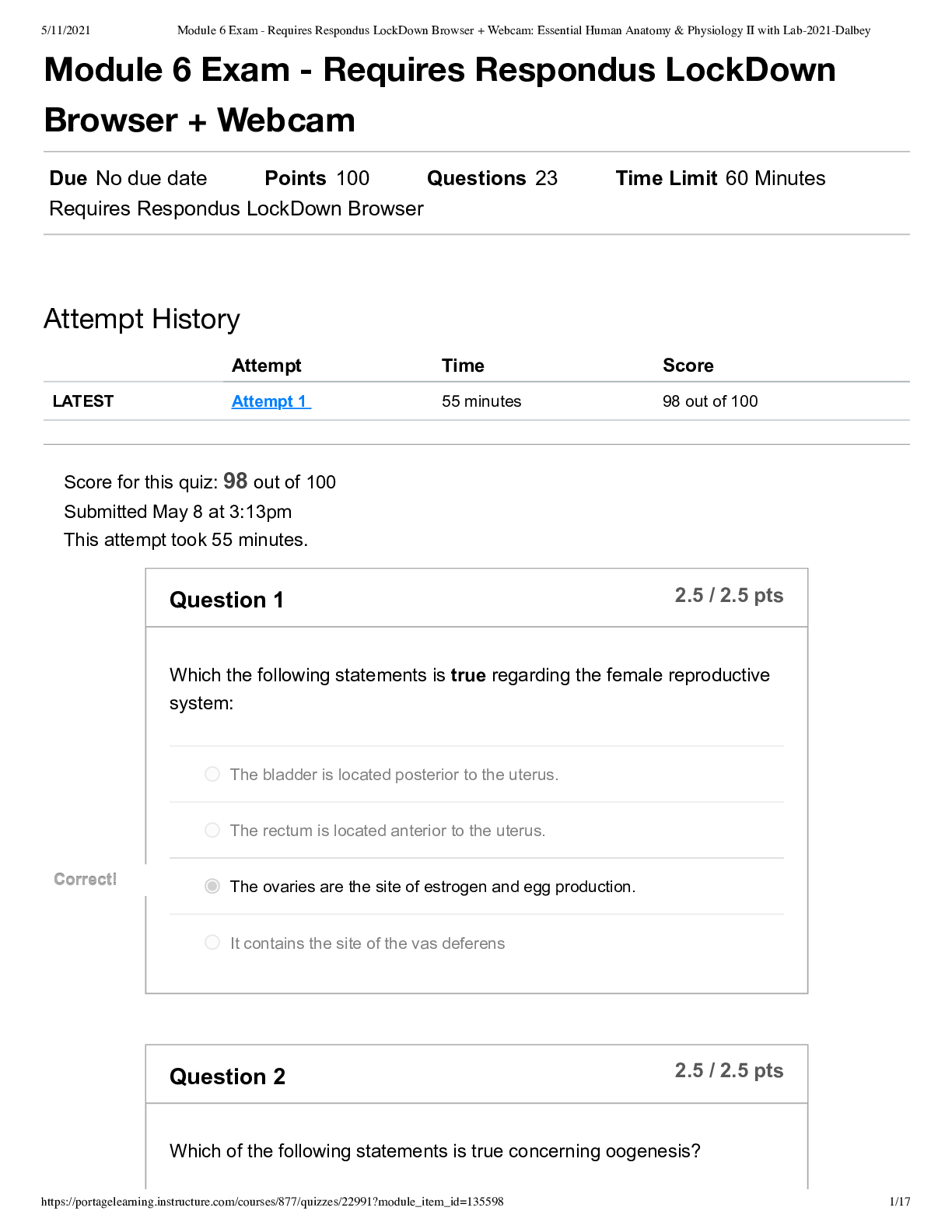
Module 6 Exam - Requires Respondus LockDown Browser + Webcam_...
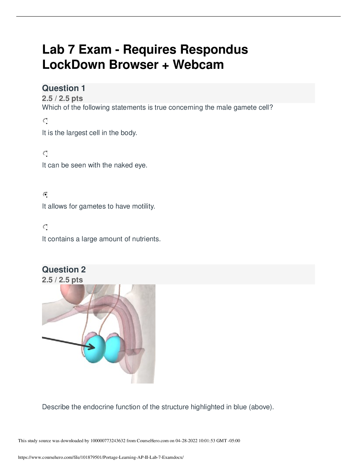
Portage Learning_ AP II Lab 7 Exam.docx. heavily discounted. 1...
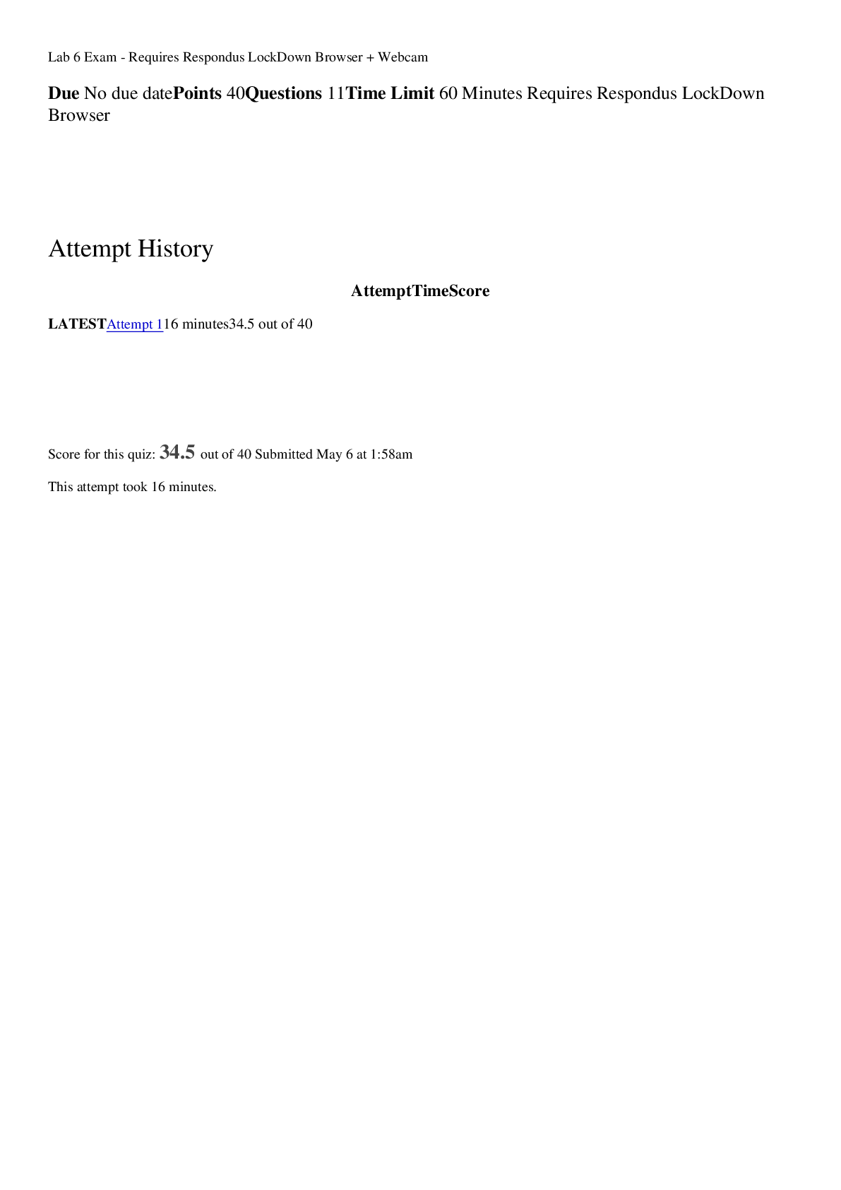
PORTAGE LEARNING BIO 152..Lab 6 Exam - Requires Respondus Lock...

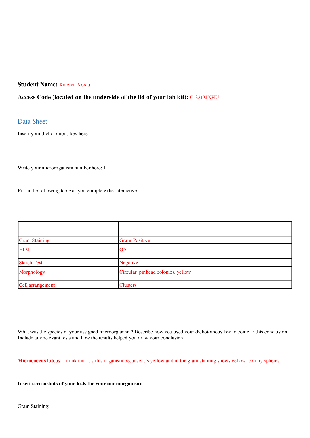
portage learning bio 152. Lab 3 Exam - Requires Respondus Lock...


.png)


.png)