Manifestations of GI disorders
I. Basic Diagnostic Studies
II. Dysphagia
III. Hiatal Hernia
IV. GERD
V. Gastritis: Acute and Chronic
VI. Peptic Ulcers: Gastric and Duodenal
VII. Dumping Syndrome
VIII. Pyloric Ste
...
Manifestations of GI disorders
I. Basic Diagnostic Studies
II. Dysphagia
III. Hiatal Hernia
IV. GERD
V. Gastritis: Acute and Chronic
VI. Peptic Ulcers: Gastric and Duodenal
VII. Dumping Syndrome
VIII. Pyloric Stenosis
IX. Gallbladder disorders
X. Jaundice
XI. Hepatitis
XII. Cirrhosis & Alcoholic Liver Disease
XIII. Pancreatitis
XIV. Chronic Inflammatory Bowel Disease: Crohn’s & Ulcerative Colitis
XV. Appendicitis
XVI. Diverticulitis
XVII. Colorectal Cancer
XVIII. Intestinal Obstruction
XIX. Peritonitis
Manifestations of Digestive Disorders
Anorexia: Loss of appetite. Often precedes Nausea / Vomiting. Non-specific (common in many disorders) Nausea: Unpleasant, subjective feeling stimulated by Distention, Irritation, Inflammation in the GI Tract Retching: Usually precedes Vomiting (nothing expelled)
Vomiting (emesis): Forceful expulsion of chyme from the stomach Activated by vomiting center in the medulla oblongata Activated by: Distention/ irritation of the GI tract
Response to unpleasant smells or tastes, or ischemia
Pain and Stress
Increased IntraCranial Pressure (ICP) “Projectile Vomiting” Stimulation of chemoreceptor trigger zone in medulla by drugs or toxins
Character of vomitus diagnositic:
Blood: (Hematemesis): “Coffee ground” Yellow/Green: Bile
Diarrhea: Excessive frequency of stools (acute or chronic)
Large-Volume diarrhea: water/infection or rapid transit
Small-volume diarrhea: contains blood, mucus, or purulence
Steathorrhea: foul-smelling, greasy, fat stools that float (fat not absorbed)
Hematochezia (Blood in stool): Occult (hidden), Frank (seen), or Melena (red-tarry stool)
Risks of Diarrhea: Dehydration
1. Hypovolemia: tissue Ischemia leads to anaerobic metabolism / lactic acidosis
2. Loss of HCO3: GI stores much of Bicarb, leading to further metabolic acidosis
Malnutrition
Burns 2011
Manifestations of Digestive Disorders
Constipation: Less frequent bowel movements resulting in hard stools
Acute or Chronic May alternate with Diarrhea in Irritable Bowel Syndrome (IBS)
Causes of Constipation:
Inadequate Dietary Fiber intake Inadequate Fluid intake
Muscle weakness and Inactivity (Failure to respond to defecation reflex)
Neurologic Disorders Obstructions: Tumors or Strictures
Medications: Opiates, Iron Supplements, Bulk laxatives (without enough water)
Chronic or Severe Constipation: May lead to….
Hemorrhoids Diverticulosis / Diverticulitis
Impaction or Obstruction Colon Cancer
Fluid Balance Disorders:
Dehydration and hypovolemia
Electrolyte Imbalances
Na+ Sodium is lost in BOTH Vomiting and Diarrhea
Cl- Chloride is lost from Gastric secretions (Vomiting, NG suction). Lost through H-Cl K+ Potassium is lost with Diarrhea
Acid-Base Imbalance: ***KNOW
Vomiting: initially, Vomiting causes Metabolic ALKALOSIS (loss of H+)
prolonged, Vomiting causes Dehydration, anaerobic metabolism, and ACIDOSIS
Diarrhea: Loss of Bicarb (HCO3) and anaerobic metabolism (dehydration) lead to Metabolic ACIDOSIS
Burns 2011
Radiographs
Basic Diagnostic Tests
X-ray (KUB (kindey, urine, bladder), Barium Swallows) CT Scan and MRI
Endoscopy (upper GI), Colonoscopy (rectum, colon), Sigmoidoscopy (rectum, colon)
Laboratory analysis of stool specimens or gastric washing
Infection (O&P, Ova & Parasites) Blood (Hemoccult, Gastroccult) Tumors
Malabsorption problems (steatorrhea in Cystic Fibrosis)
Blood Tests
Liver Function: Serum protein, coagulation, Liver enzymes (AST/ALT), Bilirubin Pancreatic Function: Pancreatic enzymes (Lipase, Amylase)
Stool analysis of enzymes and fat content Other: Tumor markers
Burns 2011
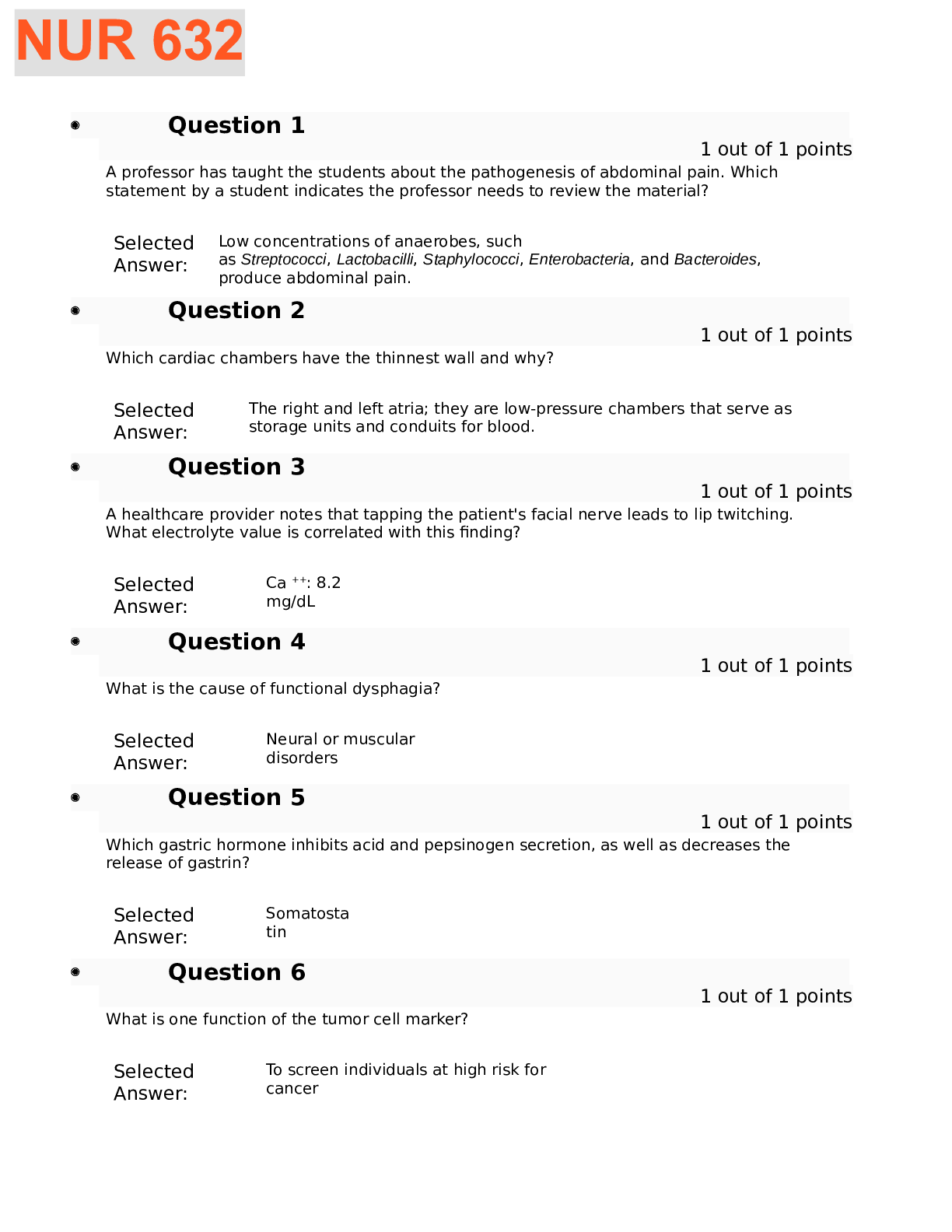
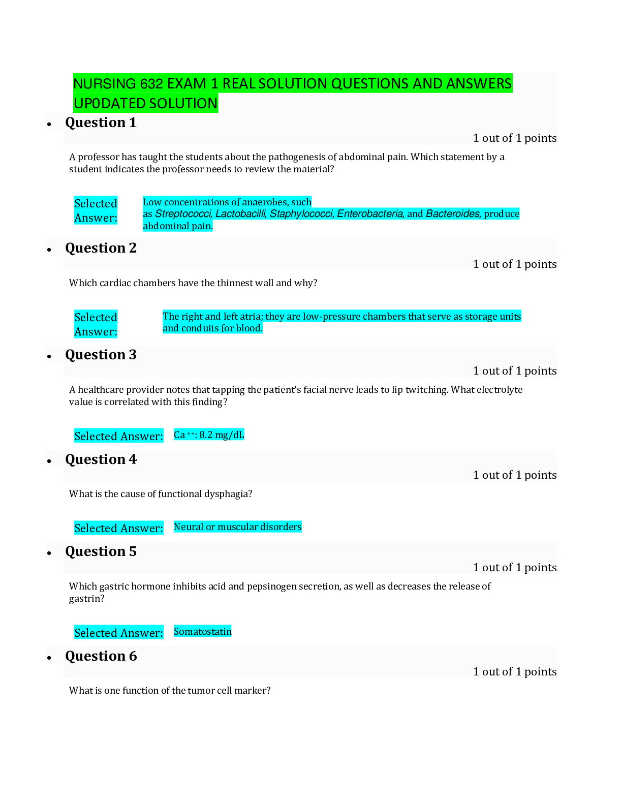
Dysphagia: Difficulty Swallowing
Causes:
Neurologic deficit
CN V, VII, IX, X, XII
Muscular disorder
Achalasia
Mechanical obstruction
Congenital atresia Congenital fisula Compression Fibrosis
Risks: Aspiration
Burns 2011
Hiatal Hernia:
Protrusion of (part of) stomach through opening (hiatus) of diaphragm
Contributing Factors:
Shortening of Esophagus Weakness of Diaphragm
Increased abdominal pressure (pregnancy)
Signs & Symptoms:
Heartburn
Frequent Belching or regurgitation
Increased discomfort with coughing, lying down after eating, or bending over
Dysphagia
Mild, persistent chest pain after meals
Two Types:
Burns 2011
GERD: Gastro Esophageal Reflux Disease
Periodic flow of gastric juice into the unprotected esophagus (past cardiac sphincter)
Often seen in conjunction with hiatal hernia Delayed gastric emptying may be a factor
Signs & Symptoms:
Heartburn 30-60 minutes after eating, or at night
Frequent reflux of gastric acid can lead to…
Inflammation and ulceration of gastric mucosa (ulcer) Eventual fibrosis and stricture of esophagus
Increased risk of esophageal cancer (Barrett’s Esophagus—precursor)
Treatment:
Type-2 Antihistamines: H-2 Blockers (Ranitidine, Cimetidine) – quick acting Proton Pump Inhibitors: PPI’s (omeprazole, desmeprazole) – takes a little longer
Burns 2011
Gastritis: Acute and Chronic
Definition: inflammation of the stomach
Acute Gastritis:
Gastritis mucosa appears inflamed, edematous.
May be ulcerated or bleeding with severe damage or poor circulation:
Infection (bacterial or viral) Allergies (shellfish or drugs)
Spicy / irritating foods Excessive Alcohol intake
Ulcerative drugs (ASA, Ibuprofen) Corrosive / toxic subtances (battery acid)
Radiation or Chemotherapy
Signs & Symptoms:
Anorexia Nausea / Vomiting Hematemesis (with ulceration) Can lead to: Dehydration Metabolic Acidosis
Chronic Gastritis: Characterized by atrophy of the mucosa, with loss of secretory glands
Achlorhydria
Lack of Intrinsic Factor (Vit B12 Deficiency Pernicious Anemia) Often Seen with:
Chronic Peptic Ulcers Alcohol Abuse Autoimmune disorders Elderly
Signs & Symptoms: *Mild Gastric Discomfort *Anorexia *Food Intolerance (spicy)
Can lead to: Peptic Ulcers Gastric Carcinoma
Burns 2011
Peptic Ulcer Disease: Gastric & Duodenal
Most Common: Proximal Duodenum
(where low-pH stomach contents empties)
Also present in the stomach or lower esophagus
Characteristics:
Single, small, round cavities with smooth margins that penetrate mucosa
Once mucosa is penetrated, acid and pepsin cause further damage to impaired gastric tissue
Ulcers can eventually erode through muscle layer causing gastric perforation!
Peritonitis, as gastric contents empty into sterile peritoneum
Burns 2011
Peptic Ulcers: continued
Pathophysiology: An area of inflammation surrounds the crater
When the erosion invades a blood vessel, bleeding ulcer occurs. Bleeding may be:
Persistent loss of small amounts
May manifest itself as Iron-deficiency anemia or occult blood in stool
Massive hemorrhage (depends on blood vessel eroded)
Etiology:
Helicobacter Pylori (H.pylori) found in most cases
Inadequate blood supply (prolonged stress, glucocorticoid secretion or administration)
Ulcerative agents (ASA, Ibuprofen, Alcohol, Steroids)
Atrophy of gastric mucosa (chronic gastritis)
Increased acid-pepsin secretions:
Increased gastrin secretion (caffeine, ETOH, Increased vagal stimulation)
More common in Men, especially in Western Countries
More Common with Blood type O
Signs & Symptoms:
Epigastric pain / burning, especially 2-3 HOURS after eating or at night Pain often relieved by ingestion of food or antacids
Heartburn / Nausea / Vomiting / Weight loss /
*Iron Deficiency Anemia or occult blood in stool
If severe, black tarry stools likely (melena)
Burns 2011
Stress Ulcers:
Peptic Ulcers: continued
Etiology Severe Trauma Massive Burns (Curling’s ulcers)
Serious systemic problems (hemorrhage or sepsis)
Pathophysiology: Multiple ulcers, usually gastric, form within hours of the precipitating event!
As blood flow is reduced to gastric mucosa from stress, there is reduced secretion of mucus and epithelial regeneration, which act as a protective barrier to the effects of acidic chyme
The acid barrier is lost; so, acid diffuses into the sensitive mucosa
Treatment: Medications administered prophylactically to decrease acid release
Proton Pump Inhibitors (-prazoles; pantoprazole, esmoprazole)
Peptic Ulcers: Healing – difficult to heal
Granulation tissue forms deep within cavity
Epithelial cell regenerates from the edges of the fragile granular tissue
Difficult due to constant exposure to irritants
Often, granular tissue broken down by the acidic chyme
Fibrous tissue forms at site, leading to scar tissue and impaired blood supply
Complications:
Hemorrhage (with blood vessel ulceration)
Bacterial peritonitis (with gastric muscle perforation into peritoneum)
Obstruction: if ulceration / fibrous tissue narrows passageway, such as duodenum
Burns 2011
Dumping Syndrome: Loss of Gastric Emptying Control
Pathophysiology:
Large amounts of ingested food is quickly “dumped” into the intestines Hyperosmolar chyme draws more fluid from the vascular compartment
Causes abdominal distention and increased motility
Fluid and electrolytes are then lost in diarrhea
Hypoglycemia results 2-3 hours after meals from “dump” loss
Signs & Symptoms:
Cramps Nausea Diarrhea
Hypovolemia: tachycardia, dizziness
Hypoglycemia: 2-3 hours after eating
Etiology:
Gastric Resection:
Loss of pyloric sphincter in surgery
(usually controls slow chyme release into duodenum Burns 2011
Pyloric Stenosis
Definition: Narrowing or obstruction of the pyloric stenosis, inhibiting free-emptying Etiology:
Developmental Defect (infants)
Acquired: Usually a result of scar tissue (ulcers)
Signs & Symptoms:
Developmental:
Episodes of Regurgitation Projectile vomiting after eating Dehydration
Small, infrequent stools
Failure to thrive / failure to gain weight Irritability (malnutrition, hunger)
Acquired:
Persistent feeling of “fullness” Increased vomiting
Contains food from recent and past meals
Burns 2011
Gallbladder Disorders: Cholecyst
Cholelithiasis: “Gallstones”
Formation of gallstones, which are masses of solid material /calculi formed in bile
Form in bile duct, gallbladder, cystic duct
Consist of cholesterol, bile pigment (bilirubin), Calcium salts
Signs & Symptoms: Often asymptomatic Often precipitated by fatty meal
Sudden waves of pain (biliary colic) in RUQ / epigastrium that
Often radiates to back / Rt shoulder
Nausea / Vomiting
Jaundice: indicates bile backed up into the liver, and thus blood (posthepatic jaundice) Leukocytosis / Fever
Pathophysiology:
Presence of stones may cause inflammation in the gallbladder wall (cholecystitis)
Infection can result from E. coli & enterococcus When a stone obstructs bile flow:
biliary colic: severe spasms
An obstruction at the sphincter of Oddi:
can cause pancreatitis
secretions backup into pancreas
cause self-digestion Burns 2011
Gallbladder Disorders: Continued
Cholelithiasis / Cholecystitis: (continued)
Etiology:
Cholesterol Stones: More common in women
Hypercholesterolemia
Obesity
Multiparity or Oral Estrogen Contraceptive use
Bile Stones: Hemolytic anemia sufferers
Alcohol Cirrhosis – effects liver
Biliary tract infection
Other Gallbladder Conditions:
Cholangitis: inflammation of gallbladder and cystic duct
Choledocholithiasis: Obstruction of the biliary tract (duct) by gallstones
Burns 2011
Jaundice: (hyperbilirubinemia “icterus”)
Definition: Yellowish color of the skin and tissues that results from high levels of serum bilirubin
Usually evident in the sclera first
Classifications of Jaundice:
Prehepatic: Etiology is “before liver”
Excessive RBC destruction (hemolytic anemias)
Liver Function is normal!
But is unable to handle workload of unconjugated bilirubin Ex: hemolytic anemias, blood reactions, newborn hemolysis
Intrahepatic: Etiology is “liver disease”
Impaired uptake of bilirubin in blood
Decreased conjugation of bilirubin by hepatocytes
Posthepatic: Etiology is “after liver”
Obstruction of bile flow into gallbladder or duodenum
Bile “traffic jam” stops liver bilirubin flow
Subsequent backup of bile into the blood
Burns 2011
Etiology:
Hepatitis: Inflammation of the liver
Idiopathic: (fatty liver)
Infection: both local and systemic
Toxicity: Drug (Acetaminophen) or chemicals (inhalants, ETOH)
Viral Hepatitis: infection by a group of viruses that specifically target the hepatocyte
Hepatitis A (HAV) Hepatitis B (HBV) Hepatitis C (HCV) Hepatitis D (HDV)
Pathophysiology: May be asymptomatic carriers (HBV, HCV, HDV)
Liver cells are damaged by: 1) Direct Action of Virus (Hepatitis C)
2) Cell-mediated immune response to the virus (Hepatitis B)
Cell injury leads to inflammation / necrosis in liver
Depending on cell damage:
1) Cells may regenerate, or…
2) May develop fibrous tissue that affects hepatic function
Chronic Inflammation with HBV, HCV, HDV (inflammation/necrosis Greater than 6 months)
Eventually causes permanent liver damage and cirrhosis
Increased incidence of hepatocellular cancer
Signs & Symptoms:
Stage 1: (Pre-Icteric) Fatigue Malaise Anorexia Malaise Sometimes RUQ pain
Stage 2: (Icteric) Jaundice Clay stools Dark urine Pruritic skin Hepatomegaly Severe Cases: Blood clotting prolonged (prothrombin / fibrinogen)
Stage 3: (Post-Icteric / “Recovery”) Marked reduction in symptoms versus Liver Failure (Icteric)
Burns 2011
Cirrhosis
Definition: Progressive destruction of liver tissue
Leads to eventual liver failure when 80-90% of liver destroyed
25,000 deaths annually
50% are Alcohol-related
Classified by the structural changes or cause of the disorder
Alcoholic Liver Disease
Biliary Cirrhosis
Postnecrotic cirrhosis
Metabolic cirrhosis
Pathophysiology: Extensive diffuse fibrosis and loss of lobular organization
Regenerated tissue can be non-functional because of vascular disorganization
Effects of cirrhosis evolve from two factors:
1) Loss of liver function 2) Interference with blood and bile flow in liver
Decreased removal and circulation of bilirubin
Decreased bile production; Impaired absorption of nutrients (especially fat-soluble vitamins) KADE
Decreased blood clotting factors (fibrinogen & prothrombin) and plasma proteins (Albumin)
Impaired glucose / glycogen metabolism (hypoglycemic risk)
Inadequate storage of Iron and Vitamin B12 (Folate)
Decreased Aldosterone & Estrogen activity
Decreased removal of toxic substances such as Ammonia and Drugs
Burns 2011
Alcohol Liver Disease:
Cirrhosis: continued
Pathophysiology: There are several steps to Alcohol Liver Disease…
Alcohol and metabolites (acetyladehyde) are toxic to the liver Malnutrition may aggravate liver damage.
First, “Fatty liver”. Accumulation of fat cells in liver Hepatomegaly. Asymptomatic Second, “Alcohol Hepatitis”. Inflammation and cell necrosis; Fibrous tissue forms Lastly, “End-Stage Cirrhosis”. Hepatitis Symptoms become exacerbated
Significant replacement of hepatocytes with fibrotic tissue
Portal Hypertension: Congestion of liver “traffic jam” causes increased pressure, edema. Impaired digestion and absorption
Signs & Symptoms:
Fatigue, anorexia, indigestion, weight loss
Portal Hypertension & low Albumin:
Ascites
General edema
Esophageal varices, hemorrhoids Decreased clotting factors and immunity
Anemia (marrow shift to RBC’s) Leukopenia / Thrombocytopenia Splenomegaly (RBC destruction) Increased bleeding / purpura
Hepatic encephalopathy: High Ammonia (CNS toxin) Tremors, confusion, coma, death
Increased Aldosterone / Estrogen / Androgen production Gynecomastia, impotence, irregular menses Fluid Retention
Jaundice (Intrahepatic): elevated unconjugated bilirubin
Pruritis Burns 2011
Cirrhosis: Summarized
Burns 2011
Acute Pancreatitis
Definition: inflammation of the pancreas from autodigestion of the tissues
May be Acute or Chronic
Acute: Medical Emergency
Pathophysiology: Premature activation of proenzyme trypinogen into trypsin
Trypsin converts other proenzymes into active pancreatic enzymes, Amylase & Lipase
Trypsin, Amylase, Lipase digest the pancreas (instead of intestinal contents) Autodigestion
Causes massive inflammation, bleeding, and necrosis
Etiology:
Gallstones Alcohol abuse
Signs & Symptoms:
Severe epigastric or abdominal pain radiating to the back.
Pain Increases when supine (flat) Signs of shock (tachycardia, hypotension) Low-grade Fever
Abdominal distention and decreased bowel sounds
Diagnostic Tests: Amylase (12-24 hours) Lipase (approx 1 week) Hypocalcemia Leukocytosis
Burns 2011
Chronic Inflammatory Bowel Disease:
Crohn’s Disease & Ulcerative Colitis
Etiology: Genetic Factor appears to be involved
Familial: More common in whites, namely Ashkenazi Jews (Eastern Europe)
Onset of Development:
Crohn’s Disease: Adolescence
Ulcerative Colitis: Young Adults (20-30)
Both characteristically have exacerbations and remissions and diversity in presentation
Crohn’s Disease
“Skip lesions” Site: Distal ileum Signs/Sx:
Soft, semi-formed stools Melena, if ulcers
RLQ pain
Anemia, Fatigue, Weight loss Complications: Hypoproteinemia Avitaminosis
Malnutrition Steatorrhea—fatty stools Adhesions (thick walls) Fistulas
Burns 2011
Colorectal Cancer
In U.S., ranked highly lethal in those Over 50
Death can be prevented by early detection and treatment of precancerous polyps
Fecal Occult Blood tests after Age 40
Sigmoidoscopy & Colonoscopy every 3-5 years after 50
Pathophysiology:
Most develop from adenomatous polyps, getting larger and dysplastic (malignant) Can be on any portion of colon
Carcinoma invades wall, mesentary, and lymph nodes causing metastasis to liver Staging based on TNM system (Tumor, Node, Metastasis)
Etiology: Primary in persons >55 year More common in Western Hemisphere
Risk Factors:
Familial multiple polyposis
Long-term ulcerative colitis
Diet
high fat
high red meat
low fiber
Burns 2011
Chronic Inflammatory Bowel Disease:
Ulcerative Colitis
Pathophysiology: Inflammation of the mucosa and submucosa at th ebase of the crypts of Lieberkuhn (mucus-secreting cells of colon)
Starts in the Rectum and advances superiorly up colon The tissue is friable and ulcerations develop
Granular tissue is susceptible to bleeding
In severe episodes: Toxic megacolon as inflammation blocks peristalsis
Obstruction and dilation of colon (usually transverse colon)
Signs & Symptoms:
Diarrhea
Frequent, watery, blood-containing Abdominal cramping
Tenesmus: rectal spasms Rectal bleeding (anemia) Fever
Weight loss
Often requires bowel resection
Burns 2011
Appendicitis
Definition: Inflammation and infection of the vermiform appendix
Incidence: Occurs in 10% of population; common in Young Adults
Pathophysiology:
Obstruction of the appendiceal lumen is commonly the initiating factor Fluid builds up inside appendix and microorganisms proliferate
Appendiceal wall becomes inflamed and exudate forms ischemia leads to necrosis
Bacteria may escape through inflamed wall and cause local bacterial peritonitis
peritonitis may spread along peritoneal membrane if not resolved
If left, the appendix will undergo necrosis and –possibly—perforation
Signs & Symptoms:
General umbilical pain
localizes to RLQ (McBurneys Point) Low-grade fever & leukocytosis Nausea / Vomiting
Rebound tenderness RLQ
Peritoneal irritation signs (Psoas / Obturator) Rupture may relieve pain temporarily Peritonitis
Boardlike abdomen
Tachycardia
Hypotension
Resolution: Appendectomy Burns 2011
Intestinal Obstruction
Burns 2011
Diverticulosis: (Diverticulitis)
Definition: Diverticulum is a herniation or “pouching out” of the mucosa through the colon wall
Frequently occurs at Sigmoid Colon
Diverticulosis: asymptomatic diverticular disease. Pouches there; no problems.
Diverticulitis: inflammation of the diverticula causing pain, cramping.
Pathophysiology: Gaps form between the bands of longitudinal muscles in colon wall In the presence of high pressure, weak areas of the wall bulge outward forming a pouch Causes: Chronic constipation Irregular bowel habits Aging
Complications: Obstruction Perforation with peritonitis Abscess formation
Signs & Symptoms:
Mild:
Abdominal pain Diarrhea
Constipation Flatulence
Acute Diverticulitis:
Statis of feces in the pouch
LLQ pain (Sigmoid colon)
Mild fever / leukocytosis
Nausea / Vomiting
May lead to obstruction / perforation
Burns 2011
Abdominal Obstruction: Dangerous Cycle
1. Obstruction Occurs
2. Fluid/gas build up
3. Distention / colic
4. Vomiting / dehydration / electrolytes imbalance
5. Wall pressure = More fluid shifts in
6. Third-spacing / hypovolemic Shock
7. Edema and Ischemia / more edema and less peristalsis
8. Increased permeability of intestinal wall allows bacteria to cross intestinal wall
9. Leads to peritonitis
10. More hypovolemic shock!
Burns 2011
Peritonitis: Inflammation of the peritoneal membrane
Caused by:
Chemical Irritation
Bacterial invasion of the peritoneal cavity (peritoneal cavity is normally sterile)
Trauma
Surgery
Appendicitis
Bowel Obstruction
Chemical Peritonitis:
Enzymes released with pancreatitis
pepsin, amylase, lipase Urine from a ruptured bladder Chyme from a perforated ulcer
Bile escaping from a perforated gallbladder Blood
Bacterial Peritonitis
Trauma Appendicitis
Intestinal Obstruction / Ischemia Abdominal surgery
Pelvic Inflammatory Disease (PID) Infection from the uterus into the fallopian tubes Provides direct-access to peritoneum
Burns 2011
[Show More]