● Ear
● Mouth, throat, sinuses
● Thorax and Lungs - Chapter 19
● Breast and Lymphatics - Chapter 20
● Heart and Neck Vessels - Chapter 20 & 21
● Peripheral Vascular System - Chapter 22
Chapter 17: Ear
Struct
...
● Ear
● Mouth, throat, sinuses
● Thorax and Lungs - Chapter 19
● Breast and Lymphatics - Chapter 20
● Heart and Neck Vessels - Chapter 20 & 21
● Peripheral Vascular System - Chapter 22
Chapter 17: Ear
Structures of the Ear
External Ear: auricle and external auditory canal
● Auricle or pinna
○ Visible part of ear that is outside of head
● External auditory canal: where you put a Q-tip
○ S-shaped structure
■ Tube running from outer ear to middle ear
● Sound waves captured by auricle are collected here and cause tympanic membrane to vibrate
■ Modified sweat glands: secrete cerumen= wax to make tympanic membrane soft
● Cerumen defends against foreign bodies
○ With age, cerumen turns darker in color
○ Cerumen sitting in person for long time→ black
● Tympanic membrane (eardrum): membrane separates middle and external ear
○ Normal: translucent, pearly gray appearance
○ Cone of light reflects to otoscope light
Middle Ear:
● Tympanic cavity: small air-filled chamber in temporal bone
● Round/ oval windows
○ Receives vibration from stapes and transmits sound to inner ear
● Auditory ossicles transmit sound waves:
○ Malleus, incus and stapes: tiny bones responsible for transmitting sound waves from the eardrum to inner ear through oval window
● Eustachian tube: equalizes air pressure on both sides of the tympanic membrane; connects middle ear to nasopharynx
○ Ex: swallow or chew gum when flying to equalize pressure when flying on airplane
Inner Ear
● Bony labyrinth: cochlea, vestibule and semicircular canals
○ Semicircular canals- responsible for balance
■ Sensory receptors: located in vestibular and semicircular canals which sense position and head movements to maintain equilibrium
○ Cochlea- “body’s microphone”; converts sound pressure impulse from outer ear into electrical impulses passes onto brain via auditory nerve
■ Organ of Corti- necessary for hearing
■ Acoustic or Vestibulocochlear nerve: connects with cochlear nerve to form cranial nerve VIII
● Detects balance issues/ equilibrium problems
Hearing
● Transmission of Hearing
○ Sound vibration travel through air are collected funneled through external ear causing eardrum to vibrate
○ Sound waves are transmitted → auditory \ossicles
○ Stapes will vibrate at oval window, sound waves enter inner ear
○ Movement of fluid stimulates hair cells in Organ of Corti and initiates nerve impulses that travel to the brain via acoustic nerve
● Conductive Hearing (mechanical): transmission of sound waves through external and middle ear
○ Conductive Loss: impacted ear wax, otitis media, foreign object, perforated eardrum (tympanic membrane), drainage in middle ear, otosclerosis
● Sensorineural Hearing (perceptive): transmission of sound waves to inner ear
○ Sensorineural Loss: related to dysfunction of Organ of Corti
■ Cranial Nerve VII or temporal lobe of brain is affected
● Ex: Presbycusis (gradual nerve degeneration)
Subjective Data: Health History
● Current level of hearing/ ear health
● Common or concerning symptoms of ear
○ Hearing loss
○ Otalgia- earache
● Ear infections, cerumen blockage, sinus infection, teeth or gum problems
● Otitis externa: infection of outer canal
○ Purulent, bloody discharge
○ Ex: swimmer’s ear
● Otitis media: infection of air-filled space behind eardrum (middle ear infection)
○ Popping sensation with purulent drainage and pain
○ Tenderness behind ear (mastoid process): suggests otitis media
○ Otorrhea- ear drainage or pain
■ Looking for blood, liquid discharge, new wax is light yellow and as ages it gets darker; purulent can be from tear in TM
● CSF- positive for glucose
○ Tinnitus- ringing of ear: ask patient if they are taking aspirin-- can cause ringing in the ear
■ Benign: unknown cause
■ Possible causes : excessive ear wax, high BP, antibiotics- aspirin
○ Vertigo- balance issues
■ “Room is spinning”
■ Disequilibrium
■ Be careful not to confuse with dizzy “pre-syncopy”
■ Vaso-vagal
■ Psychiatric disorders may cause symptoms - anxiety, panic attacks, hyperventilating
● Review of systems
○ Past history of client
■ Any deformities at birth
■ Specific problem with ear
■ Any surgeries? Or Traumas to ear?
■ Occupation - exposure to hazardous materials
■ Allergies- build up of cerumen
■ Repeated infection/ ear aches
○ Family history
■ Hx of infections, allergies, smoking (secondhand)
■ Age related hearing loss tends to run in families
○ Lifestyle habits and health practices
■ Shooting range
■ Loud music
■ Loud noises
■ Headphones vs. earbuds
■ Otitis externa “swimmer’s ear”
■ How do they clean their ears
*How loud is too loud handout* - any prolonged exposure to any noise at or above 85 decibels can cause gradual hearing loss
Objective Data: Physical Exam
● Preparation
○ Position
○ Cleaning of the ear canal
● Equipment needed
○ Use otoscope with bright light to inspect inner ear
○ Pneumatic bulb attachment (sometimes needed with infants/ young children)
○ Tuning fork to evaluate bone/ air conduction
● External ear- inspect and palpate
○ Size, shape, position (low set ears are indicative of mentally challenged)
■ Microtia- external ear not fully developed
■ Macrotia- external ear excessive enlargement
○ Skin condition ...look at piercings
○ Tenderness- tragus and mastoid
○ Discharge
● Otoscopic Examination
○ Position head and ear
■ Kid- pull ear straight down; Adults: pull ear up and back
○ Method of holding and inserting otoscope
○ External auditory canal- should be slightly pink, cerumen, hair
■ Swollen= swimmers ear
○ Tympanic membrane- appears reddish
■ Color, shape
● TM should be pearly pinkish gray, shiny, translucent
○ Elderly= cloudy ™ - normal aging process
○ Should NOT be bright red--indicates infection
● Cone of light (right ear 4-5 o’clock; left ear 7-8 o’clock) should be bright light, not dull
● TM should be flat (non-bulging)
○ Bulging--could be blood or drainage;
○ Concave- could be blockage
● Umbo- creamy colored
● Malleus- creamy colored
● White dots-- scar tissue from previous ear infections that were not treated
■ Consistency
■ Landmarks
● Handle and short process of the malleus
● Umbo
● Cone of light
● Pars flaccida and pars tensa
Hearing Acuity
● Whisper Test
○ Ask client to occlude the ear not being tested and rub tragus with finger in a circular motion
○ Start with testing the better hearing ear before the bad one
○ Stand 2 feet behind the client (so they do not see your lips move) whisper a two-syllable word like “popcorn” or football”
○ Ask client to repeat it back to you
■ If the response is incorrect the 1st time, whisper the word one more time
■ Identifying 3/6 words= passing test
● Tuning Fork Tests
○ Use 256 or 512 Hz (don’t activate on metal)
○ Weber Test
■ When client reports diminished/ lost hearing one ear
● Evaluates the difference between conductive vs. sensorineural
■ Test
● Strike a tuning fork with the back of your hand and place tuning fork midline on patient’s head (use forehead if hair is too thick)
● Ask whether the client hears the sound better in one ear or the same in both ears
○ Should hear equally on both sides: “lateralization”
● Conductive loss: hear better with poorer ear via bone conduction; sound lateralizes to impaired ear
● Sensorineural loss: hear better with ear that does not have nerve dysfunction; sound lateralizes to good ear
○ Rinne Test
■ Compares air and bone conduction sounds
■ Test
● Place vibrating tuning fork on mastoid process
○ Mastoid right over cochlea- giving direct conduction
● Ask the client to tell you when the sound is no longer heard
● Move the prongs of the tuning fork to the front of the external auditory canal
● Ask the client to tell you if the sound is audible after the fork is moved
● Normal: air conduction (AC) is heard longer than bone conduction (BC)
○ AC> BC
○ Air conduction- hearing occurs through air near ear
○ Bone conduction- hearing occurs through vibrations
● Abnormal
○ Conductive: BC >AC
○ Sensorineural: AC>BC
○ Romberg Test
■ Examining client’s equilibrium
● Testing semicircular canals in ear aka balance
■ Test
● Ask the client to stand with feet together, arms at side, have them keep their eyes open for 15 seconds then keep eyes closed for 15 seconds
● Normal
○ Client maintains position for 20 seconds without swaying or with minimal swaying
● Abnormal (Positive Romberg)
○ Client moves feet apart to prevent falls or start to fall from loss of balance
■ May indicate vestibular disorder
“Patterns of Hearing Loss” handout “Hearing Test Handout”
Aging Changes
● Outer Ear: elongated lobule with linear wrinkles, loss of flexibility of pinna, hairs stiffer, drying and thinning of tissue, cerumen of thicker consistency, tufts of wirelike hair at entrance of ear canal
● Middle Ear: decreased flexibility of the tympanic membrane, dull, retracted tympanic membrane, stiffness of the ossicles
● Inner Ear: Presbycusis- gradual hearing loss with age; hard to hear high pitches; don’t raise voice
○ Cerumen may become oxidized or hardened → conductive hearing loss
● Age-Related Disorders
○ Hearing
■ Tinnitus
■ Otosclerosis: abnormal bone growth→ hearing loss
■ Cerumen is decreased, dry, hard
○ Vestibular System
■ Dysequilibrium
■ Faintness
■ Vague lightheadedness
■ Vertigo
■ Meniere’s Disease- dysfunction of bony labyrinth --causes vertigo, nausea/vomiting, neurosensory loss; feel like you have pressure inside your ear
External Ear Normal Abnormal
Inspect auricle, tragus and lobule Ears equal in size, usually 4- 10 cm ● Ears smaller than 4 cm
○ Microtia: ear is not fully developed
● Ears larger than 10 cm
○ Macrotia
● Low-set ears may indicate chromosomal defect
● Post auricular cysts- blocked sebaceous glands
● Otitis externa- redness, swelling, itching, pain on palpation, pus
● Frostbite- pale, blue ear color
*examine UNAFFECTED ear 1st**
Auricle aligns with corner of the eye
Palpate auricle, lobule and tragus Normally all are not tender to touch ● Otitis media- tenderness behind ear
● Otitis externa or postauricualr cysts- painful auricle/ tragus
Internal Ear Normal Abnormal
Inspect auditory canal with otoscope ● Small amount of odorless cerumen
● Consistency- soft, moist, dry/ flaky ● Otitis externa- foul smelling, sticky yellow discharge
● Otitis media- purulent/ bloody discharge
● Conductive hearing loss- cerumen blocking view of external ear canal
Color/ consistency of ear canal ● Pink and smooth without nodules ● Otitis externa- reddened, swollen canals
● Polyps- block of view of eardrum
Tympanic Membrane Normal Abnormal
Inspect for color, shape, consistency ● Pearly grey, shiny, translucent
● No bulging, retraction
● Cone of light
○ Right ear: 5 o’clock
○ Left ear: 7 o’clock
● Short handle malleus and umbo are visible
→ otoscope ● Red, bulging eardrum and diminished/ absent light reflex= acute otitis media
● Yellow, bulging membrane= serous otitis media
● White spots- scarring
● Perforations- trauma
Hearing Tests Normal Abnormal
Whisper Test: gently occlude ear not being tested and rub tragus with finger in circular motion
● Stand 2 feet behind patient Able to repeat word back Unable to repeat word after 2 tries
Weber Test: evaluate conduction sound waves through bone
● Strike tuning fork and place on head or forehead Vibrations heard equally in both ears Conductive hearing loss- lateralization of sound to poor ear
● hearing louder sound in bad ear
Sensorineural hearing loss- lateralization of sound to good ear
Rinne Test: tuning fork placed at mastoid process
● Ask client to say when sound is no longer heard Air conduction heard longer than bone conduction Conductive hearing loss- bone conduction sound heard longer
Sensorineural hearing loss- damage to inner ear, air conduction heard longer
Romberg Test: client stands with eyes closed to measure balance Maintains position for 20 seconds ● Client moves/ falls
● Could mean vestibular disorder
“Patterns of Hearing Loss handout”
Abnormalities of Ear
● Sebaceous Cyst: damaged hair follicles or oil glands cause them
○ Usually harmless
● Purulent Otitis Media
○ Middle ear fluid is infected
○ Absent light reflect
○ Redness, bulging, earache, fever
● Perforation of Drum
○ Result from purulent infections of the middle ear
○ Reddened tissue surrounds eardrum, scarred eardrum, no landmarks visible, may have discharge
Chapter 17 Mouth, Nose/Throat, Sinuses:
NOSE
The nose and the paranasal sinuses constitute first part of the respiratory system
● Functions: Receiving, filtering, warming, and moistening air
● Cranial Nerve I (Olfactory Nerve) is related to the sense of smell
●
● Common or Concerning symptoms:
○ Rhinorrhea: drainage
○ Congestion: difficulty breathing
○ Epistaxis: nosebleed
○ People can have change in sense of smell
○ People can have pain
■ You would do your COLDSPA for these: (Character, Onset, Location, Duration, Severity, Pattern and Associated Factors (illness assessment)
○ Ask for any past history regarding: trauma that they have had to the nose, any difficulty smelling certains foods or odors, any surgery on the nose, see if they have done drugs through the nose (cocaine), previous nasal drainage and character of the drainage; Hx of polyps, smoking
○ Family history mostly has to deal with allergies and smoking : Nosebleeds (do they have a history of a blood disorder? and allergies (what happens and when)
○ Lifestyle and Health Practices: how do you blow your nose? (should be one nostril at a time)
SINUSES
● “Notice that the frontal sinuses are on the medial aspect, whereas the lacrimal apparatus (the tear gland) was on the lateral aspect”
● The only sinuses you are able to evaluate physically in the frontals and the maxillary
● The ethmoids and the sphenoids you have to do an X-ray to inspect
●
● Common or Concerning symptoms:
○ Nose and sinuses
■ Change in sense of smell
■ Pain in process of breathing (headache over eyebrows = frontal sinuses)
○ Need to know specifically if they have sinus infections
■ Possibility if they have chronic sinus infections
● If so.. What are they doing for it? Are they on medications for it? Did they go in and get their sinuses drained? (netti-pots)
■ Family history of sinusitis (inflamed sinuses)
MOUTH / THROAT
Entrance/Start of the digestive system
● Functions: Ingestion (receiving the food), taste, preparing food for digestion, and aiding speech
● When you are inspecting the mouth in a physical exam you are focusing on: the lips, cheeks, palates (hard and soft), tongue, teeth, gums, tonsils, and salivary glands
●
●
○ Need to know where your wharton's ducts (makes saliva) are and your stenson's ducts are (located at your upper buccal mucosa aligned with your second molar.
● Cranial Nerves assisting with mouth and throat: CN V (Trigeminal), CN VII (Facial), CN IX
(Glossopharyngeal), CN X (Vagus), CN XII (Hypoglossal)
● Common Concerning Symptoms
○ Sore Throat: Can be bacterial or viral (must be very specific in regards to documenting their symptomatology because of this)
○ Hoarseness: How long has it occured for? Remember hoarseness can occur with sore throats,
the problem how long has it been going on for! Hoarseness that has been treated with antibiotics and hasn’t gone away they must go see an otolaryngologist to see if there are polyps on the larynx which can lead to cancer
○ Lesions/Sores: Buccal mucosa The most common place is the tongue are. Remember that this can turn into cancer. You could also see candidiasis (like a white thrush)
○
○ Sore Tongue: regarding movement could be cancer
○ Bleeding Gums
○ Toothache
○ Dysphagia: Difficulty swallowing. Why is it occuring? Is it occuring due to the sore throat? Or for other reasons?
● Past History of the Client: Any dentition problems? Root canals? Teeth that has been removed? Have they ever had thrush? Problems with their tonsils? Have they been taken out? Do they use chewing
tobacco or smoke? Etc.
● Family History: Ask about past throat/mouth cancer history of the mouth or throat; allergies
● Lifestyle and personal Habits: How do they care for their mouth? Are they going to the dentist regularly. Do they brush their teeth regularly? Use floss, mouthwash etc.
AGE RELATED CHANGES
● Smell- decreases
● Taste- Decreases
● Mouth- oral mucosa is drier, gums recede
THE PHYSICAL EXAM: NOSE, SINUSES, MOUTH AND THROAT
● Equipment needed
○ Otoscope (with nasal speculum attached): Used to assess the inside of the nose.
○ Pen Light
○ Tongue Blade/ gauze pad/ gloves
● NOSE
○ Inspect and palpate the external nose
■ Look for flatness where the bone is not the cartilage see if it’s straight (look for any deviations).
■ Press and palpate the nose for any pain
○ Test Patency of nostrils
■ Have them sniff with each nostril to see if they breathe well
● Make sure they blow their nose before to rid of any exudate
○ Nasal cavity
■ Use the otoscope with the nasal speculum to view the cavity
■ You want to look for:
● Mucus membranes and hair- should be pinkish and flat. Abnormal would be red and inflamed/swollen
● Turbinates- you will always see the inferior turbinate (concave component), possibly medial, never superior
○ Within the turbinates you want to look for any polyps which will decrease their ability to breathe
● Septum- view the septum is should be midline; if holes→ ask what snorting? What ya snortin?!
● Overall look for swelling, discharge, or foreign body
Assessment Procedure Normal Findings Abnormal Findings
Inspect and palpate the external nose.
● Note the nasal color shape,
consistency, and tenderness Color same as the rest of the face; smooth and symmetrical structure; no tenderness Nasal tenderness on palpation accompanies a local infection
Check patency of airflow through the nostrils
● Occlude one nostril at a time asking the pt to sniff
● Have them blow their nose first Client is able to sniff through each nostril while other is occluded Client cannot sniff through a nostril that is occluded, nor can they sniff or blow air through the nostrils. May be a sign of swelling, rhinitis, or an obstruction of a foreign object
Inspect internal nose Nasal Nasal mucosa
Use an otoscope with nasal mucosa will be
speculum and pen light should be swollen pale
dark pink, moist, and free of exudate. Nasal septum should be intact and
free of pink, or bluish gray in pt’s with allergies. Red and swollen with upper resp.
Infection.
Exudate seen
ulcers or with
perforations infections.
Turbinates Purulent
should be discharge
dark pink, with bacterial
moist, and rhinosinusitis.
free of Any crust or
lesions. bleeding.
A deviated Ulcers. Small,
septum may pale, round,
be firm over
considered a growths or
normal masses on
finding if it mucosa
is not (polyps) seen
obstructing in clients with
the airway. chronic
allergies.
● SINUSES
■ You palpate the sinuses by pushing up (Only able to physically assess frontal and maxillary)
■
■ You can also percuss these points
■ Transillumination- you do this if the patient complains of pain after palpating the sinuses; ONLY for a “PROBLEM” GOT IT!
Assessment Procedure Normal Findings Abnormal Findings
Palpate the sinuses (frontal and maxillary) by pressing with the thumbs upward
Percuss sinuses Both sinuses are non tender to palpation and no crepitus is evident
Not tender to percussion Both sinuses are tender to palpation in clients with allergies or acute bacterial rhinosinusitis.
When there is large amounts of exudate crepitus will be felt over maxillary.
Tender upon percussion with allergies and sinus
infections
Transillumination (do this if tenderness is present)
Hold light source snugly under the eyebrows in a dark room, use the other hand to shield the light.
Transilluminate the maxillary by holding light over maxillary sinus and asking patient to open their mouth A red glow transilluminates the frontal sinuses, this indicated air filled sinus
A red glow transilluminates max sinus, red glow will be seen on the hard palate Absence of red glow which indicates sinus filled with fluid, pus, or in maxillary also could be thick mucus
● MOUTH
Physical Assessment Normal Findings Abnormal Findings
Inspect mouth for symmetry and alignment while asking client to open and close mouth Lips and surrounding tissue relatively symmetrical in net position and with smile. No lesions, swelling, or drooping Asymmetrical mouth may indicate neurological condition, tumors, infections, or dental abnormalities
Malocclusions of teeth, separation of individual teeth, or protrusion of upper or lower incisors.
Upper teeth resting on the top of the lower teeth with upper incisors slightly overriding lower
ones
Inspect and palpate lips for: color & consistency In white skin: pink
In dark skin: bluish hue or freckle like pigmentation
Moist, smooth, with no lesions Cyanotic, pale lips in shock or anemia; reddish in ketoacidosis or carbon monoxide
Dry, cracked, nodules, fissures, or lesions present.
Cheilosis- cracking in the corners (seen in riboflavin deficiencies); broken vesicles with crusting in herpes type 1; scaly nodular lesions or ulcers occur with lip carcinoma. Cleft Lip
Inspect and palpate buccal mucosa
Color &
Landmarks (Stensen’s duct) Pink (increased pigmentation often noted in dark-skinned clients)
Smooth, moist, without lesions
Stensen's duct opening are seen as small papillae located near upper second molar (seeds can plug pinhole and lead to sepsis) Pale, cyanotic, or reddened mucosa
Ulcers, dry mucosa, or white patches are present. Thick, elevated white patches (leukoplakia) that does not scrape off is precancerous.
Curdy patches that scrape off indicate thrush. Red spots over red mucosa (koplik spots) indicate measles. Canker sores.
Elevated, markedly reddened area near second upper molar
Inspect and Palpate gums for Color & Consistency Pink
Moist, clearly defined margins Pale, markedly reddened. Swollen gums that bleed are seen with gingivitis.
Periodontitis- recessed red gums with tooth loss. Bluish black gum line with lead poisoning.
Dry, edema, ulcers, bleeding, white patches, tenderness
Inspect and palpate teeth Number Position and condition color 32 teeth
Stable fixation, smooth surfaces, and edges Pearly white, and shiny Missing teeth
Loose or broken teeth, jagged edges, dental caries
Darkened, brown, or chalky white discoloration. Teeth may be yellow or brown due to staining from coffee smoking etc. Chalky white areas are seen with
beginning cavity.
Inspect protruded tongue (sticking out)
1. Color,
symmetry, & texture
2. Movement
3. Color 1. Pink moist, papillae present, symmetrical appearance; midline fissures present Common variation: fissured, geographic tongue
2. Smooth
3. Pink 1. Dry, nodules, ulcers present; papillae or fissures absent, asymmetrical. Deep longitudinal fissures seen in dehydration. Black hairy tongue (seen with conditions that cause hyposalivation). Smooth red shiny tongue seen in niacin or vitamin b12 deficiency.
Raised whitish feathery areas on the side of the tongue that cannot be scraped off suggest hairy leukoplakia
2) jerky or unilateral movement
3) markedly reddened; white patches; pale. Smokers may have brown/yellow coating on the tongue
Inspect ventral surface of the tongue and mouth floor 1)Color, consistency, lesions 2)Landmarks 3)size 1. Smooth, shiny, pink, or slightly pale with visible veins and no lesions. Slightly pale
2. Wharton's ducts (submandibular ducts) openings are located on both sides of the frenulum. Tongue is free of lesions or increased redness frenulum is centered
3. Moderate size with papillae (little protuberances)
present 1. Markedly reddened, cyanotic, or extreme pallor, lesions
2. Lesions, ulcers, nodules, or hypertrophied duct openings are present on either side of the frenulum.
A smooth reddish, shiny tongue, without papillae indicative of niacin or b12 deficiency, certain anemia, and antineoplastic therapy.
3) An enlarged tongue suggests hypothyroidism, acromegaly, or down syndromes, and angioneurotic edema of anaphylaxis, A very small tongue suggests malnutrition. An atrophied tongue or fasciculation point to cranial nerve damage.
Inspect and palpate sides of tongue for color and lesions Pink, smooth, moist; no lesions White or reddened areas, ulcerations, or induration present. Leukoplakia indicates precancerous lesions; may see canker sores.
Carcinoma of the tongue
Inspect hard and soft palates Color & Consistency Hard palate: Pale Soft palate: Pink
Hard Palate: firm with irregular transverse rugae
Soft palate: spongy texture with symmetrical elevation or phonation Extreme pallor, white patches, or markedly reddened areas
Softened tissue over hard palate; lesions present; absence of elevation with phonation. Thick white plaques are seen in candida infection; deep, purple lesions may indicate kaposi sarcoma
Common variation: palatine torus on hard palate
● THROAT
Physical Assessment Normal Findings Abnormal Findings
Inspect Oropharynx Color & Consistency Pink
Tonsillar pillars symmetrical; tonsils present (unless removed) and without exudate; uvula at midline and rises on phonation (patient says “ahh” and it rises midline cranial nerve X Markedly reddened with exudate seen in pharyngitis; yellow mucus seen with post nasal sinus drainage.
Enlarged tonsils (enlarged, red, and covered with exudate in tonsilitis); asymmetrical; uvula deviates from midline; edema, ulcers, lesions
Inspecting tonsils 0- not visible, 1+ visible, 2+ halfway to uvula, 3+ touching uvula, 4+ touching each other
Chapter 20: Breast and Lymphatics Breast cancer incidence:
● Caucasians have highest incidence rates of breast cancer
○ Followed by African Americans, Hispanics and Asians
○ Hispanics have highest mortality rate
Structure and function:
● Surface anatomy
○ Location of breasts on chest wall-lie in front of pectoralis muscle & between ribs 2-6
○ Axillary tail of Spence- projects up into the axillae
○ Nipple- tiny opening of lactiferous ducts which milk passes through
○ Areola- contains elevated sebaceous glands that secrete protective lipids during lactation
○ Cooper’s ligaments—fibrous bands that support breast tissue
● Function
○ Female:
■ Produce and store milk that provides nutrients to newborns
■ Aid in sexual stimulation
○ Male: no functional capabilities
Internal anatomy
● Glandular tissue
○ Functional part of breast, allowing for milk production (alveoli)
○ Mammary Ducts
■ Lactiferous duct that conveys milk to nipple
■ Lactiferous sinus: duct before milk reaches nipple
● Milk can be stored in these ducts until stimulated
● Fibrous tissue
○ Support for glandular tissue via Cooper ligaments
● Adipose tissue
○ Fat tissue
■ Glandular tissue is embedded in fatty tissue
■ Provides substance of breast, determining size and shape
Quadrants of the breasts:
● Four quadrants of the breast
● Map of breast and how we document masses
○ Upper outer/inner
○ Lower outer/inner
● Most common site of cancer is upper outer quadrant
Lymphatics of the breast:
● Lymphatics
○ Axillary nodes
■ Central (midaxillary)
■ Anterior (pectoral)
■ Posterior (subcapsular)
■ Lateral (brachial)
○ Drainage patterns-most of lymph drain into axillary nodes
■ From central axillary nodes, drainage flows up to the infraclavicular nodes and supraclavicular nodes
○ Lymph node may become enlarged, appearing as lumps or swelling with infection and breast cancer
■ May normally be palpable but should be normal size (pea size), non-tender and movable
■
Age-related changes of the female breast:
● Muscular and glandular tissue diminish
● Skin less elastic—sagging of breasts
Subjective data health history:
● Common or concerning symptoms
○ Lump or mass
○ Pain or discomfort- ask where are they within their ovulation cycle; are you pregnant?
○ Change in shape—symmetrical
○ Edema
○ Rashes
○ Scaling
○ Dimpling: same as retraction --indication of cancer
○ Retraction of nipple- not normal unless person is born that way.
○ Discharge
■ NO discharge should be present
● When menstruation ends—perform self breast exam
● Must have a doctor evaluate breast examination every year
● Tenderness, lump or swelling—folliculitis (infection of a hair follicle)
○ Red, increased warmth
○ Lump usually isn’t visible—you just feel something there
Subjective data: health history:
● Past Medical History
○ Trauma to the breast, history of lumps
○ When was their last mammogram (yearly)
○ Ever seen a change in breast contour
○ Fibrocystic disease (lumps and bumps)?
○ Any surgery?
○ How do they care for their breasts? Bras? Any cosmetic surgery (reduction/enhancement)?
○ Do they do self-exams (should be done 3-7 days within cycle based on when you stopped menstruating)
○ What type of birth control?
● Family History
○ Level I first degree relative cancer history- mother sister or daughter
■ Should not be put on estrogen/progesterone BC pills-- can activate/accelerate cancer markers
○ Maternal and paternal sides
○ Get BRCA 1+2 genes tested 2—ovarian cancer
○ Males—watch for GI cancer (BRCA testing)
● Lifestyle Habits—do they go for mammo?
○ Ever on progesterone/estrogen drugs—birth control pills
○ Mammographies?
● If a client comes to you with pain in their breast—you send them for a mammogram and potential ultrasound
Objective data: the physical exam:
● Preparation
○ Positions- several positions are needed to assess lumps in the breast
■ Supine position
○ Draping- only uncover areas to be examined
● Equipment needed
○ Small pillow—under head and under breast
○ Ruler marked in centimeters
○ Pamphlet or teaching aid for Breast Self Exam (BSE)
Inspection:
● Look at size, symmetry, shape, color and texture
○ Have them in sitting position with hands at side
● Even pigmentation and smooth skin
● Areolas round, Montgomery Tubercles, color
● Nipples symmetrical and no deviation
● Supernumerary nipple- “3rd nipple” along milk line
● Look for retracted tissue
Inspection- 6 Positions for Retraction/ Dimpling
1) Sitting with hands down
2) Hands on hips
3) Hands over head
4) Leaning forward: ideal for large breasts
5) Pushing palms against each other
6) Supine with pillow under shoulder
● Inspect the axillae
○ Have them lean forward and have breasts hang to inspect symmetry and shape
■ For people with large breasts or elderly
Inspection Normal Abnormal
Inspect size and symmetry ● Variety of sizes
● Breasts should be round and pendulous ● Recent increase in size → inflammation, abnormal growth
Inspect color and texture ● Texture is smooth with no edema
● Striae may be seen during and after pregnancy, weight gain or loss ● Redness is associated with breast inflammation
● Pigskin-like or orange- peel (peau d’orange) appearance
○ From edema which is seen in metastatic breast disease
○ Edema-caused by blocked lymphatic drainage
Inspect superficial venous pattern
● Observe visibility and pattern of breast vein ● Veins should radiate horizontally and toward axilla (transverse) or vertically (longitudinal) ● Prominent venous pattern may result from increased circulation due to malignancy
● Asymmetric venous pattern may be due to malignancy
Inspect areolas
● Note color, size, shape and texture ● Areolas vary from dark pink to dark brown
● Round, vary in size
● Small Montgomery tubules present ● Orange-peel skin (peau d’ orange) in areola associated with carcinoma
● Red, scaly, crusty area of nipple- Paget disease
Inspect nipples
● Size and direction
● Note any dryness, lesions, bleeding or discharge ● Nipples are nearly equal bilaterally in size and same location in each breast
● Nipple can be everted, ● Recently retracted nipple that was previously everted- malignancy
● Spontaneous discharge
inverted or flat should be referred to cytologic study
Inspect for nipple retraction
● Ask client to raise arms overhead
● Have her press hands into hips
● Have her press her hands together
● Ask client to lean forward from waist ● Client’s breasts should rise symmetrically with no sign of dimpling or retraction
● Breasts should hang freely and symmetrically ● Dimpling or retraction- malignant tumor that has fibrous strands
● Restricted breast movement or retraction of skin- fibrosis of underlying tissue, malignant tumor
Guidelines for palpating the breasts:
● Client supine- one arm overhead on of side of examination
● Small pillow under breast to be palpated
● Use finger-pads to examine
● Exam nipple last
● Use vertical method
● Start at sternum
● Include axillary line
Palpation techniques:
● Palpate all areas of the breast
○ Texture and elasticity
○ Tenderness and temperature
● Do not lift the fingers off the breast when palpating
● Use one of the methods –circular, vertical, or horizontal
○ Vertical: (preferred) and done past mid axillary line
● Have to go below the breast line
● Bimanual- for well endowed patients
Describing lumps: (from handout)
● Location- quadrants-- can make breast like a clock to identify specific location
● Size
● Shape—is it round/oval or matted--one starting to grown on another; or irregular/no shape?
● Number—how many lumps are found
● Consistency—hard (solid) or squishy
● Definition/Delineation- do you have well borders or irregular borders
● Mobility—can you pick it up? - we want moveable; fixed is more indicative for cancer
● Tenderness
● Erythema/overlying skin
● Dimpling or Retraction—concave component
● Lymphadenopathy—when you palpate lymph nodes, are you feeling them?
● 1st—say what quadrant you are in (location) describe location by diagram of a clock
Breast Cancer/ Malignancy
● Prominent asymmetric venous pattern from increased circulation
● Thickening of tissue on palpation
● Thickening of skin on breast and ridged/ dimpled skin: peau d’ orange
● Inverted nipple
● Recently retracted nipple that was previously everted
● Retracted breast tissue
● Pain or itchiness
● masses/ tumors
○ Usually found in upper outer quadrant
○ Unilateral, irregular, poorly delineated borders
○ Hard, immobile and non-tender; fixed to skin in underlying soft tissue
Palpate Normal Abnormal
Palpate texture and elasticity ● Smooth, firm, elastic tissue Thickening of tissue- malignant tumor
Palpate for tenderness and temperature ● Generalized increase in nodularity and tenderness
○ Associated with menstrual cycle or hormones ● Painful, tender breasts- fibrocystic breasts right before menstruation
● Pain- malignant tumor
● Heat- inflammation
Palpate for masses
● Location, size, shape, mobility, consistency, tenderness ● No masses should be palpated ● Malignant masses: hard, immobile, fixed, poorly defined
● Fibroadenomas- round/ oval, mobile, firm, solid, elastic, nontender (benign)
● Milk cysts- sacs filled with milk and infection
● Lipomas- collection of fatty tissue
Palpate nipples
● Note any discharge ● Nipple may become erect and areola may pucker
● Milky discharge is only normal during pregnancy and lactation ● Nipple discharge
○ Hypothyroidism, adenoma, oral contraceptives, tranquilizers
● Bloody
○ Papilloma
● Green discharge
○ Draining breast cyst
● Clear
○ Cancer (guaiac positive)
Palpate mastectomy or lumpectomy site
● Palpate scar and tissue for any redness, lesions, lumps, swelling or tenderness ● Scar is white with no redness or swelling
● No lesions, lumps or tenderness ● Redness and inflammation- infection
● Lesions, lumps, tenderness- need further evaluation
Bimanual palpation:
● Have the client sit up and palpate the breasts using two hands
● Assess for lumps or masses
Axillae:
● Palpate for Lymph Nodes
○ Central Nodes (Midaxillary)— most palpable, lay along chest wall, will feel if problem exists; stick hands into armpit
○ Pectoral Nodes (Anterior)—interior by nipple; in the crevice of arm and pec mm.
○ Subscapular Nodes (Posterior)- lateral border of the scapula, palpated deep in posterior axillary fold
○ Lateral Nodes (Brachial)—located along humerus--underneath arm (drain most of arm)
○ Infraclavicular—below clavicle
○ Supraclavicular—above clavicle
Inspect and palpate the axillary Normal Abnormal
● Hold client’s elbow with one hand and use 3 fingerpads of your other hand to palpate firmly the axillary lymph nodes
○ 1st- palpate high into axillae
○ Move downward against ribs to feel central nodes
○ Move down to posterior axillae for posterior nodes
○ Use bimanual palpation to feel for anterior axillary nodes ● No rash or infection noted
● No palpable nodes or one or two small, discrete, non- tender, movable nodes ● Redness and inflammation- infection in sweat gland
● Dark, velvet pigmentation- malignancy
● Enlarged lymph nodes- infection in hand or arm
● Large nodes are hard and fixed- malignancy
Teach breast self-examination:
● Describe correct technique
● Return demonstration
● BSE a few days after menses
● Post menopausal- same day each month
● Can feel lumps in-between mammograms
● Assess risk for breast cancer
Men:
● Do not forget about men
● Men can get breast cancer too!
● Gynecomastia—enlargement of male breast tissue-occurs with hormonal changes- puberty
● Obese male-fatty tissue not glandular tissue
● Cant have mammograms—would have to do an ultrasound
● Family history (mom/grandma) get GI checked-- can develop into GI cancer
Chapter 19 - Thorax and Lungs
Anatomy
Suprasternal Notch - you can physically feel it right at the top. Manubrium and the sternal angle (also called angle of Louis)
○ The first rib is felt along with the clavicle.
■ The first 7 ribs are attached to the sternum, 8, 9, 10 are connected at the costal margin. The distance between the costal margins is the costal angle which should be 90 degrees or less.
○ Ribs move through the costal cartilage because they are bone and can’t move themselves.
Accessory muscles are in the neck
○ Someone with “air hunger” (having difficulty breathing) will have protruding accessory muscles
Mechanics of Breathing
○ On inspiration, the costal cartilage allows the ribs to move, the diaphragm moves down so that the lungs can expand
○ On expiration, the diaphragm goes up to help push air out of the lungs.
Landmarks
Anterior Posterior
● The red line is outlining the diaphragm.
● Ribcage attaches to the thoracic vertebrae (T1 of thoracic column connects with rib 1)
● Scapula is almost right in line with the hip, you can’t hear or feel anything over it.
● You can feel two lumps in the back of your neck:
○ First: C7
○ Second: C7 and T1
Reference Lines
Anterior Midaxillary Posterior
● Midsternal - right down the middle of the sternum
● Midclavicular - middle of the clavicle
● Anterior axillary - armpit fold
● Posterior axillary line - the back of the armpit fold
● Midaxillary - straight through the armpit
● Vertebral - down spinal column
● Scapular line - starting at the TIP of the scapula - T7/T8
Lobes of the Lung
● 3 lobes on right lung (upper, middle, lower)
○ Upper lobe: from the top down to the 4th and 5th ribs.
○ Only way to listen to the middle lobe is laterally because the breast is covering it from the front and there’s no middle lobe in the back.
○ Very minimal lower lobe anterior wise
● 2 lobes on the left (upper and lower) → heart is taking up the extra space
● Lungs end at T10, when you take in a deep breath it goes to T12. They will never leave the ribcage.
● Anterior assessment- assessing upper lobes mostly
● Most of lower lobe is posterior
● Infection usually starts at the posterior lower lobe - need to evaluate to know there’s a problem
● T3 is the mark to show the end of the upper lobe posteriorly. Everything below is lower lobe.
Midaxillary
Data collection
Subjective Data
Common Symptoms Difficulty breathing ● Dyspnea (SOB) could indicate COPD, asthma, pneumonia, pneumothorax, PE, CHF, CHD, MI.
● They could feel dyspnea during sleep, with exertion, etc. (find out what they mean by SOB)
○ AT what point do they become short of breath?
○ How long has it been going on for?
○ Gradual onset is indicative of permanent lung changes
○ Sudden onset is indicative of: acute lung problems; pulmonary emboli
○ Any comorbidities ?
● Severity: Dyspnea with activity is okay if it goes away with rest.
○ If they get dyspnea with non strenuous activity, it could be indicative of lung disease or CHF
● Associated factors:
○ Dyspnea with edema or angina indicates CV issue
○ Orthopnea might mean heart failure, dyspnea could be from sleep apnea, etc.
Chest pain ● Could be true cardiac, GI, musculoskeletal, etc.
● True cardiac chest pain is substernal
● First rule out cardiac ischemia!
● There are no nerves in the lungs, only in muscles and pleura around them so if they have chest pain due to a respiratory problem it could be a late sign of pulmonary disease.
● Differentiate between different systems:
○ Cardiac will be substernal and will radiate down the arms or up the neck
○ Females experience pain going up the neck more than going down left arm
○ Musculoskeletal will be on inspiration (fractured ribs, bruised muscle)--more of a localized pain; costochondritis
○ GI tract will be because of abdominal area or lower thoracic area (GERD- hydrochloric acid starts going up into esophagus)--can be caused by high acidic foods
Cough ● Usually due to a stimulus
● Dry cough? Productive cough?
● Dry Cough: ask if pt is taking meds for hypertension;
could be due to allergies of dust or mold/ dry or cold air
● Continuous cough: acute infections.
● Early morning cough: chronic bronchial inflammation.
● Late evening coughs: exposure to irritants during the day.
● Night coughs: postnasal drip or sinusitis
● Acute: less than 3 weeks
● Subacute: 3-8 weeks
● Chronic- greater than 8 weeks
● Sputum:
○ White or gray → mucoid sputum
○ Translucent → virulent
○ Purulent → yellow, green = BAC infection
i.e. pseudomonas
○ Blood- hemoptysis→ Coming from lungs or stomach? (from stomach → hematemesis)
○ non-productive coughs mean URI or CHF.
○ Color indicates bacterial infections, blood, TB, pulmonary edema.
○ Increase in amount is usually because of irritants, chronic bronchitis, or pulmonary abscess.
○ Note the smell of the sputum-- could indicate pulm abscess
● Wheezing: CHF, asthma, or excessive secretions.
Daytime Sleepiness of Snoring Do you get very tired during day? How much sleep at night?
Do you have to nap by ⅔ pm?
Ask spouse if they snore, don’t rely on patient to know Paroxysmal Nocturnal Dyspnea - air hungry; woken up
from sleep from need for air
GI symptoms ● Acid reflux is common with asthma
Personal Health History Prior respiratory problems ● History increases risk for recurrence.
● Also trying to differentiate between disorders that present similarly (ex. asthma vs emphysema)
● Have they ever had fluid in their lungs
● TB?
● History of lung cancer?
● Have they had any diagnostic tests done?
Surgeries and trauma ● Could result in altered thorax appearance, changes in respiratory sounds, or lung tissue changes
Allergies ● Allergic reactions have respiratory symptoms
Smoking history ● Do they smoke?
● How many pack years --how many cigs do they smoke per day
Current medications/home treatments ● Antihypertensive drugs can cause a cough
Recent travel ● Travel to high risk countries may have exposed the patient to SARS - severe acute respiratory syndrome
● TB
Family history History of lung disease ● Some chronic or acute respiratory conditions are genetic
● Lung cancer?
● Asthma/COPD?
History of smoking in the home ● Secondhand smoke puts the patient at risk for COPD or lung cancer
Lifestyle and Health Practices Diet ● Severe weight loss and obesity is frequently seen in COPD
Smoking ● Use the 5 As: “ask, advise, assess, assist, arrange” to see if they are ready to try to stop smoking
● Pipes, cigars, chewing tobacco, eCigarettes
Environment ● Irritants like coal dust, insecticides, paint, air pollution, asbestos, dust
● Are they using a CPAP machine? Do they clean it?
● Using humidifier? Do they clean it often?
Stress ● Stress can cause SOB, might need to be educated on relaxation techniques
Objective Data
General Inspection
Nasal flaring/pursed lip breathing ● No nasal flaring because diaphragm and external intercostals are doing the work.
● Abdomen and lower ribs move out during inspiration, move in during expiration ● Nasal flaring means labored respirations and possible hypoxia
● Pursed lip breathing is seen in asthma, emphysema, CHF as a compensatory mechanism
● Facial expression will be panicked
● Assess capillary refill
Color of face, lips, chest ● Even colored skin tone, no discoloration ● Ruddy to purple - COPD or CHF because of polycythemia
● Cyanosis - cold or hypoxic (dark skin looks blue and dull)
Color and shape of nails ● Pink and normal shape ● Pale or cyanotic with clubbing could be hypoxia
Shape of chest ● Normal shape
● Barrel chest
● Scoliosis
● Kyphosis
● Pectus Excavatum
Inspection of Posterior Thorax (Assess Posterior first)
Position of scapulae and shape and configuration of chest wall ● Scapulae are symmetric and nonprotruding.
● Anteroposterior to transverse diameter is 1:2 (AP diameter)
● Spine is straight, thorax is symmetric, ribs slope downward ● Vertebrae that deviate laterally in thoracic area could be from scoliosis
● Ribs that look horizontal are usually due to 1:1 AP diameter which happens with COPD, emphysema (inflammation of the lungs)
Accessory muscles ● Diaphragm is major muscle at work, see the abdomen and lower chest moving ● Tripoding is indicative of COPD
Posture ● Should be sitting up and relaxed, breathing easily, arms at sides ● Tender or painful areas could indicate inflamed tissue
● Pain over intercostal spaces could be from inflamed pleurae
● Pain over ribs at costal chondral junctions could be fractured ribs
Palpation of Posterior Thorax
Tenderness or sensation ● None, temperature should be equal ● Muscle soreness from exercise or excessive work from breathing (COPD)
● Increased warmth may be related to local infection
Crepitus - when air is passing through fluid or exudate ● none ● If air escapes into subcutaneous tissue (injury, chest tube, tracheostomy)
● You might be able to feel it in areas of extreme congestion or consolidation.
Surface characteristics - lesions and masses ● none ● Refer to physician
Tactile fremitus - use the ulnar edge of your hand and feel for vibrations by having the patient repeat “ninety-nine” ● Symmetric and easily identifiable. Should fade as you move toward the base of the lungs ● Unequal fremitus means:
○ Increased: consolidation
○ Decreased: bronchial obstruction, air trapping in emphysema, pleural effusion, pneumothorax
○ Diminished: obstruction of tracheobronchial tree
Chest expansion - thumbs at T7/8 or T10 and press together ● When pt breathes in, pt’s breathing should move your thumbs 5-10 cm apart but symmetrically ● Unequal: severe atelectasis (collapse, incomplete expansion), pneumonia, chest trauma, pneumothorax
● Decreased at base: COPD because diaphragm function is impaired
Percussion of Posterior Thorax
”ladder effect”
Tone - start at apices, percuss intercostal spaces, percuss the bases
Use 3rd finger to percuss at last joint ● Resonance over lung tissue, flatness over scapulae
● Hyperresonance in cases of trapped air (emphysema, pneumothorax)
● Dullness with fluid
● Flatness heard over bone
Diaphragmatic excursion: measuring descent of the diaphragm
● In a quiet room, start on one side
● Exhale all the way out
● Percuss from top to bottom until the sound changes from resonance to dull and mark the spot - indicates the top of the diaphragm
● Inhale all the way in
● Repeat percussion until you hear dullness again and mark the spot ● 3-5 cm, well conditioned could be 7-8
● Level may be higher on the right because of the liver ● Diaphragm stays low - atelectasis of lower lobes or by emphysema, pain, abdominal changes (ascites, tumors, pregnancy)
● Uneven - inflammation from unilateral pneumonia, damage to phrenic nerve, splenomegaly)
(should pass first mark)
● Repeat on the other side, and compare results.
Auscultation of Posterior Thorax
Breath sounds:
● Use the diaphragm of the stethoscope to listen to one full inspiration and expiration
● Compare side to side
● Listen laterally
● Cant listen to bronchial from posterior ● Bronchial: over trachea, inspiration is shorter than expiration IE
● Diminished or absent - little or no air is moving because of: obstruction from secretions/mucus plug/foreign object, abnormalities of pleural space like pleural thickening, pleural effusion, pneumothorax.
● Diminished breath sounds from emphysema is because the lungs are hyperinflated
● Increased breath sounds happen with consolidation or compression that lead to denser lung area that can better transmit the sound
Adventitious sounds ● none ● crackles/rales, wheezes/rhonchi
Bronchophony - repeat ninety nine and auscultate chest wall ● Soft, muffled, indistinct, ● Words are easily understood and louder over increased density meaning consolidation from pneumonia, atelectasis or tumor
Egophony - repeat “eee” while auscultating ● Soft, muffled, but you hear the letter “E” ● Over areas of consolidation or compression the sound will be louder and sounds like “A”
Whispered pectoriloquy - whisper “one-two-three” while auscultating ● Faint and muffled, possibly inaudible ● Over areas of consolidation or compression, the sound is transmitted clearly and distinctly
Inspection of Anterior Thorax
Shape and configuration ● AP diameter is 1:2 ● AP is 1:1 resulting in barrel chest. Happens because of hyperinflation of the lungs
Inspect position of the sternum ● Sternum is positioned at midline and straight ● Sunken sternum (funnel chest) is a congenital malformation
● Forward protrusion (pigeon chest)
● Both can restrict lung expansion and decrease lung capacity
Sternal retractions ● None ● Sternal retractions with accompanying labored breathing
Slope of the ribs ● Slope downward, costal angle is 90 degrees at the most ● Barrel chest results in a more horizontal position of the ribs and a larger costal angle, usually from emphysema
Quality and pattern of respiration ● Relaxed, effortless, quiet, 10- 20 per minute ● Labored and noisy breathing is seen with asthma or chronic bronchitis
● Tachypnea, bradypnea, hyperventilation, hypoventilation, Cheyne-Stokes, Biot
Intercostal spaces ● None ● Retraction means it’s harder for the patient to breathe in. It could be because of an obstruction of the respiratory tract or atelectasis.
● Bulging could mean trapped air like in emphysema or asthma
Accessory muscles ● No use of accessory muscles ● Use of accessory muscles to help breathe in is seen with acute or chronic airway obstruction or atelectasis
● Use of accessory muscles to breathe out is seen with COPD
Palpation of Anterior Thorax
sitting or supine position
Tenderness, sensation, surface masses ● None ● Tenderness could be from exercise
● Masses could be from tumors
Crepitus - same way as in posterior ● None ● Felt in areas of extreme congestion or consolidation
Tactile fremitus - same way as in posterior --in between breast tissue ● Symmetric and easily identifiable, decreases at the bases ● Diminished: obstruction of tracheobronchial tree
● Decreased: air trapping due to emphysema
Anterior chest expansion - same way in posterior, except in the middle of the chest ● Thumbs move out symmetrically ● Unequal: severe atelectasis, pneumonia, chest trauma, pleural effusion, pneumothorax
● Decreased at the bases: COPD
Percussion of Anterior Chest
Tone
Go laterally! ● Resonance over lung tissue
● Dullness over breast tissue, heart, liver
● Tympany over stomach ● Hyperresonance in cases of trapped air like in emphysema or pneumothorax
● Dullness in areas of increased
● Flatness over bones density (consolidation, pleural effusion, tumor)
Auscultation of Anterior Thorax
Anterior breath sounds, adventitious sounds, voice sounds (egophony, bronchophony, whispered pectoriloquy) ● Normal breath sounds
● No adventitious sounds
● Normal voice sounds ● Same abnormalities as posterior
Adventitious Sounds (abnormal)
Sound What it sounds like Why it happens What is it associated with
Crackles/Rales
● Fine Discontinuous, intermittent, nonmusical, brief.
Sounds like dots in time “ ”
Popping bubble wrap, the alveoli are popping open.
High pitched short pops that don’t clear with coughing. Heard during inspiration.
Inhaled air opens up small deflated air passages that are coated with exudate
If heard on early inspiration, it is associated with COPD. If heard on late inspiration it is associated with restrictive.
● Coarse
Low pitched, bubbling, moist. Heard during early inspiration to early expiration
Inhaled air comes into contact with secretions in the large bronchi and trachea
Pneumonia, pulmonary edema, pulmonary fibrosis
Wheezes (high pitch)/ Rhonchi (low) Continuous, musical, prolonged (not necessarily persisting through respiratory cycle) Air is passing through constricted airways caused by swelling, secretions, or tumor Asthma, COPD
● Sonorous Low pitched snoring/moaning mainly during expiration and may clear with coughing Air passing through constricted airways; usually from secretions or inflammatory processes Bronchitis, single obstructions, before an episode of sleep apnea
● Sibilant High pitched musical sounds mainly during expiration
Air passing through constricted airways Acute asthma, chronic emphysema
Pleural Friction Rub: Low pitched dry, grating sound heard during inspiration and expiration Pleura are inflamed Heard with pleuritis, the patient complains of pain with inspiration
Check location (lobe), see if it clears with coughing, change position
Transmitted Voice Sounds
Sounds Normal Abnormal
Bronchophony - repeat “ninety- nine” Muffled Clear and distinct sound heard over areas of increased density, means consolidation
Egophony - repeat long eee (e changes to ay, sounds nasal)
Whispered pectoriloquy - whisper 1, 2, 3
Abnormal Findings
Configurations of the Chest
Barrel chest ● Associated with COPD
● AP = TD
● Will complain of respiratory difficulty with exercise
Kyphosis
● Exaggerated thoracic curvature
● Client complains of back pain
● Patient will have a history of osteoporosis or arthritis
● Severe deformities can result in respiratory or cardiac problems
Scoliosis
● Severe deformities of 50 degrees or more can result in respiratory problems and abdominal or cardiac problems
● Usually corrected when client rapidly develops curvature or curvature is 50 degrees or larger
● Client may be braced before 50 degrees, resulting in respiratory difficulty
Respiratory patterns **know these**
Tachypnea RR > 24 and shallow
Bradypnea RR < 10 and regular
Hyperventilation Increase in rate and depth
Hypoventilation Decrease in rate, irregular and shallow
Cheyne-Stokes Regular pattern deep rapid breathing followed by periods of apnea
Biot’s Irregular pattern with periods of apnea
Kussmaul Rapid and deep labored
Tactile fremitus
Increased Consolidation conduct sound vibration
Decreased Obstruction of air and therefore sound waves as in a pneumothorax
Aging
Changes ● Decreased elasticity - capacity for exercise decreases
● Chest wall is stiffer and harder to move
● Increased residual volume - speed of breathing out with max effort gradually decreases
● Less surface area for gaseous exchange - decreases the client’s capacity for exercise and cause the client to complain of increasing dyspnea on exertion
● Decreased ability to cough effectively
Disorders ● COPD
● Chronic bronchitis
● Emphysema
● Pulmonary tuberculosis
● Pneumonia
● Lung cancer
Chapter 21: Heart and Neck Vessels
Precordium, Heart & Great Vessels
● Large veins and arteries leading directly to and away from the heart are referred to as the great vessels
● Superior and inferior vena cava bring blood to R-atrium from upper and lower torso
● Pulmonary artery exits the R-ventricle and brings blood to the lungs
● Pulmonary veins return oxygenated blood from the lungs to the L-atrium
● Aorta transports the oxygenated blood from the L-ventricle to the body
Apex (bottom) and Base (top)
Cardiac Cycle (always talking about ventricles)
● Diastole (or filling) → ventricles are relaxed; S1 → S2
● Tricuspid valve and mitral valve are open (AV valves)
○ Tricuspid + pulmonic on the R
○ Mitral + aortic on the L
● Blood is flowing from atrium into the ventricles
● Atria contract and last amount of blood goes into ventricles
○ Pressure gradient in ventricles closes the tricuspid + mitral valves
● Tricuspid and mitral valve snap shut
○ Closure of the valves causes the sound of S1
● First heart sound = S1
○ What you hear is more closure of mitral valves than the tricuspid valve
● Systole-pressure in ventricles rises, briefly all valves are closed
● Pressure rises causing aortic valve and pulmonary valve to open (semilunar valves)
○ As the pressure rises for systole, tricuspid + mitral valves have closed and the pressure gradient in that ventricle is now going to open up the aortic and pulmonic valve
○ Now blood can either go out from the ventricles into the pulmonary or systemic circulation
● Blood goes into aorta and pulmonary artery
○ Pressure falls and pulmonary valve and aortic valve snap shut
○ Aortic + pulmonic form the sound S2 (aortic first and then pulmonic)
● Second valve close = S2
● Effect of respiration → split S2 → aortic valve closes before pulmonary valve and you can hear a split S2
○ When you listen to S2, you can actually hear 2 sounds which is the closure of the aortic and then the closure of the pulmonic
Subjective Data: Health History
Chest pain* ● If client is experiencing chest pain, use COLDSPA*
● Analyze the chest pain:
○ Is it a true chest pain occurring substernal w/ some form of radiation going down L-arm?
○ Females - up neck
○ Is it musculoskeletal bc they were hit in the chest?
■ Possibility of fractured rib
○ GERD
■ Chest pain starts in lower thoracic cavity
○ Angina
■ Chest pain that lasts only a few seconds
■ Coronary arteries may be going through a spasm (the spasm of the artery that is causing the angina)
Pain or discomfort radiating to the neck/L- shoulder/L- arm/back When someone describes chest pain, they say “I feel like an elephant is sitting on my chest”
● Crushing sensation
Nausea Occurs often when someone is having true chest pain
Diaphoresis Excessive sweating (perspiration) without having a fever
Palpitations; Skipped Beat People may state that they feel their heart is actually skipping a beat
● May occur w/ an abnormality of the heart’s conduction system (arrhythmias) or during the heart’s attempt to increase CO by increasing the HR
● Palpitations can cause client to feel anxious
Dyspnea Find out when it occurs: at rest, during exercise?
● May result from congestive heart failure
○ In CHF, you have so much fluid and your heart is not pumping properly to get rid of all the blood in the chambers so it stays in the systemic circulation
● Dyspnea could also could result from pulmonary disorders, coronary disease, myocardial ischemia, MI
● Can occur at rest, during sleep, w/ extreme exertion
Orthopnea How many pillows do you sleep with?
● 2-pillow orthopnea = you need 2 pillows in order to breathe properly when lying down
● The need to sit more upright to breathe easily due to fluid accumulation in the lungs
Paroxysmal nocturnal dyspnea In the middle of the night, you wake up, can't breathe and run to the window to get some air
● Waking up from dyspnea during the night
● Is seen with heart failure due to redistribution of fluid from the ankles to the lungs when one lies down at night
Cough Productive or nonproductive cough
● Anti-hypertensive medications can give you a cough (i.e. ACE inhibitors such as Lisinopril, Enalapril)
Edema Swelling
● Swelling on the feet → is the pt on their feet all day?
● Edema in both lower extremities at night is seen in heart failure due to a reduction of blood flow out of the heart, causing blood returning to the heart to back up in the organs and dependent areas of the body
● Anti-hypertensive meds can also cause edema
● Pitting or non-pitting edema?
○ Pitting - push thumb into skin, makes indentation
○ Non-pitting - press into skin and does not make indent
● Unilateral or bilateral?
○ Bilateral - problems w/ the heart
○ Unilateral - problems w/ arterial system
● How far does the edema come up?
○ Localized to foot
○ Spreading, coming up into leg area
● Lymphedema = non-pitting
○ Lymph system is not working properly
○ Instead, it stays in the lymphatic system and cannot go anywhere
○ Stays there and gets hard, never goes down
Nocturia Do you get up at night and go to the bathroom?
● Anti-HTN medications taken at night can cause pts to go to the bathroom during the night
● Increased renal perfusion during periods of rest or recumbent positions may cause nocturia, which occurs w/ heart failure
● For male population, do they have BPH (Benign prostatic hyperplasia)
○ An enlargement of prostate gland
○ Prostate gland goes over ureters
○ As it enlarges, it constricts so they are not completely emptying the bladder every time they go to the bathroom
Fatigue Look at fatigue in relation to what you use to do versus what you can do now
● Look at pathological problems
○ Cardiac? GI? Exercise? Respiratory? Anemia?
● May result from compromised CO
● Fatigue related to decreased CO is worse in the evening or as the day progresses, whereas fatigue seen with depression is ongoing throughout the day
● When does it happen? At rest? With physical activity?
Cyanosis/pallor Blue-ish tint color
● Seen in mucosa of mouth, around the lips
Pallor
● No ruby complexion in skin
● Pale colored
● Blood loss? Enough blood circulating?
Heartburn
SUBJECTIVE DATA: HEALTH HX - REVIEW OF SYSTEMS
PERSONAL HEALTH HX
Question Rationale
*Have you been diagnosed with:
● Heart defect or a murmur?
● HTN?
● Coronary artery disease (CAD)?
● Peripheral arterial disease (PAD)?
● Cerebral arterial disease?
● DIABETES IS BIG! Congenital or acquired defects affect the heart’s ability to pump, which decreases the O2 supply to the tissues
If diabetes is not under control, that extra sugar hangs onto those vessels
*Have you ever had rheumatic fever? Acute rheumatic fever (ARF) and rheumatic heart disease (RHD) is significant public health concern around the world
Have you ever had heart surgery or cardiac balloon interventions? Previous heart surgery could change the heart sounds when you auscultate
*Have you ever had an ECG? When was it last performed? Do you know the results? Specifically looks at how your valves are functioning:
● Prior ECG allows the health care team to evaluate for any changes in cardiac conduction or previous MI
Looking at contractility
● From atrium to ventricle
● From ventricle into systemic or pulmonary
*Have you ever had a stress test? Did they actually use treadmill or a substance to raise HR? Based on age on what you should be able to do:
● Looking for raise in HR and BP
● Checking if you get chest pain when heart is under chest
*Have you ever had a stent put in? ● Stent unclogs the arteries
● Make sure artery is linear in aspect
● Can also do a stent in the coronary artery
*Have you ever had a blood test called lipid profile? Based on the last test, do you know what Dyslipidemia presents the greatest risk for the developing coronary artery disease
the cholesterol levels were?
Elevated cholesterol linked to development of atherosclerosis
*Do you take meds or use other treatments for heart disease? How often do you take them? Why do you take them? ● Prescribed meds
● OTC meds
● Herbal supplements
Clients may have meds prescribed for heart disease but may not be taking them regularly. Clients could skip diuretics bc of frequent urination. BB may be omitted bc of adverse effects of sexual energy
FAMILY HX
*Hx of HTN, MI, CHD, DM in the family? Genetic predispositions to these risk factors could increase the clients change for developing heart disease
Hx of HTN → need an annual stress test
Hx of MI → if someone in your family had a MI before the age of 60, you have a greater chance of also getting an MI
Hx of DM (diabetes mellitus) → puts you at a greater risk of getting diabetes, if you don’t have control of the diabetes, all the extra sugar forms the lining of the vessels
LIFESTYLE AND HEALTH PRACTICES
*Do you smoke? Pk hx? Interest in quitting? Smoking narrows your vessels
● Cigarette smoking increases risk of HD
What type of stress do you have in your life? How do you cope with it? Stress increases risk of HD
Describe what you usually eat in a 24-hr period Elevated cholesterol level increases change of fatty plaque formation in the coronary vessels
*How much alcohol do you consume each day/week? Alcohol narrows your vessels
● Excessive intake of alcohol has been linked to HTN. More than 2 drinks/day for men or 1 drink for women is associated w/ high BP + other diseases
*How often do you exercise? What type of exercise do you do? Sedentary lifestyle is a known modifiable factor contributing to heart disease
Daily activities? Heart disease could impede the ability to perform daily activities
How many pillows do you use to sleep at night? Do you get up to urinate at night? Do you feel rested in the morning? If compromised heart function, CO to the kidneys is reduced during episodes of activity
At rest, CO increases as does glomerular filtration and urinary output
Orthopnea (inability to breathe while supine) and nocturia could indicate heart failure
Having both conditions could impede the ability to get adequate rest
How important is having a healthy heart to feeling good about self and appearance? A person’s feeling of self-worth could depend on the ability to perform usual daily activities and fulfill the usual roles
*How much salt do they use when cooking? ● Have pts stay away from salt or have low- sodium diet
● Anything w/ salt = no good
● Be careful of MRS. DASH
○ Mrs. Dash is used in order to flavor food
○ Low in salt, but HIGH IN POTASSIUM
○ Watch K levels
*For this chart, anything that has an asterisk and is bolded was stressed in lecture. All others were taken from textbook. So if there is anything you could take away from this chart, it is in bold w/ an asterisk!
Objective Data: Physical Exam
● Preparation
○ Position and Draping
■ Patient must be in 30 or 45 degree angle (NOT flat) *Sitting up now*
■ Expose chest
○ Room prep
● Equipment
○ Small centimeter ruler
○ Stethoscope w/ diaphragm and bell
○ Watch
○ Alcohol Swab
Healthy People 2020 Goal: Healthy People 2020 (2014) addresses the topic of cardiovascular disease as comprised of heart disease and stroke
Neck Vessels
AUSCULTATION AND PALPATION
Auscultate carotid
● Use diaphragm*
● Auscultate each side separately No blowing or swishing or other sounds are heard
May hear breathing sounds, so ask pt to hold breath for a few seconds while you listen
Bruit, blowing or swishing caused by turbulent blood flow through a narrowed vessel - indicative of occlusive arterial disease
If the artery is more than two- thirds occluded, a bruit may not be heard
Palpate carotid arteries
● Palpating each separately
● Never palpate both
→ pt will faint Pulses equally strong; 2+ or normal w/ no variation in strength from beat to beat. Contour is normally smooth and rapid on the upstroke and slower and less abrupt on the downstroke.
Strength of the pulse is evaluated on a scale of 0 to 3:
0 = absent
1+ = weak, diminished (easy to obliterate)
2+ = normal (obliterate with moderate pressure)
3+ = strong (obliterate with firm pressure)
4+ = bounding (unable to obliterate)
Arteries are elastic and no thrills noted. Pulse inequality → arterial constriction or occlusion in one carotid.
Weak pulse → hypovolemia, shock or a decreased CO.
Bounding, firm pulse → hypervolemia or increased CO
Variations in strength from beat to beat or with respiration are abnormal and could indicate many problems
Delayed upstroke → aortic stenosis
Loss of elasticity → arteriosclerosis
Thrills → narrowing of artery
INSPECTION
Observe jugular venous pulse JVP not normally visible when client sits upright. This position fully distends the vein, and pulsations may/may not be discernable. Fully distended jugular veins with the client’s torso elevated more than 45 degrees indicates increased central venous pressure that could be a result of
● R-ventricular failure
● Pulmonary HTN
● Pulmonary emboli
● Cardiac tamponade
Evaluate JVP
*Look at Jugular Venous Pressure Handout* Jugular vein should not be distended, bulging or protruding at 45 degrees or more
JVP should be 3 cm or less
JVP > 3 cm is abnormal
Distention, bulging, protrusion at 45/60/90 degrees may indicate right-sided heart failure.
Document which position you observed distention in.
Clients w/ obstructive pulmonary disease may have elevated venous pressure only during expiration.
Inspiratory increase in venous pressure, called Kussmaul sign may occur in clients w/ severe constrictive pericarditis
Heart (Precordium), Anterior Chest
Cardiac assessment points
All People Eat Turkey Meat
INSPECTION
Inspect or pulsations on anterior chest over heart
● Looking for pulsations in any of the 5 assessment points Apical pulse may or may not be visible. Should be in mitral area (left MCL, fourth or fifth ICS). Apical pulse is result of L- ventricle moving outward during systole. Heaves or lifts → enlarged ventricle from an overload of work
You will usually see heave or lift in the L-sternal border caused by pulmonic valve problems
PALPATION
Palpate apical impulse
● Apical pulse is found at 5th intercostal space Apical impulse is palpated in mitral area and may be size of a nickel (1-2 cm). Amplitude is small like a gentle tap. Duration should be brief. May not be able to palpate an apical impulse in patients with pulmonary emphysema
If apical impulse is larger than 1-2 cm, displaced, more forceful, or of longer duration → cardiac enlargement
Palpate for abnormal pulsations No pulsations or vibrations A thrill or a pulsation →
palpated in areas of the apex, L- sternal border, or base grade IV or higher murmur
Accentuated apical pulse → pressure overload; can feel the bounding coming from the apical area
Laterally displaced apical impulse → you are supposed to be slightly lateral to mid- clavicular line, so if you find the apical pulse more towards the anterior axillary line, you have a ventricular hypertrophy
AUSCULTATION
Auscultate HR and rhythm
● Remember to go through all 5 areas w/ diaphragm first then all 5 areas w/ the bell Rate should be 60-100 bpm with a regular rhythm
● Must originate from the sinus node Bradycardia (less than 60 bpm) or tachycardia (more than 100 bpm) → decreased CO
Irregular rhythms i.e. premature atrial contraction or premature ventricular contractions, A-fib, A- flutter → must evaluate further
If you detect irregular rhythm, auscultate for a pulse rate deficit
● Done to ensure the apical pulse is reaching the periphery
● 2 people needed for pulse rate deficit: One person taking the apical pulse (also the timekeeper) at the time same another person taking radial pulse Radial and apical pulse should be identical
● 70 for apical should have a 70 for radial A pulse deficit (different between apical and peripheral/radial pulse) →
A-fib, A-flutter, PVC or heart block
Auscultate S1 and S2 S1
● Closure of tricuspid and mitral valves, you will hear it louder in tricuspid and mitral areas
● Carotid pulsation and is loudest at apex of heart
S2
● Closure of aortic and pulmonic valves, you will hear it louder in aortic and pulmonic areas
● Immediately follows S1 and is loudest at base of heart
Listen to S1 Distinct sound is heart at each area but loudest at apex Accentuated, diminished, varying, or split S1 is abnormal
Listen to S2 Distinct sound is heart in each area but loudest at the base Any split S2 heard in expiration is abnormal. Abnormal split can be one of 3 types: wide, fixed, or reversed
Auscultate for extra heart sounds
Normally no sounds heart Ejection sounds or clicks (mid- systolic click associated w/ mitral valve prolapse)
Friction rub
S3 (ventricular gallop) → ischemic heart diseases, hyperkinetic states (anemia), or restrictive myocardial disease
Extra Heart Sounds: (have to do with ventricular filling; are NOT murmurs)
In Systole
● Clicks - heard during cardiac systole
In Diastole
● Opening snap
○ Due to a stenotic mitral valve
● S3 aka “ventricular gallop”
○ Dull sound heard at apex - Difficult to hear
○ Embedded into S1 and S2, but very close to S2
■ Closer to S2 bc the atrium is contracting
■ As atrium is contracting, blood flows into ventricles
■ Bc ventricles are slightly stiff, there is a vibratory component that comes through
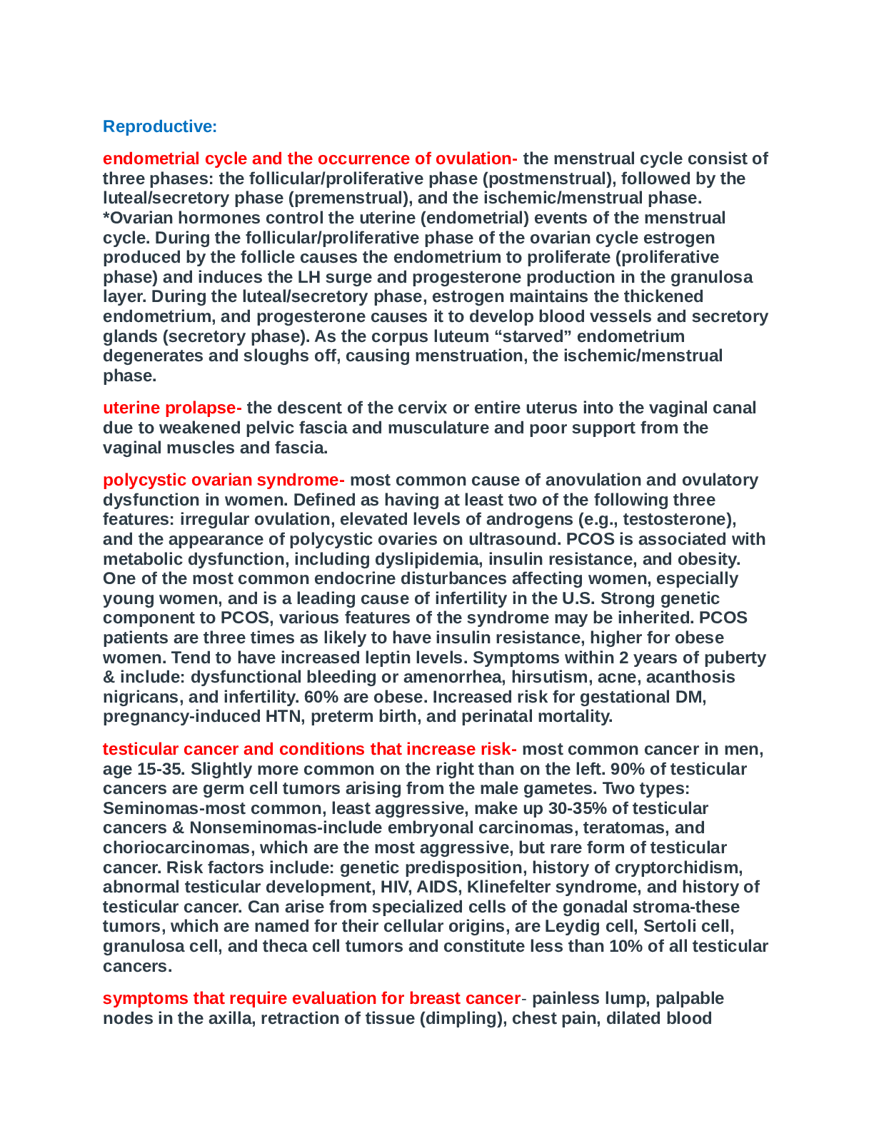
■ This is what S3 is → due to a stiff ventricle, meaning it is not compliant, it is not expanding as it is supposed to
■ So as blood comes in, it is bouncing off the walls instead of rushing in and expanding it
slightly
○ Will always occur during diastole
○ Usually occurs w/ volume overload of ventricles from:
■ Valvular heart disease
■ CHF
■ Decreased myocardial contractility
● S4 aka “atrial gallop”
○ Occurs closer to S1
■ It is the last kick out of atrium
■ In the process, it causes the vibratory sensation
■ Due to non-compliance/expandability of your ventricles
■ But only at end of when blood pushes in from the atrium
■ Usually occurs w/
● HTN
● Coronary heart disease
● Cardiomyopathy
Heart Murmurs = turbulent blood flow
● Turbulent blood flow but is making another sound (it’s own sound, heard separately from S1 and S2)
● Can occur due to:
○ Increase blood viscosity
○ Structural valve defects - valves not working properly
○ Valve malfunction - valves not working at all
○ Abnormal chamber openings - a hole between ventricles or atrium i.e. septal defect
Characteristics of Heart Murmurs:
● Timing
○ Systolic or diastolic?
○ That pause component between S1 and S2 = systole
○ Pause between S2 and S1 = diastole
● Intensity
○ Grading system used
○ Numerators:
■ 1, 2, 3 - very subjective grading systems & is based on how in-tune your ears are
■ 4, 5, 6 → always a thrill
○ Denominator will always be 6
● Pitch
○ High, medium or low; crescendo or descrescendo; same level of pitch = plateau
● Quality
○ Is it a blowing sound? Rough sound?
● Shape or pattern
○ Does it start out high and then low? Low and then high?
● Location
○ Where is it heard the loudest?
● Transmissible/radiates
○ Is it local or does it radiate elsewhere in the body?
● Ventilation and position
○ Does it change w/ position?
When charting heart murmurs:
● Timing, intensity, location, whether it is radiating or not, and if it occurs when changing positions
● I.e. picks up murmur in mitral area, and it is heard during systole, based on grading system (can’t understand her?) you have a thrill, grade 4 (loud w/ palpable thrill)
○ Grade 4/6 systolic murmur, heard loudest at mitral area, radiates to Erb’s point, does not change w/ position
● If there is no radiation or transmission, you must write that down
● On completion of auscultation:
○ *Look at Sequence of the Cardiac Examination Handout*
SYSTOLIC MURMURS
AORTIC STENOSIS
● Aortic valve is stenotic and not closing properly
● Restricting blood flow out into systemic circulation
○ Blood sitting in ventricle
○ L-Ventricular afterload
● Develop a L-ventricular hypertrophy
● Fatigue, DOE (dyspnea on exertion), chest pain or dizziness
● Heard:
○ Aortic valve, 2nd intercostal space
PULMONIC STENOSIS ● Problem w/ pulmonic valve
● Increase R-ventricular afterload
● Develop a R-ventricular hypertrophy
● Heard best:
○ Pulmonic valve, 2nd intercostal space, on the left
MITRAL REGURGITATION
● Regurgitation = backflow
● Mitral valve does not close completely
● Backflow of blood into L-atrium from the L- ventricle bc failure of mitral valve closure
● Volume overload in L-ventricle, dilatation and hypertrophy
● Loud - pansystolic murmur with a thrill
● Heard best:
○ Mitral area, 5th intercostal space
TRICUSPID REGURGITATION
● Regurgitation = backflow
● Backflow of blood into R-atrium via tricuspid valve that is not closing properly during systole
● ENGORGED NECK VEINS
● Murmur - pansystolic murmur
● Heard Best:
○ Tricuspid area, 4th intercostal space
DIASTOLIC MURMURS
AORTIC REGURGITATION
● Aortic valves not closing properly
● Backflow of blood into the L-ventricle MITRAL STENOSIS
● Mitral valve thicken, stiff
● Occurs due to rheumatic fever
● Leads to → volume overload of LV
Peripheral Vascular System & Lymphatic System Arteries in Arm
● Ulnar artery
● Radial artery
● Brachial artery
Arteries & Veins in Leg
● Femoral artery
● Popliteral artery
● Posterior tibial artery
● Dorsalis pedis artery
● Great saphenous vein = big vein in leg
Venous Flow
● Venous flow based on closures of valve
● Contraction of muscles that opens them up in order to help the blood get through
Lymphatic Flow
● Removes excess fluid + plasma proteins from tissues and returns them to the venous circulation
○ If you have lymphatic problems, it stays within the tissues
● R-lymphatic duct empties into R-subclavian and drains R-side of body
● Thoracic drains rest of body and empties into L-subclavian
SUBJECTIVE DATA: HEALTH HX - REVIEW OF SYSTEMS
PERSONAL HEALTH HX
Question Rationale
Pain in arms or legs? Peripheral arterial disease (PAD) can develop if there is 60% blockage
● Intermittent claudication is usually the 1st symptom characterized by weakness, cramping, aching, fatigue or frank pain w/ activity
Intermittent claudication?
● Pain in legs relieved by rest
Cold, numbness, pallor, hair loss? Peripheral arterial disease (PAD):
● Without the blood, peripheries are cold to touch
● Without the nutrients, peripheries lose hair
● Numbness due to not enough circulatory components
Swelling in the calves, legs or feet? Bilateral = form of edema
Unilateral = thrombophlebitis, superficial thrombophlebitis
Swelling w/ redness or tenderness? Infection of skin which is cellulitis which eventually can turn into circulatory problem
PAST HEALTH HX
Any problems w/ circulation in arms or legs? (i.e. blood clots, ulcers, coldness, hair loss, numbness, swelling or poor healing) In relationship to circulatory problems:
● Hx or thrombophlebitis or varicosity
● Changes in toenails due to PAD
FAMILY HEALTH HX
Family hx of DVT, diabetes, hypertension, CHD, intermittent claudication, elevated cholesterol or triglyceride levels? These disorders tend to be hereditary and cause damage to blood vessels
LIFESTYLE AND HEALTH PRACTICES
Exercise? Decreases risk for developing PVD
How long are you standing on your feet? Nursing, waitressing, etc.
How long are you sitting? Studying/sitting for hours, longer airplane trips are risk factors for developing PVD
● Make sure you get up every hour bc blood must be constantly circulated
Preparation: Equipment
● Paper tape measure
● BP cuff
● Stethoscope
● Doppler ultrasonic stethoscope
Objective Data: Physical Exam Arms - Inspect & Palpate
Legs - Inspect, Palpate & Auscultate
Assessment Procedure Normal Findings Abnormal Findings
Arms
INSPECTION
Skin Bilaterally symmetric w/ no edema and minimal variation in size Lymphedema results from blocked lymphatic circulation, from breast surgery
Size, Symmetry Should be even throughout Raynaud disorder is vascular disorder caused by vasoconstriction or vasospasm of fingers or toes and can have rapid change of color
INSPECTION
Temperature
● Use dorsal side of hand Warm to touch Cool extremity could be sign of arterial insufficiency i.e. cold fingers associated w/ Raynaud’s
Capillary refill time Refill time should be 2 seconds or less Refill time over 2 seconds → vasoconstriction, decreased CO, shock, arterial occlusion or hypothermia
Palpate
● Radial pulse
● Ulnar pulse
● Brachial pulse
Palpate epitrochlear lymph nodes
● Flex client’s elbow about Normally, epitrochlear lymph nodes are not palpable Enlarged epitrochlear lymph nodes could indicate infection in hand/forearm or generalized
90 degrees, use your L- hand to palpate behind elbow in groove between bicep + tricep muscle
● If nodes are detected, evaluate for size, tenderness and consistency lymphadenopathy
Perform the Allen test
● Evaluates patency of radial or ulnar arteries
● Have client make fist
● Press thumbs on radial and ulnar arteries
● Have client release the fist (notice it is still pale)
● Release pressure on ulnar artery and watch color return
● Repeat, but this time release pressure on radial artery Pink coloration returns to the palms w/in 3-5 seconds if the ulnar and radial artery is patient With arterial insufficiency or occlusion of ulnar artery or radial artery → pallor persists
Legs
INSPECTION, PALPATION & AUSCULTATION
Skin Color, no changes in pigmentation Pallor when elevated especially rubor suggests arterial insufficiency
Dark-colored toes + blisters are seen with arterial insufficiency and gangrene
Gangrene is evidence w/ ulcerations that are slow to heal, dry and shriveled skin that changes color from blue to black and sloughs off, cold and numb
Cyanosis w/ venous insufficiency
Size, symmetry Symmetrical
Hair distribution Hair covers skin on legs and appears on dorsal surface of toes Loss of hair could suggest arterial insufficiency
Temperature Warm to touch
Calf muscle
● Pain in calf is prob a form
of thrombophlebitis
○ Swelling
○ Increased heat
○ Redness (will be red on entire lower extremity)
● Homan’s sign: press on foot to check pain
○ Problem: only 50% reliable
Palpate:
● Femoral
● Popliteal tibial (behind knee)
● Dorsalis pedis (top of foot)
● Posterior tibial (near ankle)
Pretibial Edema No edema (pitting or nonpitting) Pitting edema associated w/ systemic problems like heart failure or hepatic cirrhosis, venous stasis due to insufficiency or obstruction from prolonged standing or sitting
1+ to 4+ scale is used to grade severity of pitting edema (4+ is most severe)
Position Change Test for arterial insufficiency
● Significant for arterial insufficiency to the legs
● In supine position, raise legs up, wiggles feet till they turn white, then immediately sit them up w/ legs dangling at edge of bed, feet should start to turn pink Feet pink to slightly pale in color in light-skinned individuals.
Inspect the soles in the dark- skinned client, although still difficult to see color change in darker skin.
When client sits up and dangles the legs, pinkish color returns to the tips of the toes in 10 seconds or less.
Superficial veins on top of feet fill in 15 seconds or less Pallor w/ legs elevated indicating arterial insufficiency
Return of pink color that takes longer than 10 seconds
Superficial veins that take longer than 15 seconds to fill suggest arterial insufficiency
● If you see a dusty red color
= abnormality (VERY IMPORTANT)
○ Blood trying to push its way through into the foot
Ankle-Brachial Index (ABI) Ankle pressure in a health person is the same or slightly higher than Can be used to recognize CVD
● *Look at Ankle-Brachial Index Handout*
● Taking systolic pressure in foot (posterior tibial artery or dorsalis pedis artery) and systolic pressure in upper arm (brachial artery)
● Using an arterial doppler for this
● Normal: 0.9-1.3 ABI
○ <0.9 considered to be developing PAD
○ >1.3 considered that arteries are stiff and no longer pliable the brachial pressure, resulting in an ABI of approximately 1 or no arterial insufficiency People who smoke, are physically inactive, have a BMI of >30 or are hypertensive are more likely to have an abnormal ABI, suggesting PAD
ABPI of 1.3 or more or ankle pressure at more than 300 mm Hg indicates medial calcification sclerosis → associated w/ diabetes mellitus, chronic renal failure, and hyperparathyroidism
Medial calcific sclerosis produces falsely elevated ankle pressure by making the vessels noncompressible
Reduced/absent pedal pulses, a cool leg unilaterally, lack of hair, and shiny skin on leg suggests peripheral arterial occlusive disease
Other Signs
● Homan’s Sign
● Position change test - for arterial insufficiency
● Ankle-Brachial Index
● Manual Compression
○ Compressing at top by knee and at bottom
○ *Look at Abnormalities in Arterial Pressure Handout*
● Trendelenburg test
○ Only done if pt has varicose veins
○ Used to determine the competence of the saphenous vein valves and the retrograde filling of superficial veins
Abnormalities
● Raynaud’s Syndrome
○ Microcirculation usually of fingers on hands
○ Cold hands
● Lymphedema
○ Mastectomy - used to remove all lymph nodes
○ No lymph drainage
○ Whatever was left in circulation stayed in the tissues
○ No drainage, can’t get rid of swelling
● Arterial Insufficiency
○ No blood supply getting there
○ Pallor
○ Weak pulses
● Venous Insufficiency
Age-Related Cardiovascular Changes
- Structural
★ Increase in fatty tissues in the outermost layer of the heart muscle
★ Minimal increase in the thickness of the left ventricular wall
★ Increase in collagen and elastin tissues in the heart and arteries
★ Decreased efficiency in the contractile strength of the heart muscle, decreased heart rate, stroke volume, cardiac output, ejection fraction, oxygen uptake
★ Accumulation of lipofuscin, a pigment giving a brown appearance of heart myocardium
★ Thickening of sclerosis of the valve flaps
★ Pacemaker cells decrease in number with a concomitant decrease in SA node rate
★ Veins dilate and stretch
★ Coronary arteries may become dilated, twisted, and calcified
- Functional
★ Longer recovery old heart muscle requires a longer time to recover after each heartbeat – requires a longer rest period between beats
★ Slight arrhythmias
★ Decline in cardiac output
★ Increase in atrial fibrillation and incidence in heart block
★ Changes in arteries and veins
★ Blood components volume of blood decreases as it related to lower plasma volume
REVIEW CHAPTERS
Level of consciousness - look at the “level of consciousness” handout
○ Alert
○ Lethargic - severe drowsiness where the patient can be aroused by moderate stimuli but then drifts back to sleep
○ Obtunded - lessened interest in the environment, slowed responses to stimulation and tends to sleep more than normal with drowsiness in between sleep states
○ Stupor - only vigorous shake will arouse the individual, when undisturbed they will lapse back into being unresponsive
○ Coma - unarousable unresponsiveness
● Voice and Speech Problems - look at “Disorders of Speech” handout
○ Aphonia - where you can’t speak at all
○ Dysphonia - difficulty when speaking (hoarseness)
○ Dysarthria - defect in the muscular control of speech apparatus (respiration, phonation, articulation, resonance)
○ Aphasia - total absence of ability to form speech or language
■ Wernicke’s aphasia - look at “Assessment Guidelines for Aphasia” handout
● Can speak and enunciate clearly but the sentences make no sense. “Word salad”
● Lesion in the posterior superior temporal lobe
■ Broca’s aphasia - look at “Assessment Guidelines for Aphasia” handout
● Can understand speech but have a really hard time expressing the words.
● Lesion in the posterior inferior frontal lobe
● Delirium vs. Dementia
○ Delirium- 1st clinical symptom of medical illness
○ Dementia- happens over time
○ Alzheimer’s disease
■ Repeatedly asking the same questions
■ Becomes lost/disoriented in familiar places
■ Cannot follow directions
■ Is disoriented to the date or time of day
■ Doesn’t recognize and is confused about familiar people
■ Difficulty with routine tasks like paying the bills
■ Neglects personal safety, hygiene, and nutrition
● Depression questionnaire
○ Falling asleep
■ How long does it take to fall asleep and how often does it take a long time to fall asleep
○ Sleep during the night
■ Is the patient waking up during the night and are they not able to fall back asleep
○ Waking up too early
■ Is the patient waking up before they’re supposed to and are they not able to fall back asleep
○ Sleeping too much
■ Sleeping during the night and also taking naps
○ Feeling sad
■ How often do they feel sad in the past 7 days
○ decreased/increased appetite
○ decreased/increased weight
● Ideal Body Weight
○ Female
■ 100 lb for 5ft + 5 lb for every inch over 5 ft
■ -10% for small frame
■ +10% for large frame
○ Male
■ 106 lb for 5ft + 6 lb for every inch over 5 ft
■ -10% for small frame
■ +10% for large frame
○ (actual weight/IBW) * 100 = % IBW
■ Below 70% is malnourished
■ 80-90% - concerning
■ 90-109% is normal
■ 110-119% overweight
■ 120%+ is obese
● BMI
○ 18.5-24.9 is normal
○ If they’re above 24.9, look at waist circumference to see if it’s because of fat or muscle
■ Stand straight, feet together, arms at side, relax the abdomen and take a normal breath, record at exhalation.
○ Female → 35 inches. Male → 40 inches.
● Pain
○ Definitions
■ Unpleasant sensory and emotional experiences that are primarily associated with tissue damage.
■ Whatever the person says it is
○ Physiologic responses
■ Anxiety, fear, hopelessness, sleeplessness, thoughts of suicide
■ Focus on pain, reports of pain, cries/moans, frowns/facial grimaces
■ Decrease in cognitive function, mental confusion, altered temperament, high somatization, and dilated pupils
■ Increased heart rate, peripheral, systemic, coronary vascular resistance, and blood pressure.
■ Increased respiratory rate and sputum retention, resulting in infection and atelectasis
■ Decreased gastric and intestinal motility
■ Decreased urinary output → urinary retention, fluid overload, depression of all immune responses
■ Increases hormones in the bloodstream - ADH, epi, norepi, aldosterone, glucagon, decreased insulin
■ Hyperglycemia, glucose intolerance, insulin resistance, protein catabolism
■ Muscle spasm, resulting in impaired muscle function and immobility, perspiration
○ Classification by cause
■ Nociceptive - normal response to injury of tissues such as skin, muscles, visceral organs, joints, tendons, or bones
■ Neuropathic - pain from primary lesion or disease in somatosensory nervous system
■ Inflammatory - when tissue inflammation activates nociceptive
○ Classification by duration and etiology
■ Acute pain - recent injury
■ Chronic nonmalignant pain - specific cause or injury, lasts more than 6 months
■ Cancer pain - compression of peripheral nerves/meninges or from damage to these structures following surgery, chemo, radiation, or tumor growth and infiltration
■ Intractable pain - high resistance to pain relief
○ Classification by location
■ Cutaneous pain - skin or subcutaneous tissue
■ Visceral pain - abdominal cavity, thorax, cranium
■ Deep somatic pain - ligaments, tendons, bones, blood vessels, nerves
■ Phantom pain - perceived in nerves left by a missing, amputated, or paralyzed body part
■ Radiating pain - at the source and extending to other tissue
■ Referred pain - perceived in body areas away from the pain source
○ Seven dimensions
■ Physical - patient’s perception to pain and body’s reaction
■ Sensory - quality of the pain and how severe it is
■ Behavioral - verbal and nonverbal behaviors patient demonstrates
■ Sociocultural - influence of social context and cultural background
■ Cognitive - beliefs, attitudes, intentions, and motivations related to pain and its management
■ Affective - feelings, sentiments, and emotions related to the pain experience
■ Spiritual - meaning and purpose the person attributes to the pain.
○ Use of subjective data to assess pain
■ Review past and family histories in terms of pain
■ Review lifestyle and health habits to determine how the pain interferes with the client’s life
■ Tips for collection
● Maintain quiet and calm environment that is comfortable for the client being interviewed
● Maintain client’s privacy and ensure confidentiality
● Ask questions in an open-ended format
● Listen carefully to client’s verbal descriptions and quote the terms used
● Watch for facial expressions and grimaces during interview
● Do not put words in the client’s mouth
● Ask client about past experiences with pain
● Believe the client’s expression of pain
○ Use of objective data
■ Visual analog scale (VAS) - older adult without cognitive impairment
■ Numeric rating scale (NRS) - older adult without cognitive impairment
■ Numeric pain intensity scale (NPI) - older adult without cognitive impairment
■ Graphic rating scale
■ Verbal rating scale
■ Faces pain scale
■ McCaffrey initial pain assessment tool
○ Pain assessment techniques from most reliable to least reliable
■ Self-report
● Skin
■ Search for potential causes of pain
■ Observe client behaviors
■ Surrogate reporting
■ Attempt analgesic trial
● cold/heat
● Medication
○ How to assess: Subjective Data Health History
■ Common or concerning symptoms:
● Mole = nevus (plural: “nevi”)
● Bruising = ecchymosis
● Hair loss
● Problems with nails (becoming brittle, cracking or becoming infected)
■ Client’s Personal Health History:
● What previous rashes have you had?
● Itching
● Dryness
● Callous formation
● Oiliness of the skin
● Diseases:
○ Psoriasis
○ Skin cancer
○ Eczema
○ Contact dermatitis
■ Localized (developing a rash where you touched something you may be allergic to i.e. professor allergic to timex watches, she develops a rash in the area of her wrist where the watch is located)
■ Systemic (more of an ingestion component i.e. you have congested something like medication or food)
○ Past history regarding food allergies
○ Has the patient ever had sunburn (specifically 2nd degree burns)
○ Comorbidities:
■ Diabetics - wounds will heal slower
■ Wounds on the legs or pressure ulcers - does the patient have peripheral vascular disease
○ 2 techniques to assess the skin: Inspection & Palpation
■ Inspection
● Color
○ Normal general pigmentation: an even distribution of color throughout the body
○ Abnormal pigmentation: different patches of color on skin
■ Redness
■ Vitiligo - white patches
■ Albinism - no form of melanin at all
■ Handout: Detecting color changes in light and dark skin
● Know the different color variations
■ Handout: Color variations in Light and Dark Skinned clients
● Odor
● Integrity
○ Are there any forms of breaks in the skin
○ Surgical scars
■ Once surgical scars are healed, should turn silvery in color or may not see it at all
■ New surgical scars - measure the length and keep an eye out for redness of the scar line (should eventually clear)
■ Make sure to count your staples when removing them
○ Striae = stretch marks
○ Petechiae = broken blood vessels beneath the skin
● Lesions
■ Palpate - always use dorsal surface of hand to palpate!
● Texture
○ Normal: skin should be smooth
■ Some parts of the skin are rough
● Thickness
○ Normal: Even distribution
■ Watch for calluses
■ Palms + feet are a little thicker
● Moisture
○ Normal: skin should be moist
○ Abnormal: dry or oily
● Temperature
○ Tested with the dorsal side of the hand
● Mobility and Turgor
○ Normal: skin should be mobile
○ Abnormal: taut skin
■ Tenting - pull skin up and skin will stay up
● As we get older we lose elasticity
● From dehydration
○ Turgor - pull skin up, should slowly return back
■ Best to test turgor at the clavicle
● Edema
○ Swollen area; due to an increased fluid component or when lymph is not able to leave the area (i.e. slightly swollen feet)
○ Pitting edema - excess fluid in a particular area (put thumb in to where the swelling is and see if you make and indentation, remove finger, see if skin returns back)
■ Cardiac problem could be occurring
○ Non-pitting edema - usually hard (press into swollen area and does not
cause an indentation)
■ Lymphadema - lymph is not being circulated out of the area
■ Patients with mastectomy*
● Lesions
○ Any type of cut that is in the skin (i.e. mole, abrasions, etc.)
■ Lesions
● Handout: How to Describe Lesions
● Size (include number of lesions)
○ Measure
○ Count how many lesions
● Shape or pattern
○ Round, oval, linear
○ Irregular border (associated with melanoma, but not necessarily)
● Color
○ Pustule (pus) = yellow-white
○ Vitiligo = white
○ Purpura = red to purplish
● Texture
○ Smooth, rough, scaly (usually psoriasis)
● Surface Relationships and pattern
○ Lesion + regular skin around the lesion
○ Feel the lesion, check whether it is flat/raised
■ Flat = non-palpable
■ Raised = palpable (comes up as a bump)
● Solid = hard
● Cystic = squishy
■ Depressed - atrophy, erosion, ulcer
■ Pedunculated - skin tags (use laser removal)
● Exudate
○ Any form of drainage
● Tenderness or pain
● Body location and distribution
○ Where is it specifically located
■ Evaluation of Moles/Skin Cancer
● A - Asymmetry
● B - Border
● C - Color
● D - Diameter (>6 mm is malignant)
● E - Evolving (a change in size, color, or shape)
● E - Elevated (the flatter the mole, the better! As it grows bigger, it changes elevation)
● F - Firm to palpation
● G - Growing progressively over several weeks
● Benign vs. Malignant
● Abnormal Findings: Malignant Skin Lesions
Malignant Skin Lesions Description Example
Basal cell carcinoma ● Papule with pearly borders
● “Volcano-like” (because of the depressed center)
● Occurs with light skinned individuals
○ Areas that are regularly exposed to the sun
● Age 40-80
● Least invasive of malignancies
● Most common form you will find
Squamous cell carcinoma ● Central ulcer with reddened borders
○ As it progresses, it will become elevated with many irregularities
● Second most frequently found
● Invasive skin cancer
● Occurs with light skinned individuals
○ Occurs around head + neck
○ Any areas exposed to the sun or UV light
● Age 50
Malignant melanoma ● The most invasive of skin cancers
● Use ABCDEEFG to diagnose
● Secondary Skin Lesions - when primary lesions aren’t taken care of
○ Erosion - beneath
○ Ulcer - pressure ulcers
○ Scar
○ FIssure - line that occurs
Secondary Skin Lesions Description Example
Vascular skin lesion: Petechiae Flat, red or purple macules bleeding from superficial capillaries
Lichentification Thickened skin
Keloid Excessive collagen formation; Can occur either on the surface or underneath the skin
Head and Neck
● Subjective Data
○ Common or concerning symptoms
○ Pain using COLDSPA
○ Past health history - including headaches, concussions, and possible falls (especially in elderly)
○ Lifestyle and health practices - do they use a seatbelt/helmet as needed
● Head- Inspection
○ Normal - head shape might vary depending on the person & their ethnicity
○ Usually the head is symmetric, round, erect & in midline; should be appropriate for their body size
○ Abnormal
■ Microcephaly - when the head is abnormally small
■ Acromegaly - skull & facial bones are larger and thicker
■ Acorn shaped head can indicate Paget’s disease
○ Inspection - face should be symmetric with a round oval appearance
■ Normal- symmetry as seen in earlobes
■ Abnormal - asymmetry in front of earlobes
■ Drooping, weakness, paralysis on one side of the face could be a result of stroke, CVA, or Bell’s palsy
○ Palpate temporal artery
■ Normal - elastic and non tender
■ Abnormal - thick, hard, and tender with inflammation is seen in temporal arteritis
○ Palpate the TMJ
■ Normal - should be no swelling, tenderness or crepitation
■ Abnormal - limited ROM, swelling or tenderness
● Neck- Inspection
○ Inspect movement of neck structures
■ Thyroid cartilage, cricoid cartilage should move upwards symmetrically when the patient
swallows
○ Range of motion - should be smooth & controlled
○ Palpate the trachea - should be midline
■ Abnormal - trachea pushed to unaffected side in cases of tumor/enlarged thyroid
○ Palpate thyroid gland
■ Locate key landmarks with index finger & thumb
■ Landmarks deviate from midline or are obscured because of masses or abnormal growths
■ Auscultate thyroid only if you find an enlarged thyroid gland - soft, blowing, swishing sounds is often heard in hyperthyroidism
● Different Types of Headaches
○ Sinus Headache
■ Deep, constant, throbbing pain
■ Pressure-like pain in one specific area of the face
■ Face tender to touch
■ Onset with/after a cold
■ May occur in one area of face or along the eyebrow and below cheek bone
■ Pain worsens with sudden head movement(buildup of fluid causing pressure- fluid moves causing pain), bending forward, lying down, in the morning or with sudden temperature drop
● Associated with congestion, fever, and foul smelling breath
○ Cluster Headache
■ Stabbing pain, may be accompanied by tearing, eyelid drooping, reddened eye, or runny nose
■ Has a sudden onset and may be precipitated by ingestion of alcohol
■ Localized in one eye/orbit and radiates to the facial and temporal regions
■ Typically occurs in the late evening or at night, starts when they’re sleeping.
■ Movement or walking back and forth may relieve pain
○ Tension Headache
■ Dull, tight, diffuse
■ No prodromal stage
■ May occur with stress, anxiety, depression
■ Located in the frontal, temporal, occipital region
■ Last days, months, years
■ Aching
■ Relief from local massage, heat, antidepressants, analgesics, and muscle relaxants
■ Affect women more than men
○ Migraine Headache
■ Accompanied, nausea, vomiting, and sensitivity to light
■ May have prodromal stage
■ Emotional disturbances, anxiety, depression, alcohol ingestion
■ Located around eye, temples, cheeks or forehead, may affect only one side of the face
■ Lasts up to 3 days
■ Rest brings relief
○ Tumor-related headache
■ Aching, steady
● Thyroid
■ Neurologic and mental symptoms
■ Nausea and vomiting
■ No prodromal stage, may be aggravated by coughing, sneezing, or sudden movements
■ Varies with location of tumor
■ Commonly occurs in the morning and lasts for several hours
■ Variable in intensity
■ Subsides later in the day
■ Symptoms relieved with local heat and massage.
○ Know what to do with thyroid - how to test
■ Auscultate only when you find an abnormality or if the thyroid is enlarged. Easily felt or feel a fullness in your neck.
Eyes
○ Palpate
■ Thyroid gland
● Thyroid cartilage
● Cricoid cartilage
■ Auscultate an enlarged thyroid gland
■ Palpate trachea- should be midline
○ Palpate lymph nodes of head and neck
● Preauricular
● Postauricular
● Occipital
● Tonsillar
● Submandibular
● Submental
● Superficial cervical
● Posterior cervical
● Deep cervical
● Supraclavicular
● Extraocular Muscles (EOMs)**- give eye straight and rotary movement (not responsible for names of muscles just nerves and what they innervate)
○ Inferior, Superior, Lateral, Medial Rectus - turns eye up, down, left and right
○ Superior oblique (down and inward) and inferior oblique (up and outward)
● Movement of the eye stimulated by three cranial nerves (6,4,3)
○ CN- VI - Abducens Nerve
■ Innervates lateral rectus muscle turning the eye outward away from nose
○ CN- IV- Trochlear Nerve
■ Innervates superior oblique muscle- ability to look down and inward toward nose
○ CN- III- Oculomotor nerve
■ Innervates all rest of extrinsic eye muscles, enables most movements and raises eyelid
○ Be able to know which cranial nerve may be involved in a certain deficit
● Strabismus - weakness of eye muscle; cross eyed; misalignment of the eyes, lazy eye (mostly picked up with kids)
○ Esotropia - inward turning of one eye
○ Exotropia - outward turning of one eye
● Review of Systems- Subjective Data
○ Ask patient about past eye history, family history, and lifestyle habits (wears sunglasses, contacts, eye surgery, glaucoma, surgery, cataracts, safety glasses, use of any visual aids, typical diet and vitamins)
■ Ask specifically if there is family history of:
● Cataracts- opacity that develops over the eyes, over the pupillary fills up entire cornea, decreases vision things become cloudy. Risk factors include ultraviolet light exposure.
● Glaucoma- pressure inside the eye, causes throbbing pain, and needs to come out. Genetic. May cause blind spot/affect peripheral vision.
● Macular degeneration (tested with amsler grid)- fundus is degenerating and causes vision loss
○ Lifestyle - how do you clean your eyes? How do you clean contacts?
● Objective Data
External Eye Structures (How to assess) Normal Findings Abnormal Findings
Inspect and palpate eyelids and Lashes ● Lid should overlap a little above and below the Iris
● Evenly distributed Eyelashes ● Ptosis- drooping of the upper lid
● When lid’s are retracted and a lot of sclera is showing bulging of eyes (suggests hyperthyroidism)
● REFER TO HANDOUT VARIATIONS AND ABNORMALITIES OF THE EYELIDS
● PTOSIS- Drooping of the upper lid. Causes could be myasthenia gravis, damage to oculomotor nerve, horner's syndrome(damage to sympathetic nerve supply), weakened muscle, may be congenital.
● ENTROPION- inward turning of the lid margin. This can cause irritation from the inward turning of the lashes. Asking patient to squeeze lids together and then open them may reveal an unobvious entropion.
● ECTROPION- margin of lid is turned outward, exposing palpebral
conjunctiva. This causes the eye to no longer drain effectively and tearing occurs.
● LID RETRACTION- wide-eyed stare suggests retraction. Want to look and note the rim of sclera between the upper lid and iris. Retracted lid and lid lag are often caused by hyperthyroidism.
● EXOPHTHALMOS-eyeball protrudes forward (bulges) Usually an indication of graves disease (hyperthyroidism) Unilateral exophthalmos is seen in graves disease, tumor, or inflammation i the orbit.
Ability of eyelids to close ● Lids both close easily and meet fully when closed ● Failure of lid closure puts patients at risk for corneal damage
Noting any unusual positioning of eyelids ● Lower lid is upright with no inward or outward turning ● Entropion- inverted lower lid (possible damage to cornea, pain from eyelashes)
● Ectropion - everted lower lid (exposure of drying conjunctiva)
Noting any unusual characteristics of eyelids ● No odd coloring, swelling, lesions, discharge ● Blepharitis - redness and crusting along lid margins (infection)
● Chalazion - infection of meibomian gland(located in eyelid)
● Periorbital edema- swelling of the eyelids
Position and alignment of the eyeball in the eye socket ● Eyes are symmetrically aligned in sockets without protruding or sinking ● exophthalmos - protrusion of eyeballs accompanied by retracted eyelid margins (characteristic of graves disease, hyperthyroidism)
● Sunken appearance may be seen with severe dehydration or chronic wasting illness (cachexia)
● REFER TO HAND OUT ON DYSCONJUGATE GAZE
● ESOTROPIA-turning inward of eye
● EXOTROPIA- turning outward of eye
○ These can be developmental due to imbalance in ocular muscle tone. They are classified according to direction
○ A cover-uncover test may be
helpful
○ These gaze abnormalities can also be seen due to cranial nerve abnormalities; usually seen in adulthood due to nerve injury, trauma, MS and syphilis
○ NERVES AFFECTED could be 3,4,6 (LOOK AT HANDOUT)
Inspect the bulbar conjunctiva and sclera
● Patient should keep head straight while looking from side to side then up toward the ceiling
● Want to observe clarity, color, and texture
● Gloves should be on while inspecting palpebral conjunctiva
● First inspect the palpebral conjunctiva of the lower eyelid by placing your thumbs bilaterally at the level of the lower bony orbital rim and gently pulling down to expose the palpebral conjunctiva (ask patient to look up)
● Evert the upper eyelid. Ask the client to look down with his or her eyes slightly open. (use cotton tip applicator to evert upper lid) ● Bulbar conjunctiva is clear, moist, and smooth
● Underlying structures are clearly visible (iris pupil, sclera)
● Sclera is white
● Lower and upper palpebral conjunctiva are clear and free of swelling or lesions, foreign bodies or trauma ● REFER TO HANDOUT “RED EYES”
● Conjunctivitis (pink eye)- redness of the conjunctiva due to dilation of vessels; ocular discharge (watery, mucoid, or mucopurulent); due to bacterial, viral, other infection, allergy, irritation. Usually bacterial/viral.
● Areas of dryness (could be from allergies or trauma)
● Episcleritis- local, noninfectious inflammation of the sclera
● Yellowing of sclera, usually occurs when patient is jaundice (icterus)
● Subconjunctival hemorrhage- bright red areas on sclera, breaking of blood vessels and thus trapped blood, usually harmless and fades to yellow and then goes away on own.
● Cyanosis of the lower lid suggests heart or lung disorder
● A foreign body or lesion may cause irritation, burning, pain, and/or swelling of upper lid
Inspect/palpate the Lacrimal apparatus ● No swelling or redness should appear over areas of the lacrimal gland. ● Swelling of lacrimal gland may be noticeable in upper eyelid. This could be caused by blockage or infection
● While palpating, palpate nasolacrimal duct to assess for blockage, as well as one inger to palpate inside the lower orbital rim ● Puncta should be visible with no swelling or redness
● While palpating nasolacrimal duct no drainage should be noted from puncta ● Redness or swelling around puncta may indicate infection or inflammatory condition
● Epiphora-Excessive tearing, may be indication of nasolacrimal sac obstruction
Inspect cornea and lens
● Shine light from side for eye for an oblique view
● Look through pupil to inspect the lens ● Cornea should be transparent with no opacities (opaque spots)
● Shows smooth and overall moist surface
● Lens is free of opacities ● Areas of roughness or dryness on the cornea are seen with allergic response or injury
● Cataracts are usually associated with opacities on the lens
● REFER TO OPACITIES OF THE CORNEA AND LENS HANDOUT
● CORNEAL ARCUS (arcus senilis)- thin grayish white arc around edge of cornea; could be viewed as normal with aging but also seen in younger people which suggests possible hyperlipoproteinemia
● CORNEAL SCAR-superficial grayish white opacity in the cornea, secondary to an old injury or to inflammation. (do not confuse with opaque lens of a cataract)
● PTERYGIUM- triangular thickening of the bulbar conjunctiva that grows slowly across the outer surface of cornea (usually from nasal side), may interfere with vision
● CATARACTS- opacities of the lenses visible through pupil; most common in old age.
○ NUCLEAR CATARACT- looks gray when seen by flashlight. When pupil is widely dilated the gray opacity is surrounded by a black rim. WIth ophthalmoscope, the cataracts looks black against red reflex.
○ PERIPHERAL CATARACT- spoke like shadow that points inward
Inspect Iris and Pupil
● Inspect shape and color of iris as well ● Iris should appear round, flat, and evenly colored
● Pupil should be round with ● Irregular shaped iris- shining the light on eye from side causes crescent shaped shadow on iris, indication of glaucoma
as shape of pupil
● Measure size of pupil against gauge on pen light
○ Come in from the side and quickly scan the light over the pupil. This is where we are looking and testing for direct response, then test second eye for (consensual response) PUPIL SHOULD CONSTRIC T
○ Note if they appear larger or smaller than normal as well as two different sizes regular border and centered in iris
● Pupils are equal in size (3-5mm) ● Miosis - pinpoint pupils. -possibly a result of narcotics or brain damage
● Mydriasis - Dilated fixed pupils- usually resulted from CNS injury, or circulatory collapse, or deep anesthesia
● Anisocoria -pupils of unequal size (could be considered normal)
● If difference in pupil size changes throughout pupillary response test, the inequality of size is abnormal
TEST (vision, peripheral, accommodation) Normal Findings Abnormal findings
Vision Test (distance visual acuity)
● Tools needed: snellen eye
chart, opaque card)
● Patient stands 20 feet away from chart (the denominator number) The control is the numerator
● Make sure patient wears prescribed contacts or glasses (not reading glasses) while testing ● Normal vision is between 20/20 and 20/25
● If someone's vision is 20/40 this tells us that a person who has normal visions (20/20) would be able to read what they are reading at 40 feet rather the 20 feet the patient is standing at. ● Any letters missed on 20/20 line or above
● Client cleans forward, squints,
Near visual acuity
● Testing with newspaper approx. 14 inches away from clients head ● Reads print without difficulty 14 inches away ● Client reads print by holding it closer or farther away than 14 inches
● Presbyopia- impaired near vision
● REFER TO REFRACTIVE ERRORS HANDOUT- KNOW MEDICAL TERMS FOR NEAR SIGHTEDNESS
Amsler chart (testing for macular degeneration)
● Retinal changes (macular degeneration)
● Patient wears glasses; stands 12-14 inches away from chart covering one eye. They should look at the center dot. ● No distortions, graying, blurring, or blank spots seen by patient. No changes notes from previous Amsler chart reading ● Mark the areas of distortion, graying, blurring, or blank spots seen by client. Report changes from previous baseline if they have one.
Accommodation Test
● Shows constriction by moving eyes from far to near
● Ask client to stare at an object 3-4 feet away; and move object towards clients nose ● Pupils converge and constrict as object moves in toward the nose
● Pupil responses are uniform ● Pupils do not converge or constrict.
● Pupil responses are unequal
Confrontation Test (Peripheral vision)
● Face the client at a distance of 2-3 feet; client and examiner look directly ahead and cover eye directly opposite each other. Examiner extends arm and wiggles fingers asking client if they see them. Test this is all four visual fields (inferior, superior, nasal, temporal ● Client and examiner report seeing object at the same time as it approaches from periphery ● With reduced peripheral vision client does not report seeing object at the same time as the examiner
Corneal light reflex (assessing parallel alignment of eyes)
● Ask patient to look straight ahead. Examiner holds pen light approximately 12 inches from the clients ● Reflection of light noted at same location on both eyes, which indicates parallel alignment ● Light reflections noted at different areas on both eyes occur with deviation alignment of eyes due to muscle weakness or paralysis (REFER TO
face. Shine the light toward the bridge of the nose while the client stares straight ahead.
● NOTE THE LIGHT REFLECTED ON THE CORNEAS ● Nystagmus- eye jerk, not considered abnormal
○ Abnormality would be constant twitching of the eye DYSCONJUGATE GAZE HANDOUT)
● Strabismus- constant misalignment of eyes.
● Tropia- specific type of misalignment
○ Esotropia- inward turn of the eye
○ Exotropia- outward turn of the eye
Cover Test
● Tests for abnormal eye movement using cover/uncover
● Detects deviation in alignment or strength and slight deviations in eye movement by interrupting the fusion reflex that normally keeps eye parallel.
● Patient will look ahead, covering one eye with a cover card, examiner will observe the uncovered eye for movement.
● Remove covercard and observe the covered eyes movement. ● Uncovered eye does not move when opposite eye is covered
● Covered eye does not move as cover is removed ● Uncovered eye moves to focus when the opposite eye is covered. Covered eye moves to focus when cover is removed, These findings are seen with muscle weakness and deviation in alignment of eyes.
● Phoria- term used to describe misalignment that occurs only when fusion reflex is blocked
[Show More]