Chalazion
Chalazion is a chronic sterile inflammation of the eyelid resulting from a lipogranuloma of the meibomian
glands that line the posterior margins of the eyelids (see Fig. 29-7). It is deeper in the eyelid tiss
...
Chalazion
Chalazion is a chronic sterile inflammation of the eyelid resulting from a lipogranuloma of the meibomian
glands that line the posterior margins of the eyelids (see Fig. 29-7). It is deeper in the eyelid tissue than a
hordeolum and may result from an internal hordeolum or retained lipid granular secretions.
Clinical Findings
Initially, mild erythema and slight swelling of the involved eyelid are seen. After a few days the inflammation resolves, and a slow growing,
round, nonpigmented, painless (key finding) mass remains. It may persist for a long time and is a commonly acquired lid lesion seen in
children (see Fig. 29-7).
727
Management
• Acute lesions are treated with hot compresses.
• Refer to an ophthalmologist for surgical incision or topical intralesional corticosteroid injections if the condition is unresolved or if the
lesion causes cosmetic concerns. A chalazion can distort vision by causing astigmatism as a result of pressure on the orbit.
Complications
Recurrence is common. Fragile, vascular granulation tissue called pyogenic granuloma that enlarges and bleeds rapidly can occur if a
chalazion breaks through the conjunctival surface.
Blepharitis
Blepharitis is an acute or chronic inflammation of the eyelash follicles or meibomian sebaceous glands of the eyelids (or both). It is usually
bilateral. There may be a history of contact lens wear or physical contact with another symptomatic person. It is commonly caused by
contaminated makeup or contact lens solution. Poor hygiene, tear deficiency, rosacea, and seborrheic dermatitis of the scalp and face are also
possible etiologic factors. The ulcerative form of blepharitis is usually caused by S. aureus. Nonulcerative blepharitis is occasionally seen in
children with psoriasis, seborrhea, eczema, allergies, lice infestation, or in children with trisomy 21.
Clinical Findings
• Swelling and erythema of the eyelid margins and palpebral conjunctiva
726
• Flaky, scaly debris over eyelid margins on awakening; presence of lice
• Gritty, burning feeling in eyes
• Mild bulbar conjunctival injection
• Ulcerative form: Hard scales at the base of the lashes (if the crust is removed, ulceration is seen at the hair follicles, the lashes fall out, and
an associated conjunctivitis is present)
Differential Diagnosis
Pediculosis of the eyelashes.
Management
Explain to the patient that this may be chronic or relapsing. Instructions for the patient include:
• Scrub the eyelashes and eyelids with a cotton-tipped applicator containing a weak (50%) solution of no-tears shampoo to maintain proper
hygiene and debride the scales.
• Use warm compresses for 5 to 10 minutes at a time two to four times a day and wipe away lid debris.
• At times antistaphylococcal antibiotic (e.g., erythromycin 0.5% ophthalmic ointment) is used until symptoms subside and for at least 1 week
thereafter. Ointment is preferable to eye drops because of increased duration of contact with the ocular tissue. Azithromycin 1% ophthalmic
solution for 4 weeks may also be used (Shtein, 2014).
• Treat associated seborrhea, psoriasis, eczema, or allergies as indicated.
• Remove contact lenses and wear eyeglasses for the duration of the treatment period. Sterilize or clean lenses before reinserting.
• Purchase new eye makeup; minimize use of mascara and eyeliner.
• Use artificial tears for patients with inadequate tear pools.
Chronic staphylococcal blepharitis and meibomian keratoconjunctivitis respond to oral erythromycin. Doxycycline, tetracycline, or
minocycline can be used chronically in children older than 8 years old.
[Show More]


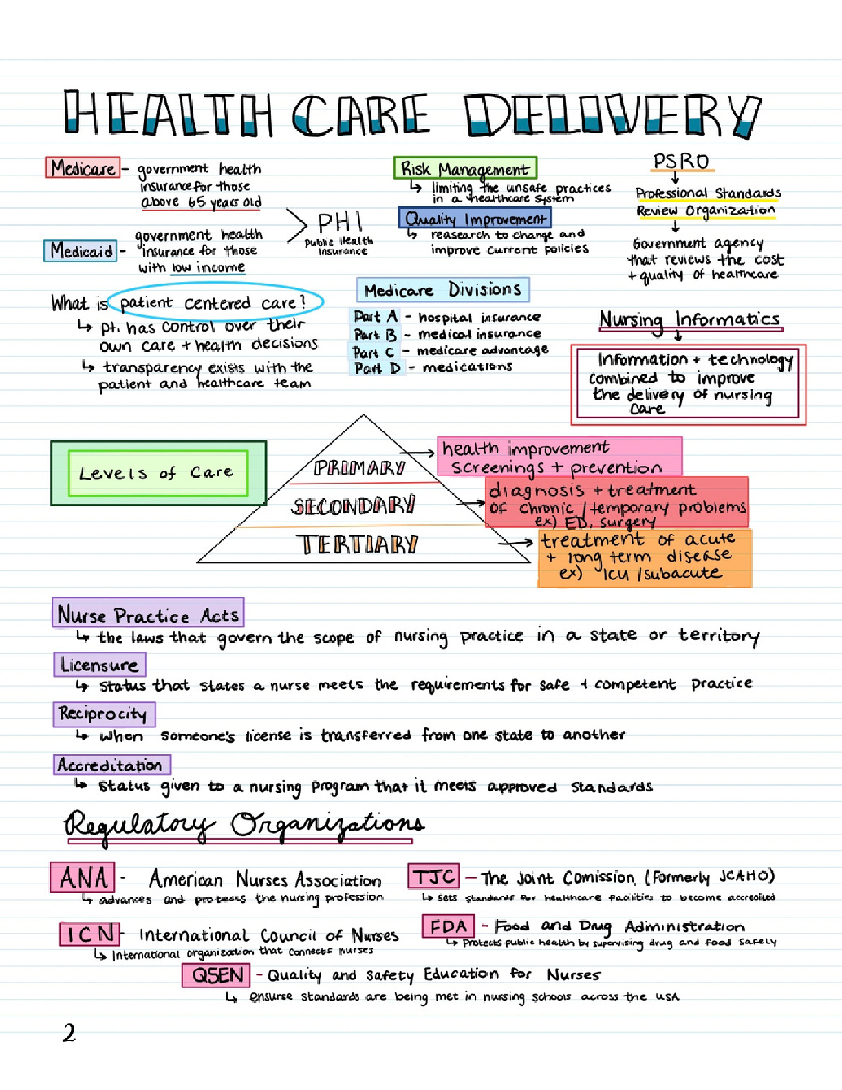
 Quiz 2 Concept Notes MY21.png)







.png)

.png)






.png)