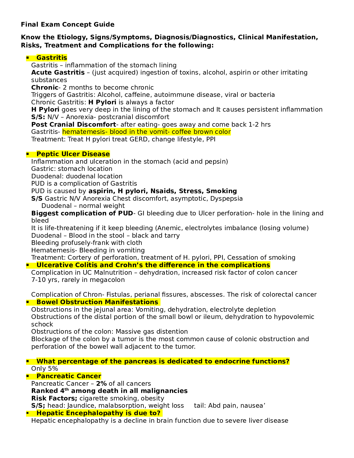
NURSE-UN 240 A&E I– FINAL EXAM STUDY GUIDE WEEK 6,7,8 & 9 Latest updated 2022
WEEK 6 – PAIN; SLEEP PAIN
What is Pain?
- “Whatever the person experiencing pain says it is”
- “Unpleasant sensory and emotional experie
...
NURSE-UN 240 A&E I– FINAL EXAM STUDY GUIDE WEEK 6,7,8 & 9 Latest updated 2022
WEEK 6 – PAIN; SLEEP PAIN
What is Pain?
- “Whatever the person experiencing pain says it is”
- “Unpleasant sensory and emotional experience associated with actual or potential tissue damage”
- Subjective: patient’s experience and self-report is essential
Nature of pain
- Involves physical, emotional, cognitive components
- Subjective & individualized
- Reduces quality of life
- Not measurable objectively
- May lead to serious physical, psychological, social and financial consequences Dimensions – psychologic, behavioral, cognitive, affective & social factors
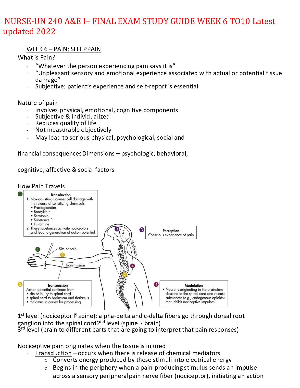
How Pain Travels
1st level (nociceptor spine): alpha-delta and c-delta fibers go through dorsal root ganglion into the spinal cord 2nd level (spine brain)
3rd level (brain to different parts that are going to interpret that pain responses)
Nociceptive pain originates when the tissue is injured
- Transduction – occurs when there is release of chemical mediators
o Converts energy produced by these stimuli into electrical energy
o Begins in the periphery when a pain-producing stimulus sends an impulse across a sensory peripheral pain nerve fiber (nociceptor), initiating an action potential
o Once transduction is complete, transmission of pain impulse begins
- Transmission – involves the conduct of the action potential from the periphery (injury site) to the spinal cord and then to the brainstem, thalamus, and cerebral cortex
o Sending of impulse across a sensory pain nerve fiber (nociceptor)
o Nerve impulses
o Pain impulses
- Perception – the conscious awareness of pain
o The point at which a person is aware of pain
o The somatosensory cortex identifies the location and intensity of pain, whereas the association cortex (primarily limbic system) determines how a person feels about it
o There is no single pain center
- Modulation – involves signals from the brain going back down the spinal cord to modify incoming impulses
o Inhibits pain impulse
o A protective reflex response occurs w/ pain reception
Physiology of pain
- Gate-control theory of pain
o Pain has emotional & cognitive components in addition to a physical sensation
o Gating mechanisms in the CNS regulate or block pain impulses
o Pain impulses pass through when a gate is open and are blocked when a gate is closed
o Closing the gate is the basis for non-pharmacological pain relief interventions
- As pain impulses ascend the spinal cord toward the brainstem and thalamus, the stress response stimulates the autonomic nervous system (ANS) – fight or flight
- Continuous, severe or deep pain typically involving the visceral organs activates the parasympathetic nervous system (PNS)
- Common indications of acute pain – clenching teeth, facial grimacing, holding or guarding the painful part, bent posture
- Chronic pain affects pt’s activity and PNS & SNS does not react like they do to acute pain
- Lack of pain expression does not indicate that a pt is not experiencing pain
Types of pain
- Acute/transient – protective (body tells you something is wrong), identifiable, short duration; limited emotional response
- Chronic/persistent noncancer – not protective, has no purpose, may or may not have an identifiable cause
- Chronic episodic – occurs sporadically over an extended duration; usually above their chronic baseline
- Cancer – can be acute or chronic
- Idiopathic – chronic pain w/o identifiable physical or psychological cause
Factors influencing pain
- Physiological – age, fatigue, genes (threshold and tolerance), neurological fxn (ie DM w/ ulcer)
o Fatigue increases perception of pain and can cause problems w/ sleep and rest
- Social – attention, previous experiences, family & social support
- Spiritual – includes active searching for meaning in situations w/ questions such as “why am I suffering?”
- Psychological – anxiety, coping style
- Pain tolerance – the level of pain a person is willing to accept
- Culture & ethnicity – meaning of pain
Critical thinking – knowledge of physiology and the many factors that influence pain help you manage a pt’s pain
Nursing Process & Pain
- Pain mgmt needs to be systematic & needs to consider pt’s QOL
- Clinical guidelines are available to manage pain – American pain society, sigma theta tau, nat’l guidelines clearinghouse
Assessment
- Through pt’s eyes – ask pt’s pain level
- Use ABCs of pain management (OLDCARTS, PQRST)
- Pain is not a #
- Pt’s expression of pain – individualistic
- Characteristics – timing, location, severity, quality, aggravating & precipitating factors, relief measures
- Effects of pain on pt – behavioral, influence on ADLs
- Concomitant symptoms – usually increases pain severity
Pain assessment tools (5th vital sign)
- COLDSPA Character, Onset, Location, Duration, Severity, Pattern, Associated Symptoms (factors increasing or relieving pain)
- OLDCARTSP – onset, location, duration, character, associated sx, radiation, timing, severity, pattern
- Simple Descriptive Pain Intensity Scale
- COMFORT scales
- FLACC scale
- Beyer Ouchers pain scale
- Wong-Bakers face scales – pediatrician
- Be aware of possible errors in pain assessment
Possible nursing
- Anxiety
- Insomnia
- Impaired social interaction
- Ineffective coping
- Impaired physical mobility
diagnoses
- Activity intolerance
- Fatigue
Planning – analyze info from multiple sources, apply critical thinking, adhere to professional standards, use a concept map, goals & outcomes, setting priorities, teamwork & collaboration, “any wound changes?”, “does pt needs before tx? How long before?”, anything pertaining to safety
Implementation – health promotion
- Maintaining wellness – helps pt understand – health literacy – pt actively participates in their own well-being whenever possible
Implementation
- Pain Treatment Principles
o Follow principles of assessment
o Treatment based on patient’s goals
o Use drug and non-drug therapies
▪ Know Patient's Previous Response to Analgesics
▪ Select Proper Medications When More Than One Is Ordered
▪ Know Accurate Dosage
▪ Assess Right Time and Interval for Administration
o Use a multidisciplinary approach
- Non-pharmacological pain-relief interventions
o CBT (cognitive and behavior approach)
o Hypnosis
o Biofeedback
o Therapeutic touch
o Animal-facilitated therapy
o Acupuncture
o Humor
o Relaxation & guided imagery (meditation techniques)
o Distraction
o Music
o Cutaneous stimulation (warm baths)
o Cold & heat application
o TENS
o Herbals
o Reducing pain perception & reception
o Massage (usually babies and elderly; really depends if pt like therapeutic touch)
- Pharmacological pain therapies
o Analgesics – “start low and go slow”
▪ Non-opioid: acetaminophen, aspirin and other salicylates, and non-steroidal anti-inflammatory drugs (NSAIDS)
▪ Opioid/narcotics (controlled substances): morphine, oxycodone, and codeine
• Affects spinal cord and brain
• Common side effects include: constipation (most common, nausea/vomiting, sedation, respiratory depression
▪ Adjuvant/co-analgesics: antidepressants, anti-epileptic drugs, and corticosteroids
• Non-pain meds that is conjunctions with pain meds to alleviate pain
o Topical analgesics – creams, ointments, patches
o Local anesthesia – local infiltration of an anesthetic medication to induce loss of sensation to a body part
▪ Regional anesthesia
o Perineural local anesthetic infusion
o Epidural analgesia – regional – administered into epidural space
Patient Controlled Analgesia (PCA)
- PCA pumps are secured computerized programmable devices that deliver small, preset dose of opioid; usually for chronic or cancer pain
- Allows pt to self-administer w/ minimal risk of overdose
- Maintains constant plasma level of analgesic
- On avg, systems are designed to deliver a specified # of doses q1-4 hr given every 5-15 mins
- Nursing Responsibilities:
o Patient teaching
o Monitoring patient responses
- Safety guidelines
o The pt is the only person who should press the button to administer pain medication
o Monitor for s/s of over sedation and respiratory depression
o Monitor for potential side effects of opioid analgesics
Implementation
- Nursing implications
o You maintain responsibility for providing emotional support to pts receiving local or regional anesthesia; numerous implications for mgmt. of epidural analgesia
▪ After administration of a local anesthetic, protect pt from injury until full sensory & motor fxn return
▪ Provide emotional support; not addressing pain # but addressing the pt overall (issues, QOL, address pain before meals, address pain before ADL)
▪ Establishing trusting nurse-pt relationship
▪ Manipulating factors affecting pain experience
▪ Initiating nonpharmacologic pain relief measures
▪ Managing pharmacologic interventions
▪ Relieving additional pain control measures (complementary and alternative relief measures)
▪ Considering ethical and legal responsibility to relieve pain
o Invasive interventions for pain relief
o Procedure pain mgmt.
o Cancer pain & chronic non-cancer pain mgmt.
o Scheduling
▪ Focus on prevention or control- Do not wait for severe pain
▪ Constant pain requires around-the-clock (ATC) administration (not PRN) – adjust dose to achieve max benefit with min side effects
▪ Allow pt as much control as possible over the regimen
▪ Fast-acting drugs for breakthrough pain
• Breakthrough pain is a superimposed pain on continuous or persistent pain that could be classified as: incident pain, end-of-dose failure pain, spontaneous pain
o ALWAYS USE 6 RIGHTS OF MEDICATION ADMINISTRATION
- Physical dependence – a state of adaptation manifested by drug class-specific withdrawal syndrome produced by abrupt cessation, rapid dose reduction, decreasing blood level of the drug and/or administration of an antagonist; body needs it to work properly and normally
- Addiction – a primary, chronic, neurobiological dz w/ genetic, psychosocial and environmental factors influencing its development & manifestations
- Drug tolerance – a state of adaptation in which exposure to a drug induces changes that result in a diminution of one or more effects of the drug over time; the more you use, the more you get used to it you need more and more to have an effect (issue for chronic pt)
- Restorative & continuing care
o Pain clinics – treat pt on an inpt or outpt basis
o Palliative care – goal is to learn how to live life fully w/ an incurable condition
o Hospices – programs for end-of-life care
o ANA supports aggressive treatment of pain & suffering even if it hastens a pt’s death
Evaluation
- Through pt’s eyes – patients help decide best times to attempt pain treatments, best judge of whether a pain-relief intervention works
- Pt outcomes – evaluate for change in severity & quality of pain
- What pt says; make sure pt is where they want to be with the agreed goal they set for themselves
Barriers of Pain Management
- Why clinicians under-treat pain:
o Inadequate skills to assess and treat pain
o Unwillingness to believe patient reports
o Lack of time, expertise, and perceived importance of pain assessments
o False concepts of addiction and tolerance
▪ Addiction is a chronic neuro-biologic disease influenced by genetic, psychosocial and environmental factors
▪ Tolerance is a state of adaptation to a drug
SLEEP
Sleep Physiology
- Sleep = rest accompanied by altered consciousness and reactive inactivity
- Rest refers to a condition in which the body is in a decreased states of activity, with the consequent feeling of being refreshed
- Sleep is part of the sleep-wake cycle
- Circadian rhythms – affected by LIGHT, TEMPERATURE, SOCIAL ACTIVITIES, WORK ROUTINES
- Biological rhythm of sleep frequently becomes synchronized w/ other body functions
- Regulated by a sequence of physiological states integrated by central nervous system activity
- Wakefulness is a time of mental activity and energy expenditure
- Sleep is a period of inactivity and restoration of mental and physical function
- Hypothalamus
- Reticular activating system (RAS)
- Homeostatic process
Adult sleep cycle
- 4 stages of NREM
- Sleep cycle lasts 90-100 mins
- Stages 1 to 2 to 3 to 4 ➔ 4 to 3 to 2 ➔ REM
Functions of sleep
- Purpose – remains unclear – physiological & psychological restoration & maintenance of biological fxns
- Dreams – occur in NREM and REM sleep – important for learning, memory & adaptation to stress
- Physical illness can cause pain, physical discomfort, anxiety, depression & sleep disturbances
o HTN
o Respiratory d/o
o Nocturia
o Restless leg syndrome (RLS)
Sleep Disorders
- Insomnia
o Adjustment sleep d/o (acute insomnia), inadequate sleep hygiene, behavioral insomnia of childhood, insomnia caused by medical condition; more tired because of no sleep affecting general overall health
- Sleep apnea
o Primary central sleep apnea, central sleep apnea caused by medical condition, obstructive apnea syndromes (overwt, obese, large neck throat closes don’t breathe for a certain pd of time loud snort start breathing stop for a pd of time choke wakes up), excessive daytime sleepiness
- Narcolepsy
o Cataplexy
o Sleep paralysis
- Sleep deprivation
o Emotional stress, medications, environmental disturbances, symptoms
- Parasomnias
o Somnambulism (sleep walking), night terrors, nightmares, nocturnal enuresis (bed-wetting), body rocking, Bruxism (grinding teeth)
Sleep & rest
- Rest contributes to (1) mental relaxation, (2) freedom from anxiety, (3) state of mental, physical and spiritual activity
- Bed rest does not guarantee that a pt will feel rested
- Sleep more when younger, sleep less when older
GROUP HOURS OF SLEEP NEEDED
Neonates 16 hr/day
Infants 8-10 at night, 15 total/day
Toddlers 12 hr/day
Preschoolers 12 hr/night
School age 9-10 hr
Adolescents Get ~7.5 hr
Young adults Get 6-8.5 hr
Middle and older adults Total # of hr declines
Factors influencing sleep
- Drugs & substances – hypnotics, diuretics, narcotics, antidepressants, alcohol, caffeine, beta-blockers, anticonvulsants
- Lifestyle – work schedule, social activities, routines
- Usual sleep patterns – may be disrupted by social activity or work schedule
- Emotional stress – worries, physical health, death, losses
- Environment – noise, routines
- Exercise & fatigue – moderate exercise and fatigue cause a restful sleep
- Food & calorie intake – time of day, caffeine, nicotine, alcohol
Critical thinking
- in the case of sleep, integrate knowledge from nursing and disciplines such as pharmacology and psychology
- Use personal experience
- Professional standards
Assessment
- Pt & family can be sources for sleep assessment
- Sleep history/diary
- Description of sleeping problems, usual sleep pattern, current life events, physical and psychological illness, emotional and mental status, bedtime routines, bedtime environment, behaviors of sleep deprivation
o Conduct a more detailed history when a pt has a sleep problem, as this ensures that you provide appropriate therapeutic care
o Open-ended questions help a pt describe a problem more fully
o Ask specific questions related to the sleep problem
o Have pts describe their normal sleep patterns
- Thorough assessment to see what is impacting their sleep if that is what needs to be addressed
Possible nursing diagnoses
- Anxiety, ineffective breathing pattern, acute confusion, compromised family coping, ineffective coping, insomnia, fatigue, sleep deprivation, readiness for enhanced sleep
Planning
- Goals & outcomes
- Setting priorities: frequent sleep disturbances are the result of other health problems
- Teamwork & collaboration
Implementation – health promotion
- Environmental controls
- Promoting bedtime routines
- Promoting safety
- Promoting comfort
- Establishing periods of rest and sleep
- Stress reduction
- Bedtime snacks
- Pharmacological approaches
- Acute care – environmental controls, promoting comfort, establishing periods of rest and sleep, promoting safety, stress reduction
- Restorative or continuing care – promoting comfort, controlling physiological disturbances, pharmacological approaches
Evaluation
- Through the pt’s eyes
- Pt outcomes
- Determine whether outcomes have been met – are you able to fall asleep within 20 mins of getting into bed? / Describe how well you slept when you exercise / Does the use of quiet music at bedtime help you relax? / do you feel rested when you wake up?
WEEK 7 – OXYGENATION, CIRCULATION & TISSUE PERFUSION; COPD
Upper Airway functions to warm, filter, humidify inspired air
- Nose (humidify, filters and warm the air), better to breathe through the nose to get better filtration, moisture and humidification
- Pharynx (filters)
- Larynx
- Epiglottis (prevent air to stomach food to bronchioles to prevent aspiration)
Lower Airway/Tracheobronchial tree functions to conduct air, mucociliary clearance and production of pulmonary surfactant
- Provides air to all our 5 lobes of the lungs alveoli fill it up gas exchange
- Components: trachea right and left mainstem bronchi segmental bronchi terminal bronchioles alveoli
- Complication: blockages from a tumor, food and fluid or inflammation
Oxygenation
- Ventilation – process of moving gases into and out of the lungs
o Respiratory muscles
o Lung compliance & elasticity; do not expand or contract easily
o Lung volume
o Pulmonary circulation; no good gas exchange
o Oxygen and carbon dioxide transport (not enough HGB/RBC)
- Perfusion – ability of CV system to pump oxygenated blood to tissues and return deoxygenated blood to the lungs
o Cardiac chambers/myocardial blood flow; arteries do not expand or contract easily atherosclerosis
o Conduction system
o Coronary blood flow; if none, MI
o Systemic circulation; pt with DM, HTN may not get adequate flow
- Diffusion – exchange of respiratory gases in the alveoli and capillaries CO = SV x HR
- Cardiac output: vol of blood pumped each minute (normal ~5L/min)
- Stroke vol: how much blood pumped out each beat/contraction
- Heartrate: the number of beats per minute
- Preload: end-diastolic pressure; the volume before you pump out
- Afterload: resistance to left ventricular ejection; what you are pumping against; if HTN, you are pushing against a lot of pressure in their BV that are stretched or can be clogged
Frank-Starling principle (“contractility) – illustrates relationship b/w cardiac output and L ventricular end diastolic volume (relationship b/w stroke volume and R atrial pressure)
- Greater stretch = greater contraction
- Pumping out the blood: the heart stretches, the more it stretches, the harder it actually pump so better to push the blood around the body (but it can only stretch so much)
- Need a good preload (stretch) to get a good contractility to pump out
Respiration physiology
- Disorders = hyperventilation, hypoventilation, & hypoxia
- Ventilation: moves gases in and out of lungs
o Respiratory muscles (diaphragm**, intercostals, sternocleidomastoid)
o Lung compliance and elasticity
o Lung volume
o Pulmonary circulation (blood exchange)
o O2 and CO2 transport
Cardiovascular physiology
- Disorders = disturbed conduction, impaired valves, myocardial hypoxia, cardiomyopathy, peripheral tissue hypoxia
- Perfusion: ability of cardiovascular system to pump oxygenated blood to tissues and return deoxygenated blood to lungs (systemic circulation)
o Cardiac chambers/myocardial blood flow – blood fueling heart because it’s a muscle too
o Conduction system – electrical impulses
o Coronary blood flow – circulation of blood vessels of heart itself
o Systemic circulation
- Left sided heart failure: left ventricle supplies most of heart’s pumping power, L is oxygenated
o Can lose it’s ability to contract – not enough force to send blood through body
o Can lose it’s ability to relax (stiffening) – heart can’t fill between beats
o Pulmonary problems (think L is from the lungs)
- Right sided heart failure: right ventricle pumps out blood to go to lungs for O2, R is not oxygenated
o Occurs as result of L sided heart failure
o Blood backs up in veins, causes peripheral edema
Balance between ventilation and perfusion
- If you have just a cardio issue or just a respiratory issue, eventually one will affect another
- Untreated resp issues can cause cardiac issues and vice versa
Cardiac conduction system
- Altered cardiac functioning
o Disturbances in condition (cardiac dysrhythmias)
o Altered CO (heart failure, L/R sides or both = biventricular)
o Impaired valvar fxn (ex: stenosis, emptying/filling uses)
o Myocardial ischemia (no O2 blood supply)
▪ Angina
▪ MI or acute coronary syndrome (ACS)
▪ MI = death of cardiac tissue, total blockage
▪ Ischemia = decrease in blood flow
Conduction system via EKG
- Normal sinus rhythm (NSR)
o Originates at the SA node, follows normal sequence through conduction system
o P wave: atrial contracting (atrium depolarization)
o PR interval: conduction from atrium to ventricular
o QRS complex: ventricular depolarization
o QT interval: ventricular repolarization
Major factors effecting oxygenation
- Physiological factor
o Pregnancy, PVD, cardiac disorders, COPD, CHF, HTN, DM, chronic dx
o Decrease O2 carrying capacity – decreased HgB, increased in RBC destruction, blood loss, inhaling CO
▪ Ex: anemia
o Decrease inspires O2 intake, narrowed airways
▪ Ex: high altitude where O2 % is lower
o Hypovolemia – not enough O2 in blood due to hemorrhage, shock, dehydration
o Increase metabolic rate – put body in hypermetabolic state which requires more O2
▪ Ex: fever or hyperthyroidism
- Developmental: size of lung, amount of O2 needed palsy, MS, congenital, less vital capacity, arteries get stiffer
- Lifestyle: smoking, general CV health
- Environmental: asbestos, occupational health, ppl in the city more exposure to pollution
- Conditions affecting chest wall movement
o Pregnancy and obesity – abdominal girth pushes on diaphragm
o Musculoskeletal abnormalities – decreases space in thoracic cage
▪ Ex: kyphosis, scoliosis, lordosis (sway in)
o Trauma (gunshot wound causes change in pressure)
o CNS/neuromuscular disease – can cause hypercapnia and hypoxemia
▪ Guillain Barre – causes ascending muscle weakness (diaphragm = muscle)
- Altered respiratory function
o Hyperventilation – fast and deep, increased removal of CO2 threatens acid base balance
o Hypoventilation – not taking in O2, slow and shallow
▪ Pneumonia, changes in mental status, etc.
o Hypoxia – decreased oxygen in tissues
▪ 1st sign is change in mental status and associated cyanosis
▪ Early sign: shows fast breathing to get more O2 in (tachypnea)
▪ Late sign: breathing slows due to fatigue (bradypnea)
▪ Central: buccal mucosa change
▪ Peripheral: extremities change
o Hypoxemia – decreased oxygen in arterial blood
Assessment of oxygenation
- Hx focus: explore factors or conditions associated with impaired oxygenation (smoking, any CV dx)
- Physical assessment: level of consciousness, general appearance, breathing pattern (tachypnea, bradypnea), etc.
o Auscultate for lung and heart sounds; PNA – crackles, COPD – ronchi, asthma – wheezing
o Assesses capillary refill, edema (aka fluid in interstitial tissues no fluid back into the heart no preload heart doesn’t stretch no good contractility no good CO), JVD
- In-depth history of a patient’s normal and present cardiopulmonary fxns
- Past impairments in circulatory or respiratory functioning
- Methods that pt uses to optimize oxygenation (masks, tubing, tank, where they got it and how they use it)
- Review of drug, food and other allergies (hypersensitivity issues)
- Physical examination
- Labs and diagnostic test
Ventilation and oxygenation diagnostic studies
- X-rays – one dimensional view, can show cardiomegaly HF, consolidation, pulmonary edema
- Blood tests: CBC/hemogram, cardiac enzymes (diagnose acute MI), serum electrolytes (especially for diuretic), cholesterol (increase risk cardiac disease)
- Bronchoscopy: visual exam of airways
- Lung CT scan (gold standard): can ID abnormal masses
- O2 saturation
- Thoracentesis: use needle to chest wall to aspirate fluid
- TB tests (PPD and QuantiFERON)
- Sputum tests: used for culture and sensitivity, can ID lung cancer, for active infection
- Cardiac tests: EKG, echo and stress test
Complete blood count
- Normal values vary with age and gender
- Determines # and type of red and white cells per mm3
- Measures RBC count, volume of red and white cells
- Concentration of hemoglobin (reflects patient capacity to carry o2)
Normal values
- HgB
o Male – 13.5 – 16.5 g/dL
o Female – 12-15 g/dL
- Hct: % of RBC in the blood
o Male – 41-50%
o Female – 36-44%
- RBC (4.5 – 5.5 x 10^6 cells/mL)
- WBC
o 4,500-11,000/mm3
Cardiac enzymes: cells break down and release enzymes
- Cardiac troponins
o Plasma cardiac troponin 1 <0.03ng/mL **results faster
▪ Evaluate 3 hours after myocardial injury
▪ Values remain elevated 7-10 days
o Plasma cardiac troponin T <0.1ng/mL
▪ Values remain elevated for 10-14 days
- Creatine Kinase (CK): CK with 50% increase between 2 samples 3-6 hours apart, peaking 12-24 hours after chest pain, or a single CK elevation (twofold) diagnosis acute MI
- Myoglobin: not specific because it’s related to ALL muscles, not just heart
o <90 mcg/L
o Early sign of damage to myocardium (no O2, wasn’t perfused cell death) in MI or reinfarction
o Values increase within 3 hours
Serum electrolytes:
- Potassium: 3.5-5 mEq/L
o Patients on diuretics = at risk for hypokalemia (and hyponatremia) because diuretics excrete K and Na unless potassium sparing
o Patients on ACE inhibitors (opposite of aldosterone) and anti-HTN meds = at risk for hyperkalemia
Cholesterol: normal levels
- Fasting < 200 mg/dL
- LDLs (bad) < 100 mg/dL
- VLDLs 7-32 mg/dL
- HDL (good)
o Male >40 mg/dL
o Female >50 mg/dL
- Triglycerides < 160 mg/dL
Possible nursing diagnoses: activity intolerance, impaired gas exchange, risk for aspiration, decreased cardiac output, impaired verbal communication, ineffective breathing pattern, fatigue, ineffective airway clearance, ineffective health maintenance
Nursing interventions/implementation
- Ambulation (good for everything!)
- Suction
- Chest PT
- Artificial airway
- Oxygenation
- Positioning for lung expansion to reduce pulmonary stasis, reduce pressure on diaphragm, and maintain ventilation & oxygenation
- Incentive spirometry to encourage voluntary deep breathing
Maintenance of Promotion of Oxygenation
- O2 therapy to prevent or relieve hypoxia
- Safety precautions
- Supply of oxygen (tanks or wall-piped system)
- Methods of O2 delivery (nasal cannula, O2 mask)
Home Oxygen Systems: needs to be properly taught (general education and safety)
- Indications – PaO2 of 55 mmHg or less OR SaO2 of 88% or less on RA at rest, on exertion, or w/ exercise
- Administered via nasal cannula or face mask
- T tube or tracheostomy collar used it pt has permeant tracheostomy
- Beneficial effects for pt w/ chronic cardiopulmonary dz
Administering Inhaled Medications
- Bronchodilators: open narrowed airways
- Nebulizers: disperse fine particles of liquid medication into deeper passages of the respiratory tract thru a mask
- Meter-dose inhalers: deliver a controlled dose of medication with each compression of the canister
- Dry powder inhalers: breath-activated delivery of medications
Types of artificial airways due to hypoxia
- Oropharyngeal and nasopharyngeal airway (temporary to stick thru nasopharyngeal airway)
- Endotracheal tube (short term, mechanically ventilated, during surgery)
- Tracheostomy tube (long term)
Bronchoscopy
- Common diagnostic procedure, can see growth and tumors
- Patient prep: nurse’s responsibility to explain procedure, verify informed consent has been obtained by MD and make sure signature is from pt, check NPO status 4-8 hrs before procedure, give sedatives/anti-anxiety PRN, check blood work results
- During procedure: monitor vitals, maintain IV access
- Follow-up care: monitor vitals for complications, bleeding, hypoxemia, injuries, punctures, check for damaged teeth and vocal cords
Thoracentesis: access interpleural space to get specimen
- Use for analysis/receive pleural effusion
- Make sure patient has proper position for procedure (tripod!)
- Make sure MD got consent
- Monitor vials during and after and for potential complications (possible pneumothorax from sticking needle into the lungs)
- Chest x ray after procedure – important to see lung puncture
- Auscultate breath sounds
- Check puncture site for bleeding and leakage
- Document procedure
Interpreting PPD: look for induration, not redness
- Considered POSITIVE if…
o It’s 15mm
o It’s 10mm AND you’re an immigrant, injection drug users, residents and employees of high-risk settings, children less than 4 years of age, infants, and adolescents, exposure to high-risk results
o It’s 5mm or more AND you have HIV/AIDS, have had organ transplants or have depressed immune system
- BCG vaccine (protects from TB, given in other countries) will show a positive reaction
Vaccinations
- Flu and pneumococcal
- Influenza vaccine administered annually (to prevent further complication like COPD) based on CDC surveillance data
- Live, attenuated nasal spray flu vax not for older adults
- Pneumococcal advised q5 yr for those >65 if advised by HCP esp for those living in long term care institutions
Evaluation
- Ask about:
o Degree of breathlessness
o If distance ambulated w/o fatigue has increased
o Rating breathlessness from 0 to 10
o Which interventions reduce dyspnea
o Frequency of cough and sputum production
- Perform
o Observe RR before, during and after any activity
o Assess any sputum produced
o Auscultate lung sounds for improvement in adventitious sounds
COPD
- Chronic obstructive pulmonary disease (COPD – refers to chronic lung disorders that result in blocked airflow in the lungs, and thus retention of CO2. The two main COPD disorders are emphysema and chronic bronchitis. Although these are separate disorders with different pathologic processes, many patients with emphysema also have chronic bronchitis at the same time.
- Usually damage is permanent and irreversible
o Emphysema – ALVEOLAR PROBLEM – Loss of lung elasticity and abnormal permanent enlargement of the air space distal to the terminal bronchioles result in dyspnea and the need for an increased respiratory rate.
▪ Loss of recoil in alveolar walls results in hyperinflation of the lungs, which flattens the diaphragm. As a result, the patient needs to use accessory muscles in the neck, chest wall, and abdomen to inhale and exhale. This increased effort increases the need for oxygen.
▪ Gas exchange is affected by the increased work of breathing and the loss of alveolar tissue. Although some alveoli enlarge, the curve of alveolar walls decrease and less surface area is available for gas exchange.
▪ Carbon dioxide is produced faster than it can be eliminated, resulting in carbon dioxide retention and chronic respiratory acidosis.
▪ The patient with late-stage emphysema also has a low arterial oxygen level because it is difficult for oxygen to move from diseased alveoli into the blood.
o Chronic bronchitis – AIRWAY PROBLEM – Chronic inflammation of the bronchi and the bronchioles caused by exposure to irritants, causing vasodilation, congestion, mucosal edema, congestion, and bronchospasm.
▪ Chronic inflammation increases the number and size of mucus glands, which produce large amounts of thick mucus. The bronchial walls thicken and impair airflow. This thickening, along with excessive mucus, blocks some of the smaller airways and narrows larger ones.
▪ Mucus provides a breeding ground for organisms and leads to chronic infection.
▪ Narrowed airways result in decreased oxygen levels, increased arterial carbon dioxide levels, and respiratory acidosis.
- Cigarette smoking is the greatest risk factor for COPD
o Consequences due to increase in proteases (protective enzymes in the lungs):
▪ Decreased ciliary activity (for airflow and dust removal)
▪ Possible loss of ciliated cells (metaplasia)
▪ Cellular hyperplasia (decreased width of bronchiole)
▪ Production of mucus
▪ Reduction in airway diameter
▪ Increased difficulty in clearing secretions
o Nicotine – acts as a stimulate to the sympathetic nervous system increased HR, peripheral vasoconstriction, increased BP and cardiac workload
- Alpha1-antitrypsin deficiency is a less common but important risk factor for COPD.
o AAT is normally present in the lungs and inhibits excessive protease activity so that the proteases only break down inhaled pollutants and do not damage lung structures.
o The production of normal amounts of AAT depends on the inheritance of a pair of normal gene alleles for this protein. The AAT gene is recessive.
- The prevalence of chronic bronchitis and emphysema in the United States has been estimated at about 15.8 million. COPD is the fourth leading cause of morbidity and mortality in the United States.
Assessment
- History – risk factors include age, gender, occupational history, family history
- Physical assessment – abnormal breath sounds (wheezes, crackles or ronchi), barrel chest, clubbing, cyanosis – late sign, dyspnea, use of accessory muscles, tripod position, unplanned wt loss
- Diagnostic tests
o CXR – not useful in early or moderate dz, used to r/o other pulmonary conditions
o Patient presentation – productive cough lasting for at least 3 months in a year for at least 2 consecutive years chronic bronchitis
- Emphysema – pink puffers
- Chronic bronchitis – blue bloaters
- Labs including:
o ABG: respiratory acidosis (pH < 7.35, HCO - >26 mEq/L), hypoxia (PaO < 80 mmHg), hypercapnia (PaCo2 > 45 mmHg)
▪ Normal ranges: pH 7.35-7.45, CO2 35-45 mmHg, HCO3 22-26 mEq/L
▪ COPD pt usually have chronic respiratory acidosis r/t hypercarbia or hypercapnia
o CBC: polycythemia, elevated WBC
o Alpha-1 antitrypsin deficiency (genetic cause of emphysema) – deficiency indicates inability to inhibit excess proteases
o Sputum analysis – for hospitalized pt w/ acute respiratory infection
o Decreased oxygen saturation – know pt’s baseline, usually lower than 90%
o Pulmonary function tests are also used to diagnose COPD, particularly FEV1.
▪ Spirometry is a method of assessing lung function by measuring the total vol of air the patient can hold and expel from the lungs after a max inhalation
▪ Spirometry readings in patients with COPD show:
• Reduced FEV1/FVC (ratio of <0.7-0.8, suggestive of obstructive lung dz): the fraction of air exhaled in the first second relative to the total volume exhaled
• Increased RV (residual vol)
• As the disease progresses, the ratio of FEV1 to FVC becomes smaller as FEV1 decreases and FVC increases.
COPD Complications
- Hypoxemia & acidosis
- Respiratory infections – can be life threatening
- Cor pulmonale (right sided heart failure) is a late-stage complication of COPD.
o Pathophysiology: Air trapping, airway collapse, and stiff alveolar walls increase the lung tissue pressure and narrow lung blood vessels, making blood flow more difficult increased pressure creates a heavy workload on the right side of the heart, which pumps blood into the lungs in response to heavy pressures, right ventricular hypertrophy occurs and leads to fatigue, increasing dyspnea, distended jugular veins, peripheral edema, and an enlarged and tender liver.
o Clinical manifestations: increased dyspnea, fatigue, distended neck veins, dependent edema, enlarged and tender liver
- Cardiac dysrhythmias COPD: Priority Nursing Diagnosis
- Hypoxemia with hypercapnia r/t alveolar-capillary membrane changes, reduced airway size, ventilatory muscle fatigue, excessive mucus production, airway obstruction, diaphragm flattening, fatigue and decreased energy
- Wt loss r/t dyspnea, excessive secretions, anorexia and fatigue
- Anxiety r/t dyspnea, a change in health status and situational crisis
- Activity intolerance r/t fatigue, dyspnea and an imbalance between oxygen supply and demand
- Risk for infection
MANAGEMENT
Drug Therapy
- Drugs used to manage COPD include bronchodilators (e.g albuterol), anti-inflammatories/steroids (e.g. Pulicort), and combination inhalers (e.g. Combivent which is albuterol & Atrovent; Advair diskus which is albuterol & steroids)
o Bronchodilators decrease airway resistance and hyperinflation, reduces dyspnea Oxygen Therapy
- Low flow O2 therapy (2-4 L/min via nasal cannula or up to 40% via Venturi mask) is used to treat hypoxemia
o Important to restrict oxygen delivery because COPD patients have a hypoxic drive (due to chronic hypercapnia desensitizing chemoreceptors)
o Goal: to bring SpO2 levels up between 88-92% (and PaO2 to 60-65%).
o Humidification also commonly used
- Chronic O2 therapy at home – improves prognosis, neuropsychologic fxn, exercise tolerance; reduces pulmonary HTN
Collaborative Care
- Placing the patient in an upright position with the head of the bed elevated can help alleviate dyspnea by increasing chest expansion and keeping the diaphragm in the proper position to contract. This position conserves energy by supporting the patient’s arms and upper body.
- Breathing techniques such as diaphragmatic breathing (to achieve maximum inhalation and slow RR) and pursed-lip breathing (prolongs exhalation).
- Controlled coughing is helpful in removing excessive mucus.
- Exercise for conditioning and pulmonary rehabilitation can improve function and endurance in patients with COPD, especially swimming; encourage pt to remain as active as possible even if O2 is needed
- Maintaining hydration (fluid intake of at least 2 L/day) may thin the thick, tenacious (sticky) secretions, making them easier to remove by coughing.
- Patient should eat 5-6 small meals to avoid feeling bloated. Avoid a high carbohydrate diet to prevent an increased CO2 load.
- Prevent respiratory infections – teach pt to avoid large crowds & importance of immunizations (flu & pna)
- Smoking cessation
o Accelerated decline in pulmonary function slows and function usually improves
o Most significant factor in slowing progression of dz!!
- Lung reduction surgery in patients with emphysema removes hyperinflated lung tissues that are filled with stagnant air containing little, if any, oxygen. Successful lung reduction results in increased FEV and decreased total lung capacity and RV.
WEEK 8 – DIABETES MELLITUS
- Glucose – efficient fuel – when metabolized in presence of O2, breaks down to form carbon dioxide and water – brain & nervous system rely almost exclusively on glucose as fuel
o Brain cannot synthesize nor store more than a few min’s supply of glucose needs continuous supply
from circulation to maintain normal cerebral fxn
o Body tissues obtain glucose from the blood – glucose that is not needed for energy is removed from the blood and stored in the liver as glycogen or converted to fat.
- Pancreas – endocrine fxn for blood glucose regulation, controlled by 2 types of islet cells
o Alpha cells – secrete glucagon
▪ Glucagon prevents hypoglycemia by triggering release of glucose from cell storage sites
o Beta cells – produce insulin
▪ Insulin prevents hyperglycemia by allowing body cells to take up, use and store carbs, fat, protein
o Need specific carrier proteins and insulin to move glucose into cells
▪ Insulin binds to insulin receptors in order to change membrane permeability to glucose
▪ Insulin stimulates glucose uptake in skeletal muscle and heart muscle; suppresses gluconeogenesis (liver production of glucose)
o Insulin levels increase as blood glucose levels increase; insulin decreases when blood glucose levels decline – insulin secreted at low levels during fasting and at increased levels after eating
Diabetes Mellitus (DM)
- DM – a group of metabolic diseases characterized by hyperglycemia resulting from defects in insulin secretion, insulin action, or both.
o In the absence of insulin, cells are prevented from using glucose for energy, so they begin breaking down fat and protein. Glucose builds up in the blood, causing high blood glucose levels (hyperglycemia).
Because glucose, like sodium, has a high pull for water, hyperglycemia causes fluid and electrolyte imbalances, leading to the classic manifestations of diabetes:
▪ Polyuria is frequent and excessive urination and results from an osmotic diuresis caused by excess glucose in the blood and urine. With diuresis, electrolytes are excreted in the urine and water loss is severe. Dehydration results, and polydipsia (excessive thirst) occurs. Because the cells receive no glucose, cell starvation triggers polyphagia (excessive eating).
▪ Dehydration with diabetes leads to hemoconcentration, hypovolemia, poor tissue perfusion, and hypoxia. Hypoxic cells do not metabolize glucose efficiently, the Krebs’ cycle is blocked, and lactic acid increases, causing more acidosis.
▪ Excess acids cause by absence of insulin increase hydrogen ion and carbon dioxide levels in the blood. These products trigger the brain to increase the rate and depth of respiration in an attempt to “blow off” carbon dioxide and acid; this type of breathing is known as Kussmaul respiration.
▪ Insulin facilitates the transport of potassium into cells, so insulin lack initially causes potassium depletion, as it is excreted in the urine. High serum potassium levels may occur in acidosis because of the shift of potassium from the inside of cells into the blood.
- Involves improper metabolism of carbohydrates, fats and proteins
- The chronic hyperglycemia of diabetes is associated with long-term damage, dysfunction, and failure of various organs, especially the eyes, kidneys, nerves, heart, and blood vessels.
DM Facts:
- Increasingly affects all age groups
- Shortens average life expectancy by 15 years
- Especially prevalent in American Indian, African and Hispanic Americans
- 34.2 million people in the US (10.5% population)
- 34.1 million diagnosed, 7.3 million undiagnosed adults
The disease is classified by the underlying problem causing a lack of insulin or its action and the severity of the insulin deficiency:
Type 1 Type 2
A.K.A. Juvenile-Onset DM Adult-Onset DM
Prevalence 10% of diabetes cases 90% of diabetes cases
Age Usually younger than 40, occurs at any age Peaks in 50s, may occur earlier
Etiology Associated with a cell-mediated autoimmune reaction (Type IV hypersensitivity): we develop autoantibodies that
destroy beta cells in the pancreas, which are responsible for the production of insulin. We don’t know what causes this. Not well understood; we become less sensitive to the action of insulin or decrease production of insulin.
Hallmark Lack of insulin and presence of islet cell antibodies (ICA) Decreased insulin production and insulin resistance
Onset Abrupt Insidious, gradual
Symptoms Weight loss (loss of calories and water in urine)
Polyuria (increased urination due to the increased glucose in the blood, creating a hypertonic solution)
Polydipsia (increased thirst due to the body’s need to dilute the hypertonic blood by pulling water out of cells, as well as to replenish what’s lost in excessive urination) Polyphagia (increased hunger due to the fact that cells are
starved for glucose) Frequently none and non-specific: Fatigue b/c brain doesn’t store glc
Recurrent infections (predosed to infection) Prolonged wound healing
Visual changes
Treatment Insulin injections Increase insulin production or increase sensitivity
of cells to insulin
- Type 1 DM:
o Autoantibodies are present for months to years before sx occur
o Manifestations develop when pancreas can no longer produce insulin, then rapid onset w/ ketoacidosis
o Necessitates insulin (exogenous insulin for life)
o Pt may have temporary remission after initial treatment
- Type 2 DM:
o Metabolic syndrome increases risk for type 2 DM (elevated glucose level, abdominal obesity, elevated BP, high levels of triglycerides, decreased levels of HDLs)
o Hyperglycemia may go many years w/o being detected
o Many times discovered with routine labs
- Risk factors
o Abdominal obesity (greatest risk factor)
o A high triglyceride level
o A low HDL cholesterol level
o High blood pressure
o High fasting blood sugar
- Checking insulin levels can distinguish between Type 1 (very low or non-existent) and Type 2 diabetes (likely high).
- 4 test to dx DM (note: 1 abnormal reading not enough to dx DM unless pt presents w/ s/s of hyperglycemia and has random plasma glucose >200 mg/dL)
o Hemoglobin A1C: measures glucose that is permanently attached to hemoglobin, which is an accurate indicator of glucose level for the past 3 months (the life of a RBC), > 6.5% could be diagnostic (pre- diabetes 5.7−6.4%); this test is the gold standard of diagnosing diabetes, and is also used to measure the success of treatment
o Fasting plasma glucose (FPG): NPO from midnight and tested the following day, > 126 mg/dL could be diagnostic
o 2-hr plasma glucose: NPO night before, come into clinic, drink a solution of 75g of glucose and take blood test two hours later to see a trend of the glucose, ≥ 200 mg/dl could be diagnostic during OGTT (oral glucose tolerance test)
• 4 options
• FPG ≥126 mg/dL (7.0 mmol/L)*
Fasting is defined as no caloric intake for ≥8 hours
• A1C ≥6.5% (48 mmol/mol)*
Performed in a lab using NGSP-certified method and standardized to DCCT assay
Diagnostic Tests
- If you have prediabetes:
o Known as impaired glucose tolerance (IGT) or impaired fasting glucose (IFG)
▪ IFG: fasting glucose lvls higher than normal (100 mg/dL < x < 126 mg/dL)
▪ IGT: 2 hour plasma glucose higher than normal (140 mg/dL < x <199 mg/dL)
o If no preventative measures taken, diabetes could develop within 10 years
- Hemoglobin A1C:
o Normal is less than 6.5% (pre-diabetes: 5.7-6.4%)
o Normal A1C reduces risk of retinopathy, nephropathy, and neuropathy
o Useful in determining glycemic levels over time
o Diagnostic and monitors success of treatment *golden standard*
- Urine tests for patients with DM:
o Ketone bodies are a product of fat metabolism, and the presence of moderate to high urine ketones (hyperketonuria) indicates a severe lack of insulin.
o Tests for kidney function are important in detecting kidney disease in diabetes. A urine microalbumin test detects very small levels of a blood protein (albumin) in the urine. Persistent albuminuria is an indicator of early-stage diabetic nephropathy.
o Urine glucose testing is an indirect measurement of blood glucose and is not accurate – this test is not used for monitoring DM management!
Acute Complications
- Hypoglycemia (Blood Glucose less than 70 mg/dl)
o Primary Causes: Receiving too much insulin, decreased food intake, alcohol intake, exercise
o Common Manifestations: NEURO – Confusion/irritability, Diaphoresis, Tremors, Hunger, Weakness and visual disturbances
▪ Can mimic alcohol intoxication
▪ Untreated can progress to loss of consciousness, seizures, coma, and death
o Treatment of Mild Hypoglycemia
▪ If alert enough to swallow, give 15 to 20 grams of simple carbohydrate, ½ cup fruit juice or, ½ cup regular soft drink; then repeat until blood sugar is > 70 mg/dL
▪ Patient should eat regularly scheduled meal/snack to prevent rebound hypoglycemia
o Treatment Severe Hypoglycemia (Blood Sugar less than 20 mg/dL)
▪ Administer 1 mg of glucagon IM or subcutaneously.
• Side effect: Rebound hypoglycemia
▪ In acute care settings
• 20 to 50 ml of 50% Dextrose intravenous (IV) push
▪ Have patient ingest a complex carbohydrate after recovery
- Hyperglycemia (Blood Glucose more than 200 mg/dL)
o Type 1: Diabetic Ketoacidosis (DKA): no insulin production in the body
▪ Blood Glucose Level >250 mg/dL
▪ CLINICAL MANIFESTATIONS: Positive ketones in the blood and urine (d/t fat breakdown), Kussmaul’s Respiration, “Fruity” breath (from presence of ketones), Nausea and abdominal pain (d/t vomiting, delayed gastric emptying and hyperosmolarity),
Clinical manifestations of Dehydration/hypovolemia d/t electrolyte depletion
• Tx: fluid replacement therapy, slow insulin administration, also watch for hypokalemia (insulin facilitates transfer of K+ into cells)
o Type 2: Hyperglycemic Hyperosmolar State (HHS): minimal amount of insulin to prevent fatty acid breakdown
▪ Blood Glucose Level > 600 mg/dL
▪ CLINICAL MANIFESTATIONS: No ketones in the blood and urine, hemoconcentrated = aggregated, thick blood = blood clots, Neurologic symptoms,
Clinical manifestations of Dehydration!
• Tx: fluid replacement therapy
Chronic Complications: decreased in ATP, decreased insulin, decreased AA affects peripheral tissues (lacking O2 and nutrients tissue death
- The chronic hyperglycemia of diabetes is associated with long-term damage, dysfunction, and failure of various organs, especially the eyes, kidneys, nerves, heart, and blood vessels. Chronic hyperglycemia thickens basement membranes, which causes organ damage, and changes in large blood vessels and small blood vessels in tissues and organs result in poor tissue perfusion and cell death.
o Macrovascular complications include coronary heart disease, cerebrovascular disease (stroke), and peripheral vascular disease.
o Microvascular complication of blood vessel structure and function lead to nephropathy (kidney dysfunction), neuropathy (nerve dysfunction), and retinopathy (vision problems).
- Glaucoma
- Cataract
- Diabetic Retinopathy
o Microvascular changes in retina
o Exam: funduscopic (dilated eye exam), fundus photography – annually
- Nephropathy
o Exam: urine for albuminuria, serum creatinine – annually
- Neuropathy
o Exam: visual examination of foot (daily), comprehensive foot exam (annually), sensory examination w/ monofilament and tuning fork (annually), palpation for pulses, temperature, callus formation (annually), avoid OTC cream, dry toes, and toe nail clipping should be done at podiatrist
o Foot injury/ulcers (due to loss of sensation in the feet) is the most common complication of diabetes leading to hospitalization. Once a failure of tissue integrity has occurred and an ulcer has developed, there is an increased risk for wound progression that will eventually lead to amputation. Therefore, patients with DM need intensive teaching about proper foot care behaviors:
▪ Inspect the feet daily
▪ Wash feet daily with lukewarm water and soap – do not soak the feet!
▪ Wear properly fitting shoes made with breathable material
▪ Wear socks to keep the feet warm
▪ Avoid walking in bare feet
▪ Trim toenails properly – straight across with the nail clipper
▪ Report non-healing breaks in the skin of the feet to the health care provider immediately – do not treat blisters, sores, or infection with home remedies
- CV Disease
o Risk factor assessment: HTN, high cholesterol, smoking, FHx of CAD and presences of albuminuria
o Exam: stress EKG test, stress echo, nuclear stress (as needed)
- Nervous System Disease
- Amputations (due to reduced feeling in the feet)
- Delayed Wound Healing
- Dental Disease
- Complications of Pregnancy
- Urinal distention
- Sexual Dysfunction
DIABETES – THE NURSING PROCESS
Assessment – subjective data (PMHx, medications, recent surgeries), ROS, objective data
Subjective PMHx, meds, SHx Subjective ROS Objective data Objective lab values
PMHx: viral infections, infection, trauma, stress, pregnancy, chronic pancreatitis, Cushing syndrome, acromegaly, FHx of DM
Meds: insulin, OA, corticosteroids, diuretics (hyperglycemia when, phenytoin (predisposes for increase glc)
Recent surgeries - Malaise
- Obesity, wt gain or loss
- Thirst, hunger, n/v
- Poor healing
- Dietary compliance
- Constipation, diarrhea
- Frequent urination, bladder infections
- Nocturia, urinary incontinence
- Muscle weakness, fatigue
- Abdomen pain, HA, blurred vision
- Numbness/tingling, pruritus
- Impotence, frequent vaginal infection
- Decreased libido
- Depression, irritability, apathy
- Commitment to lifestyle changes - Sunken eyeballs, vitreal hemorrhages, cataracts
- Dry, warm, inelastic skin (poor skin turgor)
- Pigmented skin lesions, ulcers, loss of hair on toes, acanthosis nigricans (velvety discoloration of darker tone, in the flexors of arm and neck, more in DM 2)
- Kussmaul respiration, fruity breath (DKA)
- Hypotension (d/t fluid loss)
- Weak, rapid pulse
- Dry mouth
- Vomiting
- Altered reflexes, restlessness
- Confusion, stupor, coma
- Muscle wasting (atrophy) - Serum electrolyte imbalance
- FGB > 126+
- OGTT > 200+
- Leukocytosis
- Increased BUN/creatinine
- Increased TGC and LDL, VLDL chol
- Decreased HDL
- A1C > 6.0%
- Glycosuria, ketonuria, albuminuria
- Metabolic and DK acidosis
Possible Diagnoses
- Ineffective tissue perfusion r/t interrupted blood flow secondary to development and progression of macroangiopathy and microangiopathy
- Risk for infection r/t dz process
- Risk for impaired skin integrity
- Knowledge deficit
- Ineffective therapeutic regimen management
- Imbalanced nutrition
- Risk for peripheral neurovascular dysfunction
- Risk for unstable blood glucose levels
Planning
- Overall goals and collaborative care: active pt participation, few or no episodes of acute hyperglycemic emergencies or hypoglycemic, maintain normal blood glc lvls, prevent or minimize chronic complication, adjust lifestyle to accommodate diabetes regimen, promote well-being
Nursing implementation
- Health promotion: identify, monitor and teach pt at risk, obesity: primary risk factor, routine screening for all overweight adults and those older than 45
- Acute intervention for:
o Hypoglycemia – rule of 15 (blood sugar <70 mg/dL)
▪ If alert enough to swallow give 15-20 g of simple carbohydrate, ½ cup OJ, 5-8 lifesavers, ½ cup soda
▪ Recheck blood sugar 15 mins after treatment
▪ Repeat until blood sugar above 70 mg/dL
▪ Pt should eat regularly scheduled meal/snack to prevent rebound hypoglycemia
▪ Check blood sugar again 45-60 mins after treatment
▪ SEVERE HYPOGLYCEMIA -- <20 mg/dL OR if no improvement after 2-3 doses of carbs OR pt not alert enough to swallow administer 1 mg of glucagon IM or subQ (s/e: rebound hypoglycemia)
• Acute care settings – 20-50 mL of 50% Dextrose IV push
• Have pt ingest complex carb after recovery
o DKA / HHS
- Acute illness, injury, surgery
o Blood glucose level may be elevated secondary to counter-regulatory hormones
o Frequent blood glc monitoring (ketone testing if glc > 240 mg/dL, report glc lvl > 300mg/dL for two tests or moderate to high ketone levels)
o Increase insulin for DM type 1
o DM type 2 may need insulin
- Acute illness – sick day
o Continue taking meds and insulin as usual, check and keep track of your blood glc
o Check temp every morning and evening (fever may indicate infection), weigh yourself every day (wt loss is a sign of high blood glc)
o Drink extra liquids and try to eat as you normally would
o If regular diet is not possible, supplement with carbohydrate containing fluid while continuing meds
- Intraoperative period
o IV fluids and insulin
o Frequent monitoring of blood glucose
- Ambulatory and homecare
o Overall goal to enable pt or caregiver to reach an optimal level of independence
o Use services of certified diabetes educator (CDE) or registered dietician
o Establish individualized goals for teaching
o Include family and caregivers
o Mental health check
o Assess pt’s ability to perform SMBG and insulin injection
o Assess pt/caregiver knowledge and ability to manage diet, medication, and exercise therapy
o Teach s/s and how to treat hypoglycemia and hyperglycemia
o Frequent oral care
o Foot care: inspect daily, avoid going barefoot, proper footwear, and how to treat cuts
o Travel needs: meds, supplies, food, and activity
Goals of DM management
- Reduce symptoms
- Promote well-being
- Prevent acute complications
- Delay onset and progression of long-term complications
Continuing Care Visits
- Minimum standards of American Diabetic Association
- Lab Evaluation
o HbA1c (quarterly for those not meeting goals, semiannually for stable pts)
o Lipids annually
- Annually need:
o Ophthalmology Exam
o Foot Exam (more often in those at high risk)
Pharmacologic Treatment
- Type I - Insulin
- Type II - Oral Agents (Many different classes and may progress to insulin at some point)
- Different types of insulin from immediate release to long release and individualized
Two conditions of fasting (overnight) hyperglycemia can occur. They are distinguished by whether or not they’re triggered by hypoglycemia, so to diagnose the patient would have to test their blood sugar around 2-4am:
- Dawn phenomenon results from a nighttime release of growth hormone that causes release of liver glucose resulting in blood glucose elevations at about 5-6am. It’s managed by providing more insulin for the overnight period and limiting evening snacks.
- Somogyi phenomenon is morning hyperglycemia from the counter-regulatory response to nighttime hypoglycemia resulting in release of liver glucose. It’s managed by ensuring adequate dietary intake at bedtime.
We have special concerns for older adults with diabetes, including increased risk for falls, a slower metabolism, and decreased ability to self-manage (they’re more likely to live alone).
Nonsurgical management of DM involves nutrition interventions, blood glucose monitoring, and a planned exercise program. Patients with type 1 DM require insulin therapy for blood glucose control. Drug therapy is indicated when a patient with type 2 DM does not achieve blood glucose control with diet changes, regular exercise, and stress management. Several categories of drugs are available to lower blood glucose levels. Drugs are started at the lowest effected dose and increased every 1-2 weeks until the patient reaches desired blood glucose control or the maximum dosage.
- Shorter-acting agents are preferable in older patients, those with irregular eating schedules, or those with liver, kidney, or cardiac dysfunction.
- Longer-acting agents with one-a-day dosing are better for adherence.
Continuous subcutaneous infusion of a basal dose of insulin (CSII) with increases in insulin at mealtimes is more effective in controlling blood glucose levels than other schedules. It allows flexibility in meal timing, because if a meal is skipped, the additional mealtime does of insulin is not given. CSII is given by an externally worn pump containing a syringe and reservoir with rapid-acting insulin and is connected to the patient by an infusion set. However, injection devices including a needleless system and a pen-type injector in addition to traditional insulin syringes are still commonly used.
Minimum standards for continuing care visits have been set by the American Diabetes Association and include a lab evaluation (HbA1c) quarterly for those not meeting goals or semiannually for stable patients, as well as ophthalmology exam and foot evaluation annually.
WEEK 9 – HYPERTENSION & STROKE HYPERTENSION
- Hypertension –
o Some Disparities:
▪ Incidence is highest among African-Americans
▪ Incidence is higher among men compared to women until age 45
▪ Incidence is higher among women compared to men after age 45, especially after age 54 (cause unknown)
▪ 1 in every 3 Americans has HTN
• 46% do not have it under control
• 2 out of 3 have HTN after 65 years old
▪ Uncontrolled BP increases the risk of co-morbidities:
• A 20mmHg (systolic)/10 mmHg diastolic increase in BP doubles the risk of cardiovascular disease (CVD)
o For example, a person with BP of 140/90 mmHg has twice the risk as a person with BP of 120/80 mmHg
o BP = CO x PVR (systemic arterial blood pressure is a product of cardiac output and total peripheral vascular resistance).
o CO – determined by the stroke volume (SV) multiplied by the heart rate (HR); SV is influenced by preload, afterload, and contractility:
▪ The greater the preload the greater the SV, the greater the CO, the greater the BP
• Diuretics – decreases preload
▪ The greater the afterload the greater the SV, the greater the CO, the greater the BP.
• Vasodilators – decreases afterload
▪ The greater the contractility the greater the SV, the greater the CO, the greater the BP
• Calcium channel blockers – decreases contractility
o PVR – influenced by sympathetic nervous system activity and angiotensin II.
▪ In the renin-angiotensin-aldosterone system (RAAS), the kidney produces renin (an enzyme that acts on angiotensinogen to split off angiotensin I) angiotensin I converted by enzyme in lung to angiotensin II
• Angiotensin II – strong vasoconstrictor action on blood vessels and is the controlling mechanism for aldosterone release
• Aldosterone then works on the collecting tubules in the kidneys to reabsorb sodium, which will inhibit fluid loss, thus increasing blood volume and subsequent blood pressure.
Hypertension can be classified as essential (primary) or secondary:
- Essential hypertension is the most common type (approximately 95% of cases) and is not caused by an existing health problem. However, a number of risk factors can increase a person’s likelihood of becoming hypertensive:
o Family history of hypertension
o African-American ethnicity
o Hyperlipidemia
o Smoker
o Older than 60 years old or postmenopausal
o Excessive sodium and caffeine intake
o Overweight/obesity
o Physical inactivity
o Excessive alcohol intake
o Low potassium, calcium, or magnesium intake
o Excessive and continuous stress
- Specific disease states and drugs can increase a person’s susceptibility to secondary hypertension:
o Kidney disease
o Primary aldosteronism
o Pheochromocytoma (tumor in the adrenal cortex)
o Cushing’s disease
o Brain tumor
o Encephalitis
o Pregnancy
o Drugs: estrogen (birth control pills), steroids (glucocorticoids, mineralocorticoids), NSAIDs, and cold and flu medications
Systolic BP Diastolic BP
Normal < 120 and < 80
Elevated 120-129 and < 80
Stage I hypertension 130-139 or 80-89
Stage II hypertension > 140 or > 90
Hypertensive crisis > 180 and/o r > 120
Nursing Assessment: subjective data, risk factors, objective data
Subjective PMHx, meds, SHx, FHx, ROS Risk factors Objective data Clinical Manifestations
PMHx/Meds: HTN, CV (PAD, PVD),
cerebrovascular, renal, thyroid dx, DM, pituitary disorders, obesity, dyslipidemia, menopause or hormone replacement - FHX of HTN
- Ethnicity (African-American)
- Hyperlipidemia
- Smoking
- Age > 60 yo or post-menopausal
- Excessive Na and caffeine intake
- Overweight/obesity
- Sedentary lifestyle (physical - BP readings
- Heart sounds
- Pulses
- Edema
- Body measurements (waist and air circumference - Essential/Primary HTN: Usually asymptomatic (“Silent Killer”), idiopathic
- Secondary HTN: resulting from renal diseases, sleep
ROS: salt & fat intake, wt loss or gain, nocturia, fatigue, DOE, palpitations, pain, dizziness, blurred vision, erectile dysfunction, stressful events inactivity)
- ETOH
- Stress
- DM
- Gender
- SES
- Metabolic syndrome (increase
glc, triglycerides, BP, waist circumference, decreased HDL) )
- Mental status changes apnea, thyroid problems (hyper), adrenal gland tumors, meds or illicit drugs, primary aldosteronism, Pheochromocytoma, and Cushing’s syndrome
Medications that can contribute to HTN
- Cold and flu medications
- Steroids (how to manager HTN caused by steroids that are needed)
- Birth control pills
- NSAIDs (to not be on it long term)
Effects of HTN
- Hemorrhage, stroke, dementia
- Retinopathy
- PVD
- Renal failure
- LVH, CHD, HF
Diagnostic Tests
- Must have elevated BP that is confirmed on at least 2 separate occasions for diagnosis of HTN
- Diagnostic tests:
o Kidney Disease: urinalysis, BUN/Creatinine, Glomerular Filtration Rate
o Pheochromocytoma: presence of catecholamines in the urine
o Cushing’s Disease: elevated cortisol levels in the blood and presence of 17-ketosteroids in the urine
o Routine chest radiography to recognize cardiomegaly
o ECG to show increased QRS voltage – indicative of LVH
o Echocardiogram – can show LVH and decreased ejection fraction (<55%)
o 20/10 increase in BP doubles the risk of cardiovascular disease
Nursing diagnosis
- Ineffective health mgmt
- Anxiety
- Sexual dysfunction
- Risk for decreased cardiac perfusion
- Risk for ineffective cerebral and renal perfusion
- Potential complications (stroke, MI)
Planning
- Achieve and maintain goal BP
- Follow the therapeutic plan (lifestyle changes) – including HCP appointment
- Experience minimal side effects of therapy
- Manage and cope with this condition
Implementation
- Measures to enhance compliance
o Individualize plan
o Active patient participation (involved in decision making, planning)
o Select affordable drugs
o Involve caregivers
o Combination drugs helps with compliance (take 1 combined than diff one)
o Patient teaching
Managing HTN
- Moderation of alcohol intake:
o No more than 2 drinks of ethanol (24 oz. beer, 10 oz. wine, 3 oz. 80-proof whiskey) per day in men and 1 drink for women or lighter weight person
- Exercise:
o 30 minutes of moderate activity 5 days/week
o Divide 30 minutes into shorter period of at least 10 minutes each
- DASH (Dietary Approaches to Stopping Hypertension)
o Plant-based
o Increased in dietary K+ is recommended for adults with HTN
o A diet low in fat and sodium significantly lowers BP (lean meats, cut down on dairy)
o Effect is seen in one week after starting the diet
- Medications:
o BP = CO X PVR
o Examples:
▪ Diuretics
• Thiazide (Hydrochlorothiazide) – inhibit sodium, chloride and water reabsorption in distal tubules while promoting potassium, bicarbonate and magnesium excretion
• Loop diuretics (Lasix) – inhibit sodium, chloride, water reabsorption in the ascending loop of Henle and promote potassium secretion
• Potassium sparing (spironalactone) – act on distal renal tubule to inhibit reabsorption of sodium ions in exchange for potassium, usually used in conjunction w/ another diuretic or antihypertensive drug to conserve potassium
▪ Beta Blockers (metoprolol, atenolol, carvedilol) – block beta-receptors in the heart and peripheral vessels decreases HR and myocardial contractility
▪ Angiotensin-Converting Enzyme (ACE) Inhibitors (lisinopril, enalapril) – block action of angiotensin-converting enzyme as it attempts to convert angiotensin I to angiotensin II (one of the most powerful vasoconstrictors in the body), also decreases sodium and water retention and lower PVR lowers BP
▪ Angiotensin Receptor Blockers (ARBs) drugs (losartan, valsartan) – selectively block binding of angiotensin II to receptor sites in vascular smooth muscle and adrenal tissues by competing
directly w/ angiotensin II but not inhibiting ACE
▪ Ca Channel Blockers (amlodipine) – interfere w/ transmembrane flux of calcium, resulting in vasodilation and decreased BP; also blocks SA and AV node conduction decreased HR
Anti-hypertension treatment for Older Adults
- “Start low, and go slow”
- Compliance may be low
- Medication education is very important
- Older adults more sensitive to volume depletion
HTN treatment goals (JNC 8)
- >60 yo and older w/o DM or CKD, treatment started when:
o Systolic blood pressure (SBP) reaches 150 mmHg or higher OR
o Diastolic blood pressure (DBP) reaches 90 mmHg or higher
- <60 yo OR those who have DM and CKD, treatment started when:
o SBP of 140 mmHg or higher OR
o DBP of 90 mmHg or higher
o Treat to goals below these respective thresholds.
Why patients stop taking medication:
- BP reaches target
- Side effects of meds
- Forgetting to take meds
- Fear of mixing meds with alcohol
- Cost of meds
- Ignoring need for treatment
- Using alternative treatment
- Fear of BP going to low
- Fear of mixing meds with other drugs
Evaluation
- Pt will achieve and maintain goal BP; understand, accept & implement treatment plan; report minimal side effects of therapy
STROKE
- 5th most common cause of death in the US
- Leading cause of serious, long-term disability
o About 795,000 people have a stroke each year
▪ 15-30% with permanent disability
▪ Lifelong change for survivor and family
- Risk factors:
o Age (risk doubles each decade after 55)
o Gender (more common in men, more women die)
o Ethnicity/race (higher incidence in African American)
o Family history
o Modifiable: HTN, heart dz, serum cholesterol, smoking, obesity, sleep apnea, metabolic syndrome, lack of physical exercise, poor diet, drug & ETOH abuse
Transient Ischemic Attack
- Sx last <1 hr
- 1/3 do not experience another event
- 1/3 have additional TIA
- 1/3 progress to stroke
- “not good blood flow”, small microblock for a sec (can be full obstruction for a sec and alleviated or partial obstruction)
Types of Strokes
- Ischemic – inadequate blood supply
o Thrombotic – occurs from injury to a blood vessel wall and formation of a blood clot narrowing of blood vessel
▪ Most common cause of stroke
▪ Often a/w HTN, DM, often preceded by TIA
▪ Extent of stroke depends on rapidity of onset, size of damaged area, presence of collateral circulation
o Embolic – embolus lodges (breaks off) and occludes a cerebral artery resulting in infarction and edema of area supplied by involved vessel
▪ 2nd most common cause of stroke
▪ Sudden onset with severe clinical manifestations: warning signs are less common, usually remains conscious, commonly recurs
- Hemorrhagic – bleeding
o Intracerebral – into brain tissue itself – caused by rupture of vessel
▪ HTN is the most common cause
▪ Sudden onset with progression over minutes to hours
▪ Extent of sx caries and depends on amount, location, and duration of bleeding
▪ Manifestation: neurologic deficits, HA, n/v (projectile), decreased LOC and HTN
▪ Can also be intraparenchymal hemorrhage (bleeding into brain parenchyma)
o Subarachnoid – into subarachnoid space or ventricles (intraventricular hemorrhage)
Assessment
- Primary assessment focused on cardiac status, respiratory status, neurologic assessment
- If patient is stable, obtain
o Description of current illness: pay special attention to sx onset and duration, nature and changes
o Hx of similar sx previously experienced
o Current meds
o History of risk factor and other illnesses
o FHx of stroke, aneurysm or CV dx
- FAST – face (uneven?), arm (unilateral deficit?), speech (slurred speech?), time (call emergency assistant immediately if any signs present)
- Secondary assessment include a comprehension neurologic examination: LOC (A&O x4), cognition, motor abilities, cranial nerve fxn, sensation, proprioception, cerebellar fxn, deep tendon reflexes
Diagnosis
- Increased intracranial adaptive capacity
- Risk for aspiration
- Impaired physical mobility
- Impaired verbal communication
- Unilateral neglect
- Impaired swallowing
- Situational low self-esteem
Planning
- Goals include: maintaining stable or improved LOC, ABCs, attain max physical fxn, maximize self-care abilities & skills, maintain stable body fxns, maximize communication abilities, avoid complications of stroke, maintain effective personal and family coping
Implementation
- Health promotion: promotion of healthy lifestyle, reduce incidence of stroke – focus on stroke prevention and teach how to reduce modifiable risk factors
- Acute care:
o Respiratory system (ABCs) – risk for atelectasis, risk for aspiration pna, risk for airway obstruction – may require endotracheal intubation and mechanical ventilation
o Cardiovascular (ABCs) – goal aimed at maintaining homeostasis – many stroke pts may have decreased cardiac reserves from secondary dx of cardiac dz cardiac efficiency may be compromised
▪ Adjusting fluid intake
▪ Monitoring lung sounds for cracks and wheezes (pulmonary congestion)
▪ Monitoring heart sounds for murmur
▪ Watch for orthostatic hypotension before ambulating patient for the first time
▪ Interventions: monitor VS, cardiac rhythms, calculate I&Os, regulate IV infusions
▪ After stroke, risk for venous thromboembolism (VTE): weak or paralyzed lower extremities are particularly vulnerable, related to immobility, loss of venous tone and decreased muscle pumping in the leg, most effective prevention is to keep moving
o Neurologic system – monitor closely to detect changes suggesting extension of stroke (until after 48 hours), increased ICP (probably from bleeding or swelling), vasospasm (d/t traumatized BV), recovery from stroke sx
o Musculoskeletal – maintain optimal fxn, prevent atrophy and contractures
o Integumentary – prevent skin breakdown through pressure relief & early mobility
o GI – constipation
o Urinary – incontinence, bladder retaining, catheterization
o Nutrition – IV infusions to maintain fluid & electrolyte balance, many require nutritional support
▪ 1st feeding should be approached carefully, feedings must be followed by scrupulous oral hygiene
o Communication – assess for ability to speak and understand speech therapists can help assess by
assessing and formulating a plan to support communication
o Sensory-perception alterations – r/t hemisphere of brain in which stroke occurred
▪ Visual problems may include diplopia, loss of corneal reflex, ptosis, homonymous hemianopsia
▪ R sided stroke – may have spatial deficits (difficulty judging position, distance, movement), may have behavioral changes (impulsive, impatient, deny problems r/t stroke) – responds best to directions given verbally
▪ L sided stroke – cognitive defects (slower in organization and performance of tasks; impaired spatial discrimination), may be fearful or anxious – responds well to nonverbal cues
o Coping – affects family emotionally, socially, financially, changing roles and responsibilities, explain diagnosis and therapeutic procedures, social services for pt and caregiver are helpful
- Ambulatory care/rehabilitation
o Rehab – process of maximizing pt’s capabilities and resources to promote optimal fxn
▪ Physical, mental & social well-being
o ADLs: walking, eating, toileting, transferring, bathing
o Emotional responses/coping – distract, calm environment; grief responses; communication
Evaluation
- Return to baseline fxn or as close to it as possible (mobility, ADLs, communication, sensory, sexual health, coping)
WEEK 10 DOCUMENTATION & INFORMATICS
- Health informatics – uses info and tech to communicate and coordinate pt care, manage knowledge, reduce error
- Nursing informatics (NI) is a specialty that integrates nursing science, computer science, and information science to manage and communicate data, information, knowledge, and wisdom in nursing practice.
o Informatics are used to:
▪ Communicate and coordinate patient care, manage knowledge, reduce error and support decision making
o Why do we need informatics? To err is human medical errors deadly!! (3rd leading cause of death in the US)
▪ Medical errors due to communication breakdowns, diagnostic errors, poor judgement
- The American Recovery and Investment Act of 2009: $19 billion dollars for providers to integrate EHR into their systems.
- Need to document appropriately and in a timely manner
Electronic Medical Record (EMR) – institution based
- An electronic record of health-related information on an individual that can be created, gathered, managed, and consulted by authorized clinicians and staff within one health care organization
- EMR info can be part of the EHR
Electronic Health Record (EHR) – can compare current clinical data w/ data from previous encounters – ongoing record – national (work in progress) – future cornerstone of health information systems, essential to improve pt safety and quality of care – REAL TIME PATIENT INFORMATION NECESSARY TO PROVIDE TIMELY & EFFECTIVE CARE
- An electronic record of health-related information on an individual that conforms to nationally recognized interoperability standards and that can be created, managed, and consulted by authorized clinicians and staff across more than one health care organization.
- Integrated EHR systems provide:
o Alerts about abnormal test results or other findings that need to come to the attention of a provider
o Clinical decision support tools such as linkage to protocols related to patient problems
o A centralized mechanism for access from different locations and institutions
o Efficient use of storage space for patient data
o Aggregation of data to assess quality measures Barriers to EHR
o Implementation costs
o Training of large numbers of health care professionals
o Organizational culture
o Systems funding
o Lack of planning
o Not wanting to lose patient eye contact
o Slow computers
o Inability to type quickly
o Feeling that using the computer in front of the patient is rude
o Fear of decreased critical thinking
- Information commonly entered by nurses:
o Physical assessment
o Admission nursing role
o Nursing care plan & nursing dx
o Present complaint
o PMHx
o Tests/procedures/treatment
o Discharge
o Medication administration
o Daily documentation Personal Health Record (PHR)
- An electronic record of health-related information on an individual that conforms to nationally recognized interoperability standards and that can be drawn from multiple sources while being managed, shared, and controlled by the individual.
- The IOM estimated that 72% of patient care errors were related to communication failures.
- Errors often occur at the time of a “hand off” during a shift change, transfer of a patient from one unit to another within the same institution, or between institutions.
DIKW Pathway (Data, Information, Knowledge, Wisdom)
- Process of converting raw data into wisdom
- Data – the smallest components of the DIKW pathway. Discrete or raw facts, products of observation with little interpretation, factors describing a patient, a datum with little meaning in isolation or without context
- Information – Information = data + meaning, processed to have meaning, constructed by combining different data points into a meaningful picture, giving context, answering questions of “who”, “what”, “where,” and “when”.
- Knowledge – information that has been synthesized so that relations and interactions are formalized, built meaningful information, affected by assumptions and theories, derived from discovering patterns, relationships
- Wisdom – appropriate use of knowledge to manage and solve human problems, implies ethics, knowing why and why not, clinical judgment
Documentation – 5 characteristics of quality documentation – stick to facts, avoid jargon & abbreviations, be SUCCINCT
– avoid fluff, include only essential – date & time entry
- Factual – descriptive, objective information – avoid vague terms; only subjective data included is what pt states
- Accurate
- Complete
- Current
- Organized
Notetaking Methods for Nurses
- SBAR-R (Identify yourself, Situation, Background, Assessment, Recommendations, Readback and Document the Response)
- SOAP(IE) (Subjective, Objective, Assessment, Plan, Interventions, Evaluation)
- DAR (Data, Action, Response)
- ADPIE (Assessment, Diagnosis, Plan, Intervention, Evaluation)
- Focus Note- Organized narrative
- Narrative- Just writing the note
o In a narrative note, nurses often chart by exception, meaning they only document positive results (so readers can assume everything else was negative).
- When documenting, it’s important that nurses do:
o Confirm correct chart
o Reflect nursing process
o Write legibly and watch grammar, spelling, and punctuation
o Converse with health care provider
o Record time patient was provided care
o Tell the whole story without garnishing it
o Properly note a late entry
- When documenting, it’s important that nurses don’t:
o Alter patient’s record
o Write unacceptable abbreviations
o Write imprecise descriptions
o Chart ahead of time
o Include opinions
Computerized Physician Order Entry (CPOE)
• Computer program that allows practitioners to input patient orders in the hospital or the long term care setting.
• Most health care facilities are beginning to use computer provider order-entry (CPOE) systems.
• CPOE systems allow the prescribing provider to enter medication orders in a standard format.
• CPOE systems guide the prescriber in complete, accurate, and appropriate prescribing. The computer sends the prescription directly to the pharmacy and enters the prescription into the patient’s permanent record. This prevents any guessing when handwriting is illegible or drug names are similar
• Safe practice dictates that a nurse follows only a written or typed order, or an order entered into a computer order- entry system because these types of orders are less likely to result in error or misunderstanding.
Verbal Orders
• The only circumstance in which an attending physician, nurse practitioner, or house officer may issue orders verbally is in a medical emergency.
• Ex.- When the physician or nurse practitioner is present but finds it impossible, owing to the emergency situation, to write the order.
• The order must be given directly by the physician or nurse practitioner to a registered professional nurse or registered professional pharmacist, who receives, reads back, documents, and executes the order.
• In “read-back,” the recipient reads back the message as he or she heard and interpreted it.
• The person giving the order then confirms that such recording and interpretation of the order is correct.
Privacy and Confidentiality
• Discussing patient information in any public area where those who have no need to know the information can overhear
• Leaving patient medical information in a public area
• Leaving a computer unattended in an accessible area with medical record information unsecured
• Failing to log off a computer terminal
• Sharing or exposing passwords
• Copying or providing data, either on paper or in machine-readable form, for yourself, coworkers, or any other party, except as required to fulfill job responsibilities
• Improperly accessing, reviewing, or releasing birth dates and addresses of friends or relatives, or requesting another person to do so
• Improperly accessing, reviewing, or releasing the record of a patient out of concern or curiosity, or requesting another person to do so
HIPAA (1996)
• Health Insurance Portability and Accountability Act (HIPAA): a patient's private health information is any information that relates to the person's past, present, or future physical or mental health.
• HIPAA is part of federal legislation that addresses actions for how PHI is used and disclosed.
• Protecting and maintaining patient privacy and confidentiality are basic obligations
• Includes not only specific details such as a patient's name or picture but also information that gives enough details that someone may be able to identify that person.
• If a health institution wants to release a patient’s health information (PHI) for purposes other than treatment, payment, and routine health care operations, the patient must be asked to sign an authorization.
Communication
• The Joint Commission included “managing handoff communications” among its 2009 National Patient Safety Goals.
• The Institute for Healthcare Improvement (www.ihi.org) is promoting the ISBAR communication technique as a framework for communication between members of the health care team about a patient’s condition:
• Identity/Introduction: Communicate who you are, where you are, and why you are communicating.
• Situation: Communicate what is occurring and why the patient is being handed off to another department or unit.
• Background: Explain what led up to the current situation and put in context if necessary.
• Assessment: Give your impression of the problem.
• Recommendation: Explain what you would do to correct the problem.
Shift Change Communication
• The IOM estimated that 72% of patient care errors were related to communication failures.
• Errors often occur at the time of a “hand off” during a shift change, transfer of a patient from one unit to another within the same institution, or between institutions.
Bedside Nursing Care Rounds: Two Advantages
1. Nursing personnel can actually see the patient as a report of care is given, and the patient and family can participate in discussions of patient-centered care.
2. Nurses should use language the patient can understand when holding discussions at the bedside.
Purposeful Rounding (every hour)
• A proactive, systematic, nurse-driven, evidence-based intervention that helps nurses anticipate and address patient needs
• Address four Ps: pain, personal needs (toileting), positioning, fall prevention.
• Ask “Is there anything else I can do for you before I go? I have time.”
• Promotes patient safety, encourages team communication, and improves staff ability to provide efficient patient care.
• Reduces anxiety, contributes to efficiency to accomplish scheduled tasks.
Interdisciplinary Rounds (IDR)
• Each discipline plays a vital role in the management of the patient.
• Participation in IDRs demonstrate taking a step toward inter-professional collaboration
Nursing Informatics
• Informatics drives innovation that is defining future approaches to information and knowledge management in biomedical research, clinical care, and public health.
• informatics has been around for centuries.
• Many have documented that the first informatics nurse (IN) was Florence
• Nursing informatics (NI) is a specialty that integrates nursing science, computer science, and information science to manage and communicate data, information, knowledge, and wisdom in nursing practice.
• NI supports consumers, patients, nurses, and other providers in their decision making in all roles and settings. (ANA, 2015)
• FN compiled and processed data to improve sanitation conditions in military hospitals during the Crimean War in the 1850s
• This is brought about because you need to communicate and coordinate patient care in the digital age
• We’re hoping that we will be able to manage the knowledge and reduce error as well as support your decision making in the field
HITECH (Health Information Technology)
• Health Information Technology for Economic and Clinical Health (HITECH) Act, has allocated billions of dollars to stimulate the adoption of quality health information technology (IT) systems or EHRs that demonstrate meaningful use
• Meaningful Use - the use of certified electronic health record technology to achieve health and efficiency goals, with a financial incentive from Medicare and Medicaid to
– Improve quality, safety, efficiency, and reduce health disparities
– Engage patients and family
– Improve care coordination and population and public health
– Maintain privacy and information
• It is hoped that the meaningful use compliance will result in:
– Better clinical outcomes
– Improved population health outcomes
– Increased transparency and efficiency
– Empowered people
– More robust research data on health
– Systems security of patient health
• Ultimately, it is hoped that the meaningful use compliance will result in: Better clinical outcomes Improved population health outcomes Increased transparency and efficiency Empowered people More robust research data on health systems
Security and Privacy of Electronic Data
• The term used to describe the ability to share patient data across health care systems is interoperability
• This means that all individuals, their families and health care providers should be able to send, receive, find and use electronic health information
KEEP DATA SAFE:
• Never give your personal password or computer signature to anyone, including another nurse in the unit, a float nurse, or a doctor.
• Don’t leave a computer terminal unattended after you have logged on. Know and follow the correct protocol for correcting errors.
• Don’t leave information about a patient displayed on a monitor where others may see it.
• Keep a log that accounts for every printout of a computerized file that you’ve generated from the system.
• Never use e-mail to send protected health information unless it has been encrypted to protect it from unauthorized access.
• Follow the facility’s confidentiality procedures for documenting sensitive material, such as a diagnosis of acquired immunodeficiency syndrome (AIDS) or human immunodeficiency virus (HIV) infection.
• In the paper-and-pen era, the chart was in only one physical location; a limited number of people had access to the chart, and you typically needed to fill out a request form to access it from Medical Records for review. Today, with data residing in electronic systems connected to networks, available from virtually anywhere with an Internet connection, the patient’s data now becomes much easier to access by many more people—some without a justified need
TeleHealth
• Telemedicine-the use of telecommunications technologies to support the delivery of all types of medical, diagnostic, and treatment-related services, usually by physicians or nurse practitioners.
• Examples include: conducting diagnostic tests, monitoring a patient’s progress after treatment or therapy, and facilitating access to specialists that are not located in the same place as the patient.
• Telemedicine involves only remote clinical services
• Telehealth is being used to improve care within all health care settings.
• Simple technologies, such as videoconferencing equipment, provide patients with a live, real-time interaction with a specialist across town or across the country.
• Telepsychiatry- being explored within skilled nursing facilities located in communities where psychiatrists and other mental health providers are limited.
[Show More]