ACLS 2022 Exam Questions and
answers, 100% Accurate, rated A+
Targeted temperature management - adults. - ✔✔32 - 36 C (89.6 - 95.2F).
Titrate inspired O2 to... - ✔✔the lowest level required to achieve arterial O2 sat
...
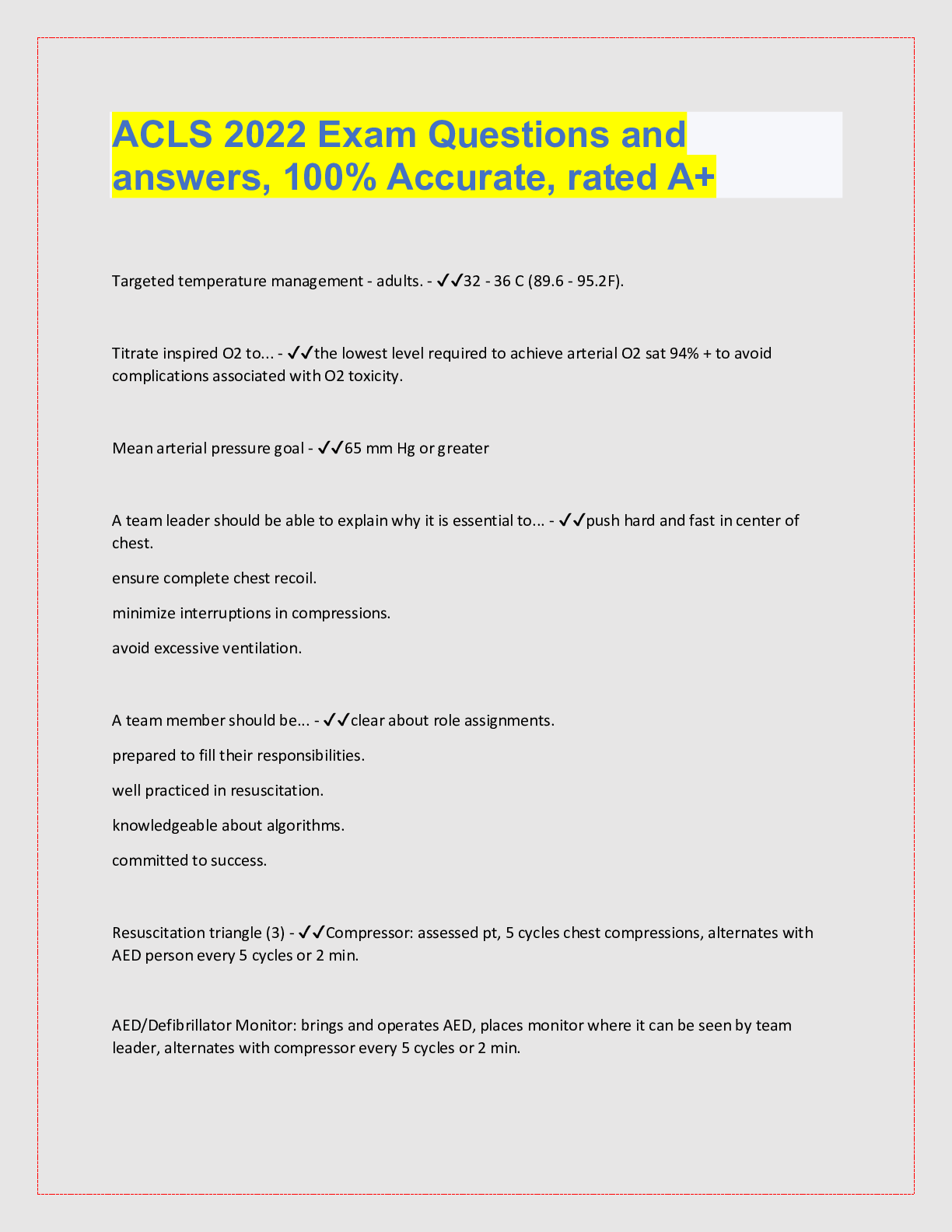
ACLS 2022 Exam Questions and
answers, 100% Accurate, rated A+
Targeted temperature management - adults. - ✔✔32 - 36 C (89.6 - 95.2F).
Titrate inspired O2 to... - ✔✔the lowest level required to achieve arterial O2 sat 94% + to avoid
complications associated with O2 toxicity.
Mean arterial pressure goal - ✔✔65 mm Hg or greater
A team leader should be able to explain why it is essential to... - ✔✔push hard and fast in center of
chest.
ensure complete chest recoil.
minimize interruptions in compressions.
avoid excessive ventilation.
A team member should be... - ✔✔clear about role assignments.
prepared to fill their responsibilities.
well practiced in resuscitation.
knowledgeable about algorithms.
committed to success.
Resuscitation triangle (3) - ✔✔Compressor: assessed pt, 5 cycles chest compressions, alternates with
AED person every 5 cycles or 2 min.
AED/Defibrillator Monitor: brings and operates AED, places monitor where it can be seen by team
leader, alternates with compressor every 5 cycles or 2 min.
Airway: Opens airway, provides bag-mask ventilation, inserts airway adjuncts as appropriate.
Leadership roles (3) - ✔✔Team leader: every team needs one. assigns roles to team members, makes
treatment decisions, provides feedback, assumes responsibility for roles not assigned.
Meds: Initiates IV/IO access, administers meds.
Time recorder: records time of interventions & medications and announces when next are due, records
frequency and duration of interruptions in compressions, communicates to the team leader.
Should you start CPR when you are unsure about a pulse? - ✔✔Yes, unnecessary compressions are less
harmful than failing to provide compressions when needed.
Agonal gasps - ✔✔A sign of cardiac arrest!
May be present in first minutes.
Looks like pt is drawing in air quickly.
Occur at a slow rate.
May be forceful or weak.
Time passes between gasps.
Snort, snore, groan.
BLS assessment - ✔✔Check responsiveness.
Shout for help.
Get AED/send someone.
Look for breathing - chest 5-10 sec.
Check pulse at same time. 5-10 sec.
No pulse in 10 sec, start chest compressions.
If pulse, rescue breathing 1 breath q 5-6 sec. Check pulse q 2 minutes.
Defibrillation: check for shockable rhythm, shock, follow with compressions.
Minimize interruptions - ✔✔No longer than 10 sec!
Avoid...
prolonged rhythm analysis.
frequent/inappropriate pulse checks.
taking too long to give breaths.
unnecessary moving the pt.
Coronary Perfusion Pressure (CPP) - ✔✔Aortic relaxation (diastolic) - right atrial relaxation (diastolic)
pressure.
Correlates with both myocardial blood flow and return of spontaneous circulation.
ROSC does not occur unless it is 15 mm Hg or +
If < 20 improve chest compressions and vasopressor therapy.
Quality compressions - ✔✔compress 2 in (5 cm).
Rate 100-120 BPM.
Allow complete recoil.
single rescuer CPR - ✔✔Cardiac arrest: Call for help, get AED, return to pt, start CPR.
Hypoxia (drowning): give 2 mins CPR before activating emergency response system.
Primary Assessment - ✔✔Assess before action!!!
Airway, Breathing, Circulation, Disability (Alert, Voice, Painful, Unresponsive), Exposure (remove clothing
to examine).
Secondary Assessment - ✔✔Focused medical history and physical exam.
SAMPLE
Signs and sx.
Allergies.
Medications (including last dose taken).
Past medical hx.
Last meal consumed.
Events.
H&Ts - ✔✔Common reversible causes of cardiac arrest.
Hypovolemia, hypoxia, hydrogen ion (acidosis), Hypo/hyperkalemia, hypothermia.
Tension pneumo, tamponade (cardiac), toxins, thrombosis (coronary or pulmonary).
2 most common causes of PEA - ✔✔Hypoxia and hypovolemia.
PEA hypovolemia - ✔✔rapid, narrow-complex tachycardia (sinus tachy).
Increased diastolic, decreased systolic pressure.
BP drops.
Narrow QRS.
Common causes: occult internal hemorrhage, severe dehydration.
Consider volume infusion.
acute coronary syndrome - ✔✔sudden symptoms of insufficient blood supply to the heart indicating
unstable angina or acute myocardial infarction
Tx for PE - ✔✔Fibrinolytics.
Tx for cardiac tamponade - ✔✔pericardiocentesis.
tx for Tension pneumo - ✔✔needle aspiration and chest tube placement.
Drug overdose/toxic exposure - ✔✔may lead to peripheral vascular dilation and/or myocardial
dysfunction with hypotension.
Tx: prolonged basic CPR, etracorporeal CPR, intra-aortic balloon pumping, renal dialysis, IV lipid
emulsion, digoxin, glucagon, bicarbonate, transcutaneous pacing, correction of electrolyte imbalances.
Normal respiratory rate.
Normal tidal volume. - ✔✔12-16/min.
8-10 ml/kg.
***<6 = assisted ventilation with BVM or advanced airway with 100% O2.
Respiratory Distress - ✔✔abnormal respiratory rate or effort.
Sx: nasal flaring, retractions, use of accessory muscles, hypoventilation, bradypnea, stridor, wheezing,
grunting, tachycardia, cyanosis, changes in LOC, agitation, use of abdominal muscles.
Mild respiratory distress sx - ✔✔mild tachypnea and mild increase in respiratory effort with changes in
airway sounds.
Severe respiratory distress sx - ✔✔tachypnea, increased respiratory effort, deterioration in skin color,
changes in mental status.
Can be indicitive of respiratory failure.
Respiratory Failure - ✔✔inadequate oxygenation, ventilation or both.
Often the end stage of respiratory distress.
Confirm dx with objective measures (pulseOx, ABGs).
Sx of respiratory failure - ✔✔marked tachypnea, bradypnea, apnea (late), increased/decreased/no
effort, poor/absent distal air movement, tachycardia (early), bradycardia (late), cyanosis, stupor, coma
(late).
Causes of respiratory failure - ✔✔upper or lower airway obstruction.
Lung tissue disease.
Disordered control of breathing.
Rise in arterial CO2 (hypercapnia).
Drop in blood O2.
Respiratory arrest - ✔✔the cessation of breathing.
Caused by drowning or head injury.
Tx: tidal volume 500-600ml (visable chest rise).
***Pts with airway obstruction or poor lung compliance may require higher pressures.
Complications of excessive ventilation - ✔✔gastric inflation.
regurgitation.
aspiration.
Intrathoracic pressure increase --> decreased venous return to heart --> low cardiac output.
cerebral vasoconstriction --> decreased blood to brain.
The most common cause of upper airway obstruction in an unconscious pt - ✔✔loss of tone in the
throat muscles --> tongue occluding airway.
Basic airway opening - ✔✔head tilt-chin lift.
neck injury: jaw thrust maneuver. (may do HTCL if this is not effective).
Oropharyngeal airway (OPA) - ✔✔used in unconscious patients who have obstruction risk from tongue.
***check for gag/cough reflex***
J shaped device.
Holds tongue away from the posterior wall of pharynx.
also used when suctioning intubated pts to prevent them from biting and occluding the ET tube.
Too large or too small causes airway obstruction.
OPA insertion - ✔✔clear mouth/pharynx with suctioning.
select proper size: flange is at corner of mouth, tip at mandible.
Insert so it curves upward toward the hard palate.
Insert at 90 or 180 degrees and turn.
Nasopharyngeal Airway (NPA) - ✔✔Used in pts who need a basic airway management adjunct.
Soft rubber/plastic uncuffed tube that allows airflow between nares and pharynx.
May be used in conscious, semi-conscious or unconscious pts.
Indicated when OPA is technically difficult or dangerous (gag reflex, trismus, massive mouth trauma,
wired jaws), neurologically impaired pts with poor pharyngeal tone/coordination.
NPA Insertion - ✔✔Select proper size: tip of nose to earlobe. (some providers use diameter of pt's
smallest finger as a guide).
Lubricate airway.
Insert.
If resistance: slightly rotate or attempt placement in other nares.
***Caution with facial trauma: risk of misplacement into the cranial cavity through a fractured
cribriform plate.
OPA and NPA precautions - ✔✔Always check for spontaneous respirations immediately after insertion.
Absent respirations: PPV.
if no adjuncts available start mouth-to-mask barrier ventilation.
Suctioning - ✔✔Force of 80-120 mmHg.
Soft catheters - ✔✔used in the mouth or nose.
Come in sterile wrappers and can be used for ET tube deep suctioning.
Rigid catheters - ✔✔Ex: Yankauer.
Used in the oropharynx.
Better for suctioning thick secretions.
3 EKG categories - ✔✔ST segment elevation suggesting ongoing acute injury.
ST segment depression suggesting ischemia.
Nondiagnostic/normal EKG.
Drugs to relieve ischemic discomfort, dissolve clots and inhibit thrombin/platlets - ✔✔O2, aspirin, nitro,
opiates (morphine), fibrinolytics, heparin.
Additional: B-blockers, ADPs (clopridogrel, prasugrel, ticagrelor), ACEs, Statins, glycoprotein IIb/IIa
inhibitors.
Common causes of ACS deaths - ✔✔VF or pulseless VT.
***VF likely to develop in first 4 hrs after sx onset.
Priority in managing ACS - ✔✔obtaining a 12 lead EKG.
***should be done in pt's first 10 min of arrival.
Most common sx of myocardial ischemia and infarction - ✔✔retrosternal chest discomfort.
Also, pressure or tightness.
Sx of ACS - ✔✔uncomfortable pressure/fullness, squeezing pain in center of chest for several mins.
chest discomfort spreading to shoulders, neck, arms, jaw, between shoulder blades.
chest discomfort with light-headedness, dizziness, fainting, sweating, n/v.
Unexplained SOB with or without chest discomfort.
Other causes of chest discomfort - ✔✔Aortic dissection.
PE.
Acute pericardial effusion with tamponade.
Tension pneumo.
When to administer O2 - ✔✔Dyspneic, hypoxemic, obvious sx of HF, arterial sat < 90%, or sat is
unknown.
Titrate > 90%.
When to administer Aspirin (Acetylsalicylic acid) - ✔✔160-325 mg non-enteric coated medication causes
platelet inhibition. This reduces coronary reocclusio
[Show More]