*NURSING > STUDY GUIDE > MS1 Comprehensive Final Study Guide (All)
MS1 Comprehensive Final Study Guide
Document Content and Description Below
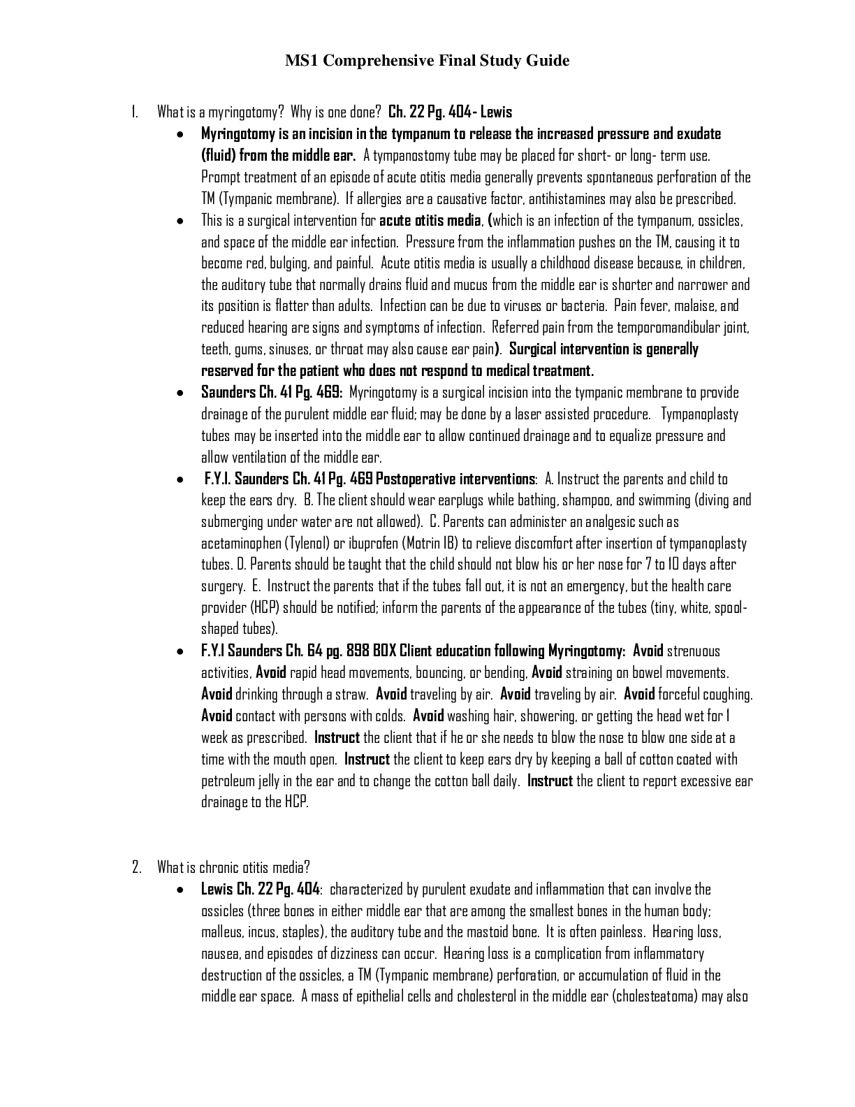
MS1 Comprehensive Final Study Guide 1. What is a myringotomy? Why is one done? Ch. 22 Pg. 404- Lewis • Myringotomy is an incision in the tympanum to release the increased pressure and exudate (f ... luid) from the middle ear. A tympanostomy tube may be placed for short- or long- term use. Prompt treatment of an episode of acute otitis media generally prevents spontaneous perforation of the TM (Tympanic membrane). If allergies are a causative factor, antihistamines may also be prescribed. • This is a surgical intervention for acute otitis media, (which is an infection of the tympanum, ossicles, and space of the middle ear infection. Pressure from the inflammation pushes on the TM, causing it to become red, bulging, and painful. Acute otitis media is usually a childhood disease because, in children, the auditory tube that normally drains fluid and mucus from the middle ear is shorter and narrower and its position is flatter than adults. Infection can be due to viruses or bacteria. Pain fever, malaise, and reduced hearing are signs and symptoms of infection. Referred pain from the temporomandibular joint, teeth, gums, sinuses, or throat may also cause ear pain). Surgical intervention is generally reserved for the patient who does not respond to medical treatment. • Saunders Ch. 41 Pg. 469: Myringotomy is a surgical incision into the tympanic membrane to provide drainage of the purulent middle ear fluid; may be done by a laser assisted procedure. Tympanoplasty tubes may be inserted into the middle ear to allow continued drainage and to equalize pressure and allow ventilation of the middle ear. • F.Y.I. Saunders Ch. 41 Pg. 469 Postoperative interventions: A. Instruct the parents and child to keep the ears dry. B. The client should wear earplugs while bathing, shampoo, and swimming (diving and submerging under water are not allowed). C. Parents can administer an analgesic such as acetaminophen (Tylenol) or ibuprofen (Motrin IB) to relieve discomfort after insertion of tympanoplasty tubes. D. Parents should be taught that the child should not blow his or her nose for 7 to 10 days after surgery. E. Instruct the parents that if the tubes fall out, it is not an emergency, but the health care provider (HCP) should be notified; inform the parents of the appearance of the tubes (tiny, white, spool-shaped tubes). • F.Y.I Saunders Ch. 64 pg. 898 BOX Client education following Myringotomy: Avoid strenuous activities, Avoid rapid head movements, bouncing, or bending, Avoid straining on bowel movements. Avoid drinking through a straw. Avoid traveling by air. Avoid traveling by air. Avoid forceful coughing. Avoid contact with persons with colds. Avoid washing hair, showering, or getting the head wet for 1 week as prescribed. Instruct the client that if he or she needs to blow the nose to blow one side at a time with the mouth open. Instruct the client to keep ears dry by keeping a ball of cotton coated with petroleum jelly in the ear and to change the cotton ball daily. Instruct the client to report excessive ear drainage to the HCP. 2. What is chronic otitis media? • Lewis Ch. 22 Pg. 404: characterized by purulent exudate and inflammation that can involve the ossicles (three bones in either middle ear that are among the smallest bones in the human body; malleus, incus, staples), the auditory tube and the mastoid bone. It is often painless. Hearing loss, nausea, and episodes of dizziness can occur. Hearing loss is a complication from inflammatory destruction of the ossicles, a TM (Tympanic membrane) perforation, or accumulation of fluid in the middle ear space. A mass of epithelial cells and cholesterol in the middle ear (cholesteatoma) may also develop. The cholesteatoma enlarges and can destroy the adjacent bones. Unless removed surgically, it can cause extensive damage to the ossicles and impair hearing. Otoscopic examination of the TM may reveal changes in color and mobility or a perforation. Culture and sensitivity tests of the drainage are necessary to identify the organisms involved so that appropriate antibiotic therapy can be prescribed. The audiogram may demonstrate a hearing loss as great as 50 to 60 dB if the ossicles have been damage or separated. Sinus x-rays, MRI, or a computed tomography (CT) scan of the temporal bone is done to assess for bone destruction and the presence of a mass. • Nursing care: The aims of treatment are to clear the middle ear of infection, repair any perforations and preserve hearing. Otic and systemic (oral and IV) antibiotic therapy is started based on the culture and sensitivity results. In many cases of chronic otitis media, antibiotic resistance is present. The patient may need to undergo frequent evacuation of drainage and debris in an outpatient setting. Often chronic TM perforations do not heal with conservative treatment, and surgery is necessary. Tympanoplasty (myringoplasty) involves reconstruction of the TM and/or the ossicles. A mastoidectomy is often performed with a tympanoplasty to remove infected portions of the mastoid bone. Removal of tissue stops at the middle ear structures that appear capable of conducting sound. Sudden pressure changes in the ear and postoperative infections can disrupt the surgical repair during the healing phase or cause facial nerve paralysis. Table 22-9 COLLABORATIVE CARE CHRONIC OTITIS MEDIA DIAGNOSTIC COLLABORATIVE THERAPY • History and physical examination • Otoscopic examination • Culture and sensitivity of middle ear drainage • Ear Irrigations • Otic, oral, or parental antibiotics • Analgesics • Antiemetics • Surgery • Tympanoplasty (see eTable 22-2) • Mastoidectomy • F.Y.I Postoperative INFO: Impaired hearing is expected during the postoperative period if there is packing in the ear. A cotton ball dressing is used for the incision made through the external auditory canal (endaural). Instruct the patient to change the cotton packing as needed. If a postauricle (behind the ear) incision is used and a drain is in place, place a dressing over the mastoid area (sits behind the ear). A small gauze pad is cut to fit behind the ear, and soft dressing material is applied over the ear to prevent the outer circular head dressing from placing pressure on the auricle. Monitor the tightness of the dressing to prevent tissue necrosis and assess the amount and type of drainage. Keep the suture line dry. Also from Saunders pg. 899: Keep dressing clean and dry. Keep client flat as prescribed, with the operative ear up for at least 12 hours. Administer antibiotics as prescribed. Mastoid disease? • Saunders ch. 64 pg. 899 (this book had more about this than our text book): Mastoid disease (Mastoiditis) may be acute or chronic and results from untreated or inadequately treated chronic or acute otitis media. The pain is not relieved by myringotomy. • Assessment: swelling behind the ear and pain with minimal movement of the head. Cellulitis on the skin or external scalp over the mastoid process. A reddened, dull, thick, immobile tympanic membrane, with or without perforation. Tender and enlarged postauricular lymph nodes. Low-grade fever. Malaise, and Anorexia. • Intervention: Prepare the client for surgical removal of infected material. Monitor for complications. Simple or modified radical mastoidectomy with tympanoplasty is the most common treatment. Once infected tissue is removed, the tympanoplasty is performed to reconstruct the ossicles and tympanic memebrane in an attempt to restore normal hearing. • Complications: Damage to the abducens and facial cranial nerves. Damage is exhibited by inability to look laterally (cranial nerve VI, abducens) and a drooping of the mouth on the affected side (cranial nerve VII, facial). Meningitis. Brain abscess. Chronic purulent otitis media. Wound infections. Vertigo, if the infection spreads into the labyrinth. • Postoperative interventions: Monitor for dizziness. Monitor for signs of meningitis, as evidenced by a stiff neck and vomiting. Prepare for a wound dressing change 24 hours postoperatively. Monitor the surgical incision for edema, drainage, and redness. Position the client flat with the operative side up as prescribed. Restrict the client to bed with bedside commode privileges for 24 hours as prescribed. Assist the client with getting out of bed to prevent falling or injuries from dizziness. With reconstruction of the ossicles via a graft, take precautions to prevent dislodging of the graft. Ossiculitis? (Not in the book) • Inflammation of the ossicle( the middle ear malleus, incus, and stapes). Otosclerosis? Lewis Ch. 22 Page 405 • A hereditary autosomal dominant disease. It is the most common cause of hearing lost in young adults. Spongy bone develops from the bony labyrinth, preventing movement of the footplate of the stapes in the oval window. This reduces the transmission of vibrations to the inner ear fluids and results in conductive hearing loss. Although, otosclerosis is typically bilateral, one ear may show faster progression of hearing loss. The patient is often unaware of the problem until the loss becomes so severe that communication is difficult. Collaborative Care: Otosclerosis Diagnostic Collaborative Therapy History and physical examination Hearing aid Otoscopic examination Microdrill or laser surgical treatment involves opening the footplate (Stapedotomy) or replacing the stapes with a metal or Teflon substitute. Surgery (Stapedectomy or stapes prosthesis) Rinne Test Weber Test Audiometry Tympanometry Drug Therapy: Sodium fluoride with Vitamin D Calcium Carbonate • Otoscopic examination: may reveal a reddish blush of the tympanum (Schwartz’s sign) cause by the vascular and bony changes within the middle ear. Tuning fork tests and an audiogram demonstrate good hearing by bone conduction but poor hearing by air conduction (air-bone gap). Usually a difference of at least 20 to 25 dB between air and bone conduction levels of hearing is seen in otosclerosis. The hearing loss associated with otosclerosis may be stabilized by the oral administration of sodium fluoride with vitamin D and calcium carbonate. These medication retard bone resorption and encourage the calcification of bony lesions. Amplification of sound by a hearing aid can be effective because the inner function is normal. Surgical procedures are usually performed with patient under conscious sedation. The ear with poorer hearing is repaired first, and the other ear may be operated on within a year. Patient often reports immediately after surgery significant improvement in hearing in the operative ear. Because of the accumulation of blood and fluid in the middle ear during the postoperative period, the hearing level decreases initially, but it improves gradually with healing. • Nursing management: Use Gelfoam on the incision flap to limit bleeding. Place a cotton ball in the ear canal, and cover the ear with a small dressing. The patient may experience dizziness, nausea, and vomiting as a result of stimulation of the labyrinth during surgery. Some patients demonstrate nystagmus because of disturbance of the perilymph fluid. The patient should take care to avoid sudden movements that may bring on or exacerbate vertigo. Actions that increase inner ear pressure, such as coughing, sneezing, lifting, bending, and straining during bowel movements, should be avoided. • F.Y.I. Saunders book Ch. 64 pg. 899-900 Description: • A. Otosclerosis is a genetic disorder of the labyrinthine capsule of the middle ear that results in a bony overgrowth of the tissue surrounding the ossicles. B. causes the development of irregular areas of new bone formation and causes the fixation of the bones. C. Stapes fixation leads to a conductive hearing loss. D. If the disease involves the inner ear, sensorineural hearing loss is present. E. Bilateral involvement is common, although hearing loss may be worse in one ear. F. It is thought to be a hereditary autosomal dominant disorder and is most commonly seen in young woman. G. Nonsurgical intervention promotes the improvement of hearing through amplification. H. Surgical intervention involves removal of the bony growth causing the hearing loss. I. A partial stapedectomy or complete stapedectomy with prosthesis (fenestration) may be performed. • Assessment: A. slowly progressing conductive hearing loss. B. Bilateral hearing loss. C. A ringing or roaring type of constant tinnitus. D. Loud sounds heard in the ear when chewing. E. Pinkish discoloration (Schwartze’s sign) of the tympanic membrane, which indicates vascular changes within the ear. F. Negative Rinne Test. G. Weber’s test shows lateralization of sound to the ear with the greatest degree of conductive hearing loss. 3. What would be priority nsg (nursing) dx for a pt. with TB? Lewis Book Pg. 532- 533 Nursing Diagnoses for patient with TB may include, but are not limited to the following • Ineffective breathing pattern related to decreased lung capacity • Ineffective airway clearance related to increased secretions, fatigue, and decreased lung capacity • Noncompliance related to lack of knowledge of disease process, lack of motivation, long-term nature of treatment, and lack of resources • Ineffective self-health management related to lack of knowledge about the disease process and therapeutic regimen • F.Y.I the overall goals are that the patient with TB will 1. Comply with therapeutic regimen, 2. Have no recurrence of disease, 3. Have normal pulmonary function, and 4. Take appropriate measures to prevent the spread of the disease. • F.Y.I Pg. 531 Non adherence is a major factor in the emergence of multidrug resistance and treatment failures. Many individuals do not adhere to the treatment program in spite of understanding the disease process and the value of treatment. 4. What medications are used to treat severe acne? What pre cautions are needed? Lewis Ch. 24 page 438-439 • Isotretinoin (Accutane) is used for sever nodulocystic acne to possibly provide lasting remission. Pregnancy tests, Monitoring of liver function, cholesterol, triglycerides, and depression essential. DRUG ALERT: Isotretinoin (Accutane) • Can cause serious damage to fetus • Blood donation prohibited for those taking the drug and for 1 month after treatment ends • Contraindicated in women who are pregnant or who are intending to become pregnant while on the drug • Linked to liver function test abnormalities F.Y.I Saunders Pg. 586-587 • 1. Severe acne is usually treated with Isotretinoin (Amnesteem or Claravis). Derivative of vitamin A (Vitamin A supplements should be discontinued during therapy); in addition, the use of tetracycline can increase the risk of adverse effects and should be discontinued before use of isotretinoin. 2. Used to treat severe cystic acne; reserved for persons who have not responded to other therapies, including systemic antibiotics. 3. Side effects include nosebleeds; inflammation of the lips or eyes; dryness or itching of the skin, nose, or mouth; pain, tenderness, or stiffness in the joints, bones, or muscles; and back pain. 4. Less common side effects include rash, hair loss, peeling of the skin, headache, and reduction in night vision. 5. Causes sensitization of the skin to UVL. 6. The medication elevates triglyceride levels, which should be measured before and during therapy; alcohol consumption should be eliminated during therapy because alcohol could potentiate elevation of serum triglyceride levels. 7. The medication may cause depression in some clients; if depression occurs, the medication should be discontinued. • Isotretinoin is highly teratogenic and can cause fetal abnormalities. If prescribed, the client needs to follow strict rules of iPLEDGE Program. It must not be used if the client is pregnant. iPLEDGE Program: • 1. A risk management program that ensures that no woman starting isotretinoin is pregnant and that no woman taking this medication becomes pregnant. 2. Access to the medication is controlled through a central automated system. 3. Strict rules must be followed by the client, HCP prescribing the medication, pharmacist dispensing the medication, and the wholesaler of the medication to ensure safety and to ensure that no woman is pregnant on initiation of therapy or becomes pregnant while taking the medication. 5. What post op nursing actions can be delegated? What cannot be delegated? (see box pg. 354) • In the PACU, patients require frequent assessment and intervention by RNs. On the clinical units, the RN is responsible for assessment, development of an individualized plan of care, evaluation of the patient, and discharge teaching. Much of the care can be delegated to LPN/LVNs and UAP. Registered Nurse (RN) duties PACU • Assess patient’s initial airway, breathing, and circulation status. • Evaluate for the return to consciousness, ability to maintain airway and breathing. • Provide ongoing assessments for postoperative problems (e.g., airway obstruction, hypoventilation, hypotension or hypertension, dysrhythmias, emergence delirium). • Evaluate patient’s readiness to transfer to clinical unit or be discharged from ambulatory surgery. • Provide oral and written report about patient status when transferring patient to RN on the clinical unit. • Provide discharge teaching for patient and caregiver after ambulatory surgery. Clinical Unit • Assess patient on initial admission to the clinical unit • Assess for postoperative complications (e.g., atelectasis, hemodynamic instability, cognitive dysfunction, pain, fluid and electrolyte disturbance, fever or hypothermia, nausea and vomiting, urinary retention, wound infection). • Develop an individualized plan of care based on identification of patient risk factors and potential complications. • Develop and implement individualized patient and caregiver education, including discharge teaching. Licensed Practical/Vocational Nurse (LPN/LVN) Duties PACU • Administer and titrate O2 based on agency protocols. • Administer analgesics and IV fluids (consider state nurse practice act and agency policy) Clinical Unit • Titrate O2 administration according to prescribed parameters. • Monitor pain level and administer prescribed analgesics. • Administer medications (consider state nurse practice act and agency policy for medications). • Provide wound care, including dressing changes. • Use bladder ultrasound to check for urinary retention. • Insert catheter as prescribed for urinary retention. Unlicensed Assistive Personnel (UAP) Duties PACU • Assist with positioning of patients in the “recovery” position • Obtain vital signs and pulse oximetry; report abnormal levels to RN. • Assist patient with elimination needs. • Assist in transfers of patient to clinical unit Clinical Unit • Monitor vital signs, pulse oximetry, and intake and output. Report abnormal levels to RN. • Assist patient with deep breathing and coughing exercises. • Report complaints of pain to RN or LPN/LVN. • Reposition and ambulate patients • Provide hygiene, including oral care • Assist with nutrition and elimination needs. 6. What are the s/s otitis media? • Saunders Nclex Pg. 469. Otitis Media(this is not in our book only acute and chronic) is an inflammatory disorder usually caused by an infection of the middle ear occurring as result of a blocked Eustachian tube, which prevents normal drainage; can be acute or chronic. It’s a common complication of an acute respiratory infection (most commonly from respiratory syncytial virus or influenza). Infants and children have Eustachian tubes that are shorter, wider, and straighter, which makes them more prone to otitis media. S/Sx: Fever, Acute onset of ear pain, crying, irritability, lethargy, loss appetite, rolling of head from side to side, pulling on or rubbing the ear, purulent ear drainage may be present, red, opaque, bulging, immobile tympanic membrane on otoscopic examination, signs of hearing loss (indicative of chronic otitis media). • F.Y.I Acute otitis media(chronic otitis media is listed in Q 1) Lewis Pg. 404: infection of the tympanum, ossicles and space of the middle ear. Swelling of the auditory tub from colds or allergies can trap bacteria, causing a middle ear infection. Pressure from the inflammation pushes on the TM (Tympanic Membrane), causing it to become red, bulging, and painful. Pain, fever, malaise, and reduced hearing are signs and symptoms of infection. Referred pain from the temporomandibular joint, teeth, gums, sinuses, or throat may also cause ear pain. • F.Y.I Otitis Media with Effusion pg. 404 Lewis: inflammation of the middle ear with collection of fluid in the middle ear space. S/S: feeling of fullness of the ear, a “plugged” feeling or popping, and decreased hearing. Patient does not experience pain, fever, or discharge from ear. It is common to have otitis media with effusion for weeks to months after an episode of acute otitis media. It usually resolves without treatment but may recur. Otitis externa? Lewis Pg. 403 • Involves inflammation or infection of the epithelium of the auricle and ear canal. Swimming may alter the flora of the external canal because of chemicals and contaminated water. This can result in an infection often referred to as “swimmer’s ear”. Trauma from picking the ear or using sharp objects (e.g., hairpins) frequently causes the initial break in the skin. Piercing of cartilage in the upper part of the auricle also places the patient at higher risk for infection. S/S: Ear pain (otalgia) is one of the first signs of external otitis. Even mild cases, the patient may experience significant discomfort with chewing, moving the auricle, or pressing on the tragus. Swelling of the ear canal an muffle hearing. There may be serosanguineous (blood-tinged fluid) or purulent (white to green thick fluid) drainage. Fever occurs when the infection spreads to surrounding tissue. • Pg. 898 Nclex: Assessment findings: pain, itching, plugged feeling in the ear, redness and edema, exudate, hearing loss. Otitis interna? • Pg. 900 Nclex book. This is also known as Labyrinthitis an infection of the labyrinth that occur as a complication of acute or chronic otitis media. May result from growth of a cholesteatoma, a benign overgrowth of squamous cell epithelium in the middle ear. S/S: hearing loss that may be permanent on the affected side. Tinnitus. Spontaneous nystagmus to the affected side. Vertigo. Nausea and vomiting. What does a normal tympanic membrane look like? • Pg. 378 Lewis: Tympanic membrane (eardrum) is shiny, translucent, pear-gray membrane is composed of epithelial cells, connective tissue, and mucous membrane. It serves as partition and an instrument of sound transmission between the external auditory canal and the middle. • Pg. 283 Nclex: The normal Tympanic membrane is intact, without perforations, and should be free from lesions. The tympanic membrane is transparent, opaque, pearly gray, and slightly concave. A fluid line or the presence of air bubbles is not normally visible. If the tympanic membrane is bulging or retracting the edges of the light reflex will be fuzzy (diffuse) and may spread over the tympanic membrane. 7. What are s/s of neurotoxicity that can occur with chemo? • By looking in the index for neurotoxicity it list Table 16-11page 266-267 in the Lewis book. I believe this is just the changes that can occur with chemo therapy such as: Nausea and vomiting, diarrhea, alopecia, fatigue… Please review the table on page listed. Too long to type…. 8. What is candidiasis? How is it cured? What pt teaching is needed? • Pg. 436 table 24-6 Lewis book: Candidiasis is caused by candida albicans. Also known as moniliasis. 50% of adult’s symptom-free carriers. Appears in warm, moist areas such as groin area, oral mucosa, and submammary folds. HIV infection, chemotherapy, radiation, and organ transplantation related to depression of cell-mediated immunity that allows yeast to become pathogenic. Clinical manifestations: Mouth: White, cheesy plaque, resembles milk curds. Vagina: vaginitis with red, edematous, painful vaginal wall, white patches. Vaginal discharge. Pruritus. Pain on urination and intercourse. Skin: Diffuse popular erythematous rash with pinpoint satellite lesions around edges of affected area. • Treatment: Microscopic examination and culture. Azole antifungals (e.g, fluconazole, ketoconazole) or other specific medication such as vaginal suppository or oral lozenge. Sexual abstinence or use condom. Skin hygiene to keep area clean and dry. Powder is effective on nonmucosal surfaces of skin to prevent recurrence. ............Continued [Show More]
Last updated: 3 years ago
Preview 1 out of 43 pages
Buy this document to get the full access instantly
Instant Download Access after purchase
Buy NowInstant download
We Accept:

Reviews( 0 )
$23.50
Can't find what you want? Try our AI powered Search
Document information
Connected school, study & course
About the document
Uploaded On
Feb 08, 2021
Number of pages
43
Written in
All
Additional information
This document has been written for:
Uploaded
Feb 08, 2021
Downloads
0
Views
53


