NR 602 Quiz 2
Restrictive Processes
Restrictive disease is less common in pediatric patients and is characterized by decreased lung compliance with relatively normal flow rates.
Examples of causative factors include n
...
NR 602 Quiz 2
Restrictive Processes
Restrictive disease is less common in pediatric patients and is characterized by decreased lung compliance with relatively normal flow rates.
Examples of causative factors include neuromuscular weakness, lobar pneumonia, pleural effusion or masses, severe pectus excavatum, or
abdominal distention. Key findings of restrictive lung disease are rapid respiratory rate and decreased tidal volume/capacity (Carter and
Marshall, 2011).
Defense Systems
The respiratory defense system includes mechanical and biologic processes. Mechanical defenses include:
• Filtering of particles
• Warming and humidifying of inspired air
• Clearing of airway through mucociliary and coughing actions
• Spasm and breathing changes
Approximately 75% of inspired air is warmed as it passes through the nose, paranasal sinuses, pharynx, larynx, and upper portion of the
trachea. Final warming and humidifying of the airstream take place in the trachea and large bronchi. Heat and moisture are removed during
the expiratory phase of respiration. The nose has a large surface area on which particles larger than 5 mm are trapped and filtered to prevent
them from entering the lower airways. The trachea and bronchioles are lined with various defensive cells and mucus glands. Goblet cells
secrete the mucous layer that lies on the tip of cilia. Particles entering the conducting airway are quickly cleared by the mucociliary defenses.
Coughing is a reflex mechanism that has three phases: (1) inspiratory, (2) compressive, and (3) expiratory. Through forceful expiration FBs
and other materials can be removed from the airways; coughing propels particles. Young infants and children cannot effectively expectorate
mucus, so they swallow it. Loss of the cough reflex leads to aspiration and pneumonia. Temporary breathing cessation, reflex shallow
breathing, laryngospasm, and even bronchospasm are compensatory efforts aimed at stopping foreign matter from further entry into the lower
respiratory tract. 797However, these respiratory efforts offer limited protection and have significant drawbacks.
Biologic processes that protect the respiratory system include:
• Phagocytosis
• Absorption of noxious gases in the vasculature of the upper airway
• Absorption of particles by the lymph system
Phagocytosis, aided by the secretory IgA plus interferon, lysozyme, and lactoferrin, is the principal antimicrobial defense. Particles
reaching the alveoli can be phagocytized by alveolar macrophages and polymorphonuclear (PMN) cells, cleared from the lung by the
mucociliary system, or carried by lymphocytes into regional nodes or the blood. These particles can take days to months to clear.
The respiratory defense system is at risk for compromise from numerous environmental factors. Damage to epithelial cells is caused by a
variety of substances and gases, such as sulfur, nitrogen dioxide, ozone, chlorine, ammonia, and cigarette smoke. Hypothermia, hyperthermia,
morphine, codeine, and hypothyroidism can adversely alter mucociliary defenses. Dry air from mouth breathing during periods of nasal
obstruction, tracheostomy placement, or inadequately humidified oxygen therapy results in dryness of the mucous membrane and slowing of
the cilia beat. Cold air is also irritating to the lower airways.
Phagocytic ability is also reduced by many substances, including ethanol ingestion and cigarette smoke. Hypoxemia, starvation, chilling,
corticosteroids, increased oxygen, narcotics, and some anesthetic gases also impair phagocytosis. Recent acute viral infections can reduce
antibacterial killing capacity. Damage from infection and chemical irritants may or may not be reversible.
Recurrent respiratory infections in children merit investigation for immunodeficiency or other underlying diseases, such as primary ciliary
dyskinesia or CF. The mnemonic SPUR (Bush, 2009) can help determine which children need further workup:
Severe infection
Persistent infection and poor recovery
Unusual organisms
Recurrent infection
Immunodeficiencies should be considered if the child has four or more new ear infections in a year, two or more serious sinus infections,
two or more pneumonias in a year, persistent oral candidiasis, failure to thrive, two or more deep seeded skin abscesses, 2 or more months on
antibiotics without improvement, and/or the need for intravenous (IV) antibiotics to clear infections. Also consider immunodeficiencies if
there is a family history of immunodeficiency or two or more deep skin infections (Modell et al, 2014).
Assessment of the Respiratory System
The history provides valuable information about the causes, progression, and potential complications of a child's respiratory condition. The
physical examination and diagnostic testing allow the provider to determine the extent of respiratory distress.
History
History of the present illness can be assessed using the mnemonic PQRST:• Promoting, preventing, precipitating, palliating factors
• Contacts: Are any family members or close contacts (e.g., day care, school) ill with similar signs and symptoms?
• Prevention: Do you give your child any medications or supplements (include any herbs, botanicals, or vitamins) to try to prevent a
cold? What are your hand washing practices? Do you encourage fluids when your child has a URI? Are the child's immunizations up
to date?
• Progression: Are the respiratory signs or symptoms increasing in severity, lessening, or about the same? Is the child easily fatigued,
less active, having trouble sleeping, or working harder to breathe?
• Treatment: Have any OTC, prescription drugs, herbs, supplements, or botanicals been used? Have any other treatment modalities been
used, including folk cures or home remedies?
• Quality or quantity
• How severe are the symptoms? Is the illness interfering with school attendance or play? Are breathing problems affecting the child's
ability to sleep and eat?
• Region or radiation
• Does the child complain of chest pain?
• Severity, setting, simultaneous symptoms or similar illnesses in the past
• Key signs and symptoms: Has the child had symptoms or signs of a daytime or nighttime cough, fever, vomiting, malaise, rhinorrhea,
sore throat, lesions in the mouth, retractions, cyanosis, dyspnea, or increased respiratory effort? Table 32-1 lists key characteristics
and causes of cough.
TABLE 32-1
Key Characteristics of Cough, Common Causes, and Questions to Ask in a Pediatric History
Key Characteristics to
Consider Description and Questions to Ask
Age factor Infants have a weak, nonproductive cough.
Quality
Staccato-like (Chlamydia trachomatis in infants); barking or brassy (croup, tracheomalacia, habit cough); paroxysmal or inspiratory
whoop (pertussis or parapertussis); honking (psychogenic).
Is the cough wet or dry?
Duration
Acute (most causes are infectious and last less than 2 weeks), subacute (cough lasts from 2 to 4 weeks); recurrent (associated with
allergies and asthma), or chronic (lasting greater than 4 to 8 weeks [e.g., CF, asthma]).
Is the cough continuous or intermittent?
Productivity Mucus producing or nonproductive?
Timing During the day, night (associated with asthma), or both?
Effect on parent and
child
Are parents frustrated with the cough? Is it causing them to lose sleep and work time? Are they concerned that the child may have
something serious?
Associated symptoms Fever: May indicate bacterial infection (pneumonia).Key Characteristics to
Consider Description and Questions to Ask
Rhinorrhea, sneezing, wheezing, atopic dermatitis: Associated with asthma and allergic rhinitis.
Malaise, sneezing, watery nasal discharge, mild sore throat, no or low fever, not ill appearing: Typical of URI.
Tachypnea: Pneumonia or bronchiolitis in infants (infants may not have a cough).
Exposure to infection
or travel
Has the child been out of the country (tuberculosis)? Is there a member of the household being treated for “bronchitis” or another
cough illness?
Causes
Congenital anomalies Tracheoesophageal fistula, vascular ring, laryngeal cleft, vocal cord paralysis, pulmonary malformations, tracheobronchomalacia,
congenital heart disease
Infectious agent Viral (RSV, adenovirus, parainfluenza, HIV, metapneumovirus, human bocavirus), bacterial (tuberculosis, pertussis, Streptococcus
pneumoniae), fungal, and atypical bacteria (C. and M. pneumoniae)
Allergic condition Allergic rhinitis, asthma
Other FB aspiration, gastroesophageal reflux, psychogenic cough, environmental triggers (air pollution, tobacco smoke, wood smoke, glue
sniffing, volatile chemicals), CF, drug induced, tumor, congestive heart failure
CF, Cystic fibrosis; FB, foreign body; HIV, human immunodeficiency virus; RSV, respiratory syncytial virus; URI, upper respiratory infection.
Adapted from Chang AB: Cough, Pediatr Clin North Am 56(1):19–31, 2009; Cherry JD: Croup (laryngitis, laryngotracheitis, spasmodic croup,
laryngotracheobronchitis, bacterial tracheitis, and laryngotracheobronchopneumonitis). In Cherry J, Kaplan S, Demmler-Harrison G, et al, editors: Feigin
& Cherry's textbook of pediatric infectious diseases, ed 6, vol 1, Philadelphia, 2009, Saunders/Elsevier, pp 254–268.
• Associated symptoms: Has there been a decrease in appetite or feeding? Any rashes, headaches, or abdominal pain?
• Similar illnesses in the past: Does the child have a history of respiratory tract infections, allergies, or asthma? How many similar
infections has the child had (e.g., croup, pneumonia, rhinosinusitis, streptococcal tonsillopharyngitis, or frequent colds)?
• Temporal factors
• When did the illness begin?
• Was the onset acute or insidious or proceeded by the common cold?• How long has it lasted? How has it changed over time?
• Family history
• Do others in the family have a history of allergies or asthma?
798
• Is there any family history of immunodeficiency, ear-nose-throat, or respiratory problems?
• Does anyone in the family have genetic diseases, such as CF or alpha 1-antitrypsin deficiency?
• Are other family members ill?
• Review of systems
• Note any infections, constitutional diseases, or congenital problems that might have a respiratory component.
• Environment
• Does anyone in the family or in the day care setting smoke? Does the child live or attend school in an urban or industrial area subject to
air pollution (e.g., near a major highway, industrial plant, or bus terminal)? Has the child or a family contact traveled recently and
where?
Physical Examination
When determining respiratory distress, think about the total presentation and not just individual isolated findings. Consider the anxiety level,
respiratory rate and rhythm, use of accessory muscles, color, breath sounds, grunting, and pulse oximetry results. Information pertinent to the
physical examination of a child with suspected respiratory disease includes the following:
• Measurement of vital signs and observation of general appearance:
• A normal respiratory rate is age dependent and, if elevated, is a key indicator of lower respiratory involvement.
• The level of anxiety, nasal flaring, and position of comfort are useful indicators of respiratory distress. 799Changes in skin color may
be subtle or obvious, depending on the level of deoxygenation. Grunting is a sign of small airway disease.
• Inspection of:
• Nose: Look for rhinorrhea—clear, mucoid, mucopurulent; FBs, erosion, polyps, lesions, bleeding, septal position, and color of the
mucous membrane.
• Throat, pharynx, and tonsils: Look for lesions, vesicles, exudate, enlargement of any structure, or other abnormalities. If epiglottitis is a
consideration, do not inspect the mouth or attempt to elicit a gag reflex (see discussion later in this chapter).
• Chest: Look at the depth, ease, symmetry, and rhythm of respiration. These are key indicators of lower respiratory tract involvement.
The use of accessory muscles and the presence of retractions should be noted. A prolonged expiratory phase is associated with
respiratory obstruction in the lower airways.
• Palpation or percussion of:
• Chest: Percuss for signs of dullness or hyperresonance caused by consolidation, fluid, or air trapping.
• Auscultation of the chest:
• Upper tract: Pathology frequently causes noisy breathing, snoring, stridor, and musical or wheezing tracheal breath sounds and can be a
source of referred breath sounds (Bohadana et al, 2014).
• Lower tract: Pathology is suggested by fine crackles, coarse crackles, rhonchus, pleural friction rub, wheezing, and bronchial breath
sounds (Bohadana et al, 2014).
Upper Respiratory Tract
The upper respiratory tract includes the nostrils, nasopharynx, larynx, upper part of the trachea, eustachian tubes, and sinuses.
Air is warmed and humidified as it travels through the nasal passages, and coarse nasal hairs filter out particles. The nasal
passages are lined with lysozymes, secretory immunoglobulin A (IgA), and immunoglobulin G (IgG) in nasal mucosa to defend
against microbial invasion. The nasal mucosa is continuous and similar to the sinus mucosa except that the nasal mucosa is
thicker with more glands. A blanket of mucus covers the surface epithelium of the nasal and sinus mucosa.
The mucosal lining of the sinuses is composed of pseudostratified, ciliated columnar epithelium interspersed with mucus
producing goblet cells (Rose et al, 2013). The mucociliary action of the paranasal epithelium moves secretions from the sinuses to
the nasal cavity. The frontal, maxillary, and anterior parts of the ethmoid sinuses drain to the middle meatus of the nose,
whereas the sphenoid and posterior parts of the ethmoid sinuses drain to the superior meatus of the nose. Secretions need to be
able to move through patent ostia into the nose. The quality of secretions and normally functioning cilia are key factors in the
movement of secretions into the nose. Inflammation of nasal mucosa frequently causes edema and disruption of the sinus
secretions. If there is significant swelling of the ostia due to a URI or allergic inflammation, or mechanical or local obstruction,
ostial obstruction results and obstruction of the sinus secretions occurs. Cilia movement and mucus flow allow the sinuses to be
free of pathogens.
The maxillary sinuses are present by the second trimester of gestation but are not fully pneumatized until a child is about 4
years old. Ethmoid sinuses develop by the fourth month of gestation and form the thin lateral walls of the orbit of the eye. They
are pneumatized at birth and can be visualized on plain radiographs when the child is 1 to 2 years old. The sphenoid sinuses
start to form in the first 2 years of life but remain rudimentary until age 6, which is when they become visible on radiographs.By 12 years old, they reach their permanent size, but not shape. As a result, the nasal cavity and paranasal sinuses reach adult
proportion by age 12 (Cherry and Shapiro, 2009). The sinuses become clinically significant sites of infection at the following
ages:
• Maxillary and ethmoid sinuses: As early as late infancy
• Sphenoid sinuses: Around 3 and 4 years old
• Frontal sinuses: Around 6 to 10 years old
The epiglottis deflects swallowed material toward the esophagus to protect the larynx. The vocal cords form a V-shaped
opening known as the glottis.The subglottic space is beneath the vocal cords, and its walls converge toward the cricoid ring to
form a complete ring of cartilage around the larynx. In children younger than 2 to 3 years old, the cricoid ring is the narrowest
part of the airway; in older children and adults, the glottis is narrowest. The rings of tracheal cartilage support the trachea and
the mainstem bronchi.
795
The trachea and airways of the infant and young child are more compliant than those of an adult. Hyperextension of the neck
can constrict the airway of infants. Consequently, changes in intrapleural pressure lead to greater changes in an infant's or
young child's airway compared with the effect that such changes would exert on adult airways, thereby causing an increased
risk of airway collapse. Similarly, increased chest wall compliance in young infants makes them more vulnerable to adverse
events, and their respiratory muscles cannot effectively handle sustained, intense respiratory workload that occurs during
severe pulmonary illnesses.
Bronchiolitis
Bronchiolitis is also called infectious asthma, asthmatic bronchitis, wheezy bronchitis, or virus-induced asthma. Bronchiolitis is a disease that
causes inflammation, necrosis, and edema of the respiratory epithelial cells in the lining of small airways, as well as copious mucus
production (Ralston et al, 2014). Bronchiolitis is characterized by the insidious onset of URI symptoms over 2 to 3 days that progresses to
lower respiratory symptoms that last as long as 10 days (Da Dalt et al, 2013). It is a communicable disease found primarily in infancy to 2
years old (Teshome et al, 2013) that accounts for 10% of visits to a primary provider the first 2 years of life (Schroeder and Mansbach, 2014).
Bronchiolitis is a common diagnosis used for an infant seen with wheezing for the very first time and is the leading cause of hospitalizations
for infants. The most common age for severe disease occurs in infants between 2 to 3 months due to the natural postnatal nadir in maternal
immunoglobulins received via the placenta during the last trimester (Da Dalt et al, 2013). More than 80% of the cases of bronchiolitis occur
in infants younger than 1 year of age with a male-to-female ratio of 1.5 : 1 (Welliver, 2009). In mild cases, symptoms can last for 1 to 3 days.
In severe cases, cyanosis, air hunger, retractions, and nasal flaring with symptoms of severe respiratory distress within a few hours may be
seen. Apnea can occur with a wide range of prevalence reported (Ralston et al, 2014) and may require mechanical ventilation.
Newer understanding of the pathophysiology in bronchiolitis points to airway obstruction as a result of epithelial and inflammatory
cellular debris due to infiltration of the virus into the small bronchiole epithelium and alveolar epithelial cells (AEC), types I and II.
Membranous pneumatoceles, or AEC type I, are dominant and cover 96% of the respiratory tree. Their role is in gas exchange, whereas AEC
type II are important to surfactant production (Chuquimia et al, 2013). It is a disease of the small bronchioles that are 2 mm in size. There is a
sparing of basal cells in the bronchiole. The main lesion is epithelial necrosis, which leads to a dense plugging of the bronchial lining. This
results in increased airway resistance, atelectasis, hyperinflation, and increased mucus production (Teshome et al, 2013).
Bronchiolitis is a viral illness predominantly caused by RSV, especially in outbreaks (Da Dalt et al, 2013; Welliver, 2009). Recent data
suggest that up to 30% of infants with severe bronchiolitis are co-infected with two or more viruses (Mansbach et al, 2012). In descending
order after RSV, rhinovirus, parainfluenza, adenovirus, and mycoplasma are causes (Teshome et al, 2013). Metapneumovirus was discovered
in 2001 and is a cause of bronchiolitis 7% of the time. Human bocavirus is a common co-infecting virus with RSV and is found up to 80% of
the time (Teshome et al, 2013). RSV-specific immunoglobulin E (IgE), eosinophils, and chemokines may play a role in the pathogenesis of
bronchiolitis (Welliver, 2009). Adenovirus and RSV can cause long-term complications. The incubation period for RSV is 2 to 8 days and
typically occurs from November through March with virtually no outbreaks in the summer (Teshome et al, 2013; Welliver, 2009). Fever
tends to be higher with adenovirus versus RSV (Teshome et al, 2013).
Respiratory viruses are spread by close contact with infected respiratory secretions or fomites and can live on 818surfaces for up to 30
minutes (Teshome et al, 2013). The most frequent mode of transmission is hand carriage of contaminated secretion. The source of infection is
an older child or adult family member with a ―mild‖ URI. Older children and adults have larger airways and tolerate the swelling associated
with this infection better than infants do. Most cases of bronchiolitis resolve completely, but recurrence of infection is common, and
symptoms tend to be mild.
Infants who are at higher risk of severe RSV include children with major chronic pulmonary disease, such as CF, neuromuscular
disorders, or bronchopulmonary dysplasia; premature birth before 35 weeks of gestational age; and infants with significant hemodynamically
difficulties due to congenital heart disease (Teshome et al, 2013). Other risk factors for severe RSV disease are male gender, crowded
household, lack of breastfeeding, smoke exposure, day care attendance, having siblings, birth during the winter months, and
immunodeficiency (Da Dalt et al, 2013).
Clinical Findings
History
The following are reported:• Initial presentation: Typically the illness begins with URI symptoms of cough, coryza, and rhinorrhea and progresses over 3 to 7 days
(Smith, 2011).
• Gradual development of respiratory distress marked by noisy, raspy breathing with audible expiratory wheezing.
• Low-grade to moderate fever up to 102° F (38.9° C).
• Decrease in appetite.
• No prodrome in some infants; rather they have apnea as the initial symptom.
• Usually the patient's course is the worst by 48 to 72 hours after the wheezing starts and then the patient starts to improve. If the child has a
bacterial illness, the child will continue to worsen with a high fever.
Physical Examination
Findings include the following:
• Upper respiratory findings
• Coryza
• Mild conjunctivitis in 33% (Welliver, 2009)
• Pharyngitis
• Otitis media in up to 15% (Welliver, 2009)
• Lower respiratory findings (Teshome et al, 2013)
• Tachypnea (approximately 40 to 80 breaths per minute)
• Substernal and/or intercostal retractions
• Heterophonous expiratory wheezing
• Fine or coarse crackles may be heard throughout the breathing cycle
• Varying signs of respiratory distress and pulmonary involvement (e.g., nasal flaring, grunting, retractions, cyanosis, prolonged
expiration)
• Abdominal distention
• Palpable liver and spleen, pushed down by hyperinflated lungs and a flattened diaphragm
Diagnostic Studies
A diagnosis of bronchiolitis should be based on the history and physical examination (Ralston et al, 2014). Overuse of diagnostic testing
persists in clinical practice despite available guidelines on the diagnosis and management of bronchiolitis (Librizzi et al, 2014; Ralston et al,
2014; Turner et al, 2014). The routine use of chest radiographs in previously healthy infants with mild RSV bronchiolitis is not indicated.
Evidence-based guidelines from the AAP and the Scottish Intercollegiate Guidelines Network (SIGN) are strongly against routine chest
radiography, including those in previously healthy infants with mild RSV bronchiolitis (Ralston et al, 2014; SIGN, 2006). In severe illness, a
chest x-ray may be ordered to rule out pneumonia or pneumothorax, but its use must be weighed against the dangers of radiation exposure.
The findings of chest radiography can vary, and even with severe illness the x-ray can be clear with a flattened diaphragm and an increase in
anteroposterior diameter. Areas of atelectasis can appear like a pneumonitis, but true pneumonia is uncommon (early bacterial pneumonia can
be difficult to detect and cannot be ruled out by radiographs).
Routine virologic testing is not recommended. In selected situations (hospitalization or if an infant has received monthly palivizumab
[Synagis]), enzyme-linked immunosorbent assays or fluorescent antibody techniques to look for RSV are the diagnostic procedures of choice
in most laboratories. Viral culture of nasal washings can be done in severe cases to confirm RSV, parainfluenza viruses, influenza viruses,
and adenoviruses. PCR is helpful in deciding about isolation of cohorts with the same infection in the hospital setting. The cost of the
diagnostic viral testing may outweigh the clinical usefulness of knowing which virus is infecting the patient.
Hematologic testing is not recommended in the latest guidelines. If a CBC is done for another reason, a mild leukocytosis may be seen
with 12,000 to 16,000/mm3. Routine laboratory tests are usually not required to confirm the diagnosis, because they lack specificity.
However, young infants pose a diagnostic dilemma, because they are at greater risk of a serious bacterial infection (SBI) and, therefore, blood
cultures and CBC with differential are done with a higher rate of antibiotic use in infants who had these blood tests (Librizzi et al, 2014).
Urine cultures actually have a higher rate of positive results in the young febrile infant (up to 2.3% in a bronchiolitis study conducted by
Librizzi and colleagues).
Differential Diagnosis
The diagnosis of bronchiolitis can be confused with asthma, but there are some differences that may be helpful. Asthma is an acute process
due to airway hyperreactivity and inflammation, whereas the onset of bronchiolitis is insidious. The response to the usual asthma therapies of
beta agonist and 819steroids is poor in infants with bronchiolitis. In contrast, certain viral illnesses in young children can induce wheezing
that will respond to a β-agonist with good results.
FB aspiration is discussed in greater detail later in this chapter, but this is usually a toddler with a history of choking who then develops
focal areas of wheezing. Although children with congestive heart failure can wheeze, they also show symptoms of sweating and the signs of
failure to thrive with a murmur and an S4 gallop rhythm. Other differentials include airway irritants, gastroesophageal reflux, pneumonia,
allergic pneumonitis, vascular rings, lung cysts, and lobar emphysema (Teshome et al, 2013; Welliver, 2009).
Management
Evidence-based guidelines published by the AAP no longer support a trial of bronchodilators as an option for infants and children with
bronchiolitis because of the risk associated with its use and the lack of evidence of an effect (Ralston et al, 2014; Schroeder and Mansbach,2014). The use of epinephrine is also not recommended for infants and children. Administration of nebulized hypertonic saline to infants in
the emergency department is not recommended; however, nebulized hypertonic saline can be administered to infants and children diagnosed
with bronchiolitis and hospitalized. Systemic corticosteroids should not be administered in the treatment of bronchiolitis in infants; chest
physiotherapy is contraindicated in infants and children. Antibiotics have no place in the treatment of a viral disease (such as, bronchiolitis),
unless there is a concomitant bacterial infection or strong suspicion. Most infants with mild signs of respiratory distress can be treated as
outpatients if their oxygen level is within a normal range (Ralston et al, 2014; Schroeder and Mansbach, 2014):
• Supportive care consists of adequate hydration and use of antipyretics.
• The need for supplemental oxygen administration is based on oxyhemoglobin saturation levels. If an infant's or child's oxyhemoglobin level
is greater than 90%, the decision to administer oxygen is left up to the provider (Ralston et al, 2014).
• Transcutaneous oxygen saturation monitoring (continuous pulse oximetry) is also an individual provider's choice (Ralston et al, 2014).
• Fluid intake is strongly recommended to prevent dehydration.
• Nasal suctioning to clear the upper nasal passages is recommended.
The inpatient management of bronchiolitis may include using heated, humidified, high-flow oxygen via nasal canula. The mechanism of
action is to improve mucous ciliary clearance and avoid nasal dryness. The high flow delivers positive airway pressure to keep the alveoli
open and reduce ventilation perfusion mismatch and small airway microatelectasis (Da Dalt et al, 2013). This method needs more research
but is being regularly used in the inpatient basis (Schroeder and Mansbach, 2014; Teshome et al, 2013).
Hypertonic saline (3%) is being used to treat bronchiolitis in hospitalized infants and children. The mechanism of action is due to
decreasing mucus viscosity, thus improving airway clearance. It is not recommended for outpatient use and does not reduce hospital
admission in patients being treated in the emergency department. However, its use does reduce length of hospital stay (Da Dalt et al,
2013; Zhang et al, 2008). Research on this method is ongoing.
The use of deep airway suctioning is avoided, though continuing to keep the nasal airway clear on a regular basis may improve airflow.
This intervention is intuitive and does not need a randomized trial to show its benefit (Schroeder and Mansbach, 2014).
As stated previously, there is no evidence for the routine use of antibiotics, β-agonist, or corticosteroids. Ribavirin is no longer
recommended routinely and is presently only used in infants with severe illness due to underlying immunodeficiency, chronic lung disease,
or hemodynamically unstable cardiac conditions (Da Dalt et al, 2013). Although leukotriene levels are high in bronchiolitis, the use of
antileukotriene inhibitors has not been adequately studied and, thus, is not recommended. A recent review showed an increased risk of
bronchiolitis with low cord blood vitamin D level (Belderbos et al, 2011). At present, there is no evidence to show any pharmacologic
therapy is clearly superior.
Parents caring for infants and children at home need to understand:
• The management of rhinitis (use of saline drops and suctioning of nares)
• Indications for the use of antipyretics
• The use of home oxygen
• Signs of increasing respiratory distress or dehydration that call for hospitalization
• Guidelines for feeding an infant with signs of mild respiratory distress (amount of fluid needed per 24 hours; smaller, more frequent
feedings; monitoring of the respiratory rate; and guarding against vomiting)
• Education that infants and children with bronchiolitis typically have symptoms for 2 to 3 weeks
Infants younger than 2 months old and older infants with signs of severe respiratory distress should be hospitalized. Signs that suggest
increasing respiratory distress include the following (Smith, 2011):
• Progressive stridor or stridor at rest
• Apnea
• Increasing respiratory rate (sleeping rate of greater than 50 to 60 breaths per minute)
• Restlessness, pallor, or cyanosis
• Hypoxia recorded by either blood gas (partial pressure of oxygen [PO2] less than 60 mm Hg) or pulse oximetry (less than 92% on room air)
• Rising partial pressure of carbon dioxide (PCO2) (recorded by blood gas)
• Inability to tolerate oral feedings
• Depressed sensorium
820
• Presence of chronic cardiovascular or immunodeficiency disease
• Parent unable to manage at home for any reason
In-hospital management focuses on supportive care, focusing on suctioning of nares, humidified supplemental oxygen, and elevation of
the child to a sitting position at a 30- to 40-degree angle. IV hydration (or in infants nasogastric hydration) is needed when respiratory distress
interferes with nursing or bottle feeding.
Occasionally a hospitalized child is not able to be quickly weaned back to room air. Home management of these infants requiring oxygen
is sometimes difficult and may require a team approach, including involvement of a pediatric health care provider and home care nursing
visits. Strict outpatient follow-up is mandatory for as long as the child is receiving home oxygen.Complications
The first 48 to 72 hours after the onset of cough are the most critical. Apneic spells are common in infants. The child is ill-appearing and
toxic but gradually improves. The fatality rate associated with bronchiolitis is about 1% to 2%. Infants younger than 12 weeks old and those
with underlying cardiorespiratory or immunodeficiency are at risk for severe disease.
Prolonged apnea, uncompensated respiratory acidosis, and profound dehydration secondary to loss of water from tachypnea and an
inability to drink are the factors leading to death in young infants with bronchiolitis. In some children, bronchiolitis can cause minor
pulmonary function problems and a tendency for bronchial hyperreactivity that lasts for years. RSV bronchiolitis has been associated with the
development of asthma, but its role in the causality of asthma is still debated. Recurrent episodes of wheezing can be seen during childhood
in patients with a history of bronchiolitis. This persists into adolescence with 10% of the children still wheezing. However, this figure may
not be different from the general population (Welliver, 2009).
Prevention
Palivizumab (Synagis) is an RSV-specific monoclonal antibody used to provide some protection from severe RSV infection for high-risk
infants (see Chapter 24 for guidelines). Educate caregivers about decreasing exposure to and transmission of RSV, especially those with
high-risk infants. Advice should include limiting exposure to child care centers whenever possible; use of alcohol-based hand sanitizers if
available or hand washing if the alcohol-based hand sanitizer is not available (Ralston et al, 2014); avoiding tobacco smoke exposure; and
scheduling RSV prophylaxis vaccination, when indicated.
Foreign Body Aspiration
The symptoms and physical findings associated with aspiration of an FB depend on the nature of the material aspirated, plus the location and
degree of the obstruction. The cough reflex protects the lower airways, and most aspirated material is immediately expelled with coughing.
Onset of a sudden episode of coughing without a prodrome or signs of respiratory infection should make the provider suspicious of FB
aspiration.
Objects that are either too large to be eliminated by the mucociliary system or cannot be expelled by coughing eventually lead to some
form of respiratory symptomatology. Obviously a large FB occluding the upper airway can cause suffocation. A small object in the lower
respiratory tree may not produce symptoms for days to weeks. Obstruction results from either the FB itself or edema associated with its
presence. Hot dogs are one of the most common causes of fatal aspiration. Although toddlers commonly aspirate FBs, aspiration occurs in
children of all ages.
Laryngeal Foreign Body
Clinical Findings
History.
A rapid onset of hoarseness and the development of a chronic croupy cough with aphonia are reported. Be suspicious of an FB aspiration in
children with sudden episode of cough, unilateral wheezing, and or recurrent pneumonia.
Physical Examination.
The child can present with cough, unilateral wheezing, clinical signs of pneumonia, hemoptysis, dyspnea, and cyanosis.
Diagnostic Studies.
Expiratory or lateral decubitus chest radiographs should be ordered. Because most FBs are not radiopaque, radiographs may not be useful in
the diagnosis. However, if a chest radiograph does not reveal an FB but shows local emphysema—an area that does not inflate or deflate—
suspect FB aspiration (Carter and Marshall, 2011). If the history suggests FB aspiration, bronchoscopy must be undertaken. Direct
laryngoscopy might reveal the presence of foreign matter.
Tracheal Foreign Body
Clinically the child has a history of a brassy cough, hoarseness, dyspnea, and possibly cyanosis. The most characteristic signs of tracheal FB
aspiration are the homophonic wheeze and the audible slap and palpable thud sound produced by the momentary expiratory effect of the FB
at the subglottic level.
Bronchial Foreign Body
Most objects are aspirated into the right lung. A careful medical history may reveal a forgotten episode of choking.
Clinical Findings
History.
An initial episode of coughing, gagging, and choking is described. Some objects are inhaled with no choking (e.g., a spear of grass). Bloodstreaked sputum may be expectorated, but hemoptysis rarely occurs as an early symptom. On rare occasions hemoptysis does occur as aninitial symptom months or years after the aspiration event took place. Children aspirating a metallic object often complain of a ―metallic
taste‖ in their mouths.
821
Physical Examination.
The initial clinical findings are similar to those seen in either tracheal or laryngeal FB aspiration. If the object is nonobstructive and
nonirritating, few or no initial symptoms may be seen. The child may have limited chest expansion, decreased vocal fremitus, atelectasis, or
emphysema-like changes with resulting hyporesonance or hyperresonance. Diminished breath sounds are often found. A small object can act
as a bypass valve, and homophonic wheezes can be heard. Crackles, rhonchi, and wheezes can be present if air movement is adequate. If the
acute episode is missed or not appreciated, a latent period of mild ―wheezing‖ or cough may be evident.
Diagnostic Studies.
Clinical suspicion is the clue to this diagnosis. Inspiratory and forced expiratory chest radiographs and chest fluoroscopy are
useful in identifying radiolucent FBs (Fig. 32-1).
Management
The patient should be referred to a pulmonary specialist for bronchoscopy. If the object is removed via bronchoscopy before permanent
damage occurs, recovery is usually complete. Secondary lung infections and bronchospasms should be treated as suggested in the section on
management of pneumonia in this chapter and asthma in Chapter 25.
Complications
If the FB is vegetable matter, vegetal or arachidic bronchitis can occur. This severe condition can be characterized by sepsis-like fever,
dyspnea, and cough. If the material has been there for a long time, suppuration can occur. Lobar pneumonia, intractable wheezing, status
asthmaticus can develop. Emphysema or atelectasis can also occur as the result of a large obstruction caused by a bronchial FB.
Prevention.
Anticipatory education regarding prevention of FB aspiration should be part of well-child supervision guidance. Parents should be cautioned
about high-risk foods (e.g., whole carrots, nuts, popcorn, and hot dogs). Young children need to be supervised closely as they put small
objects into their mouths as well as when they cry, shout, run, and play with food or other objects in their mouths.
Nonbacterial and Bacterial Pneumonia
Pneumonia is a lower respiratory tract infection associated with fever and respiratory symptoms involving the parenchyma of the lung
(Gereige and Laufer, 2013). It can be lobar, interstitial, or bronchopneumonia. Lobar pneumonia involves infection of the alveolar space that
results in consolidation; it is described as ―typical‖ pneumonia. Atypical pneumonia describes patterns of consolidation that are not localized.
In interstitial pneumonia, cellular infiltrates attack the interstitium, which makes up the walls of the alveoli, the alveolar sacs and ducts, and
the bronchioles; this type of pneumonia is typical of acute viral infections but may also be a chronic process. Viral infection affects the lung
defenses by altering normal secretions, inhibiting phagocytosis, modifying the normal bacterial flora, and disrupting the epithelial layer.
Many childhood viruses set the stage for secondary bacterial infection. Children with immunologic problems or chronic illnesses are prone to
primary bacterial pneumonia and experience recurrent pneumonias or fail to clear the initial infection completely. Bronchial pneumonia is
associated with bacterial infection 824with multiple areas of consolidation involving one or more pulmonary lobules. Pneumonitis is a
general term used to describe lung inflammation that may or may not be associated with consolidation. Community-acquired pneumonia is
acquired in the community as opposed to hospital-acquired or nosocomial pneumonia.
In neonates, the risk factors for early onset pneumonia include prolonged rupture of membranes, maternal amnionitis, premature delivery,
fetal tachycardia, or maternal intrapartum fever. The risk factors for late onset pneumonia include having anomalies of the airway, severe
underlying disease, prolonged hospitalization, neurologic impairment, or nosocomial infection from poor hand washing or overcrowding.
Risk factors for childhood pneumonia include male gender, coming from a lower socioeconomic class, poor nutrition, lack of breastfeeding,
exposure to cigarette smoke (either passive or active), alcohol use, drug use, having underlying cardiopulmonary disease or neuromuscular
disease, gastroesophageal reflux, tracheoesophageal fistula, or congenital and acquired immunodeficiency (Gereige and Laufer, 2013).
Bacterial pneumonia occurs as a primary infection caused by organisms that spread from the nasopharynx or as a secondary complication
of a viral pneumonia (Bradley et al, 2011). Primary bacterial pneumonia is less common in childhood than secondary bacterial infection after
a viral infection. S. pneumoniae is the leading cause of bacterial pneumonia in all age groups except newborns (Gereige and Laufer, 2013).
Certain bacterial pneumonias have a specific pattern of disease: S. pneumoniae causes a lobar pneumonia, whereas community-associated
methicillin-resistant Staphylococcus aureus(MRSA) is associated with empyema and necrosis. It has been associated with influenza A; this
virus may enhance the transmission of S. aureus (Geriege and Laufer, 2013). The identification of the infecting organism is difficult and,
particularly in young children, leads to overuse of antibiotics.
Viral pneumonia is the most common pulmonary infection, especially in children younger than 2 years old, and can result in serious
illness in a young infant (Bradley et al, 2011). It often involves both the conducting airways and the alveoli. Viral infection affects the lung
defenses by altering normal secretions, inhibiting phagocytosis, modifying the normal bacterial flora, and disrupting the epithelial layer.
Many childhood viruses set the stage for secondary bacterial infection. Children with immunologic problems or chronic illnesses are prone to
primary bacterial pneumonia and experience recurrent pneumonias or fail to clear the initial infection completely. The onset of viral
pneumonia is gradual over a 1- to 2-day period of coryza, respiratory congestion, fever, cough, and increasing fretfulness (Boyer, 2009).Atypical bacterial pneumonia is caused by M. pneumoniae, Chlamydophila (formerly Chlamydia) pneumoniae, and C. trachomatis and is
called walking pneumonia. M. pneumonia (or primary atypical pneumonia) is the most common cause of pneumonia in children older than 5
years through the young adult years. M. pneumoniae, an organism without a cell wall, is transmitted from one symptomatic patient to another
by droplet spread. The incubation period is 2 to 3 weeks, and asymptomatic carriage after infection can last for weeks. Wheezing in a child
over 5 years of age without a history of wheezing may point to an atypical pneumonia. This disease is usually mild and self-limited.
C. trachomatis pneumonia is a characteristic pneumonia resulting from the transmission of C. trachomatis from the infected genital tract
of the mother to the infant. It does not become apparent until the infant is 2 to 19 weeks old. Due to prenatal screening, the incidence of C.
trachomatis infection of the newborn has decreased but should be considered in a mother with inadequate or no prenatal care. C.
trachomatis is an organism that has many subtypes within the species. Approximately 50% of infants born to infected mothers acquire this
infection, but only 5% to 20% of these infants develop C. trachomatis pneumonia with a typical onset between 1 and 3 months of age.
Age influences the clinical manifestations of pneumonia and differing infectious agents cause different presentations and symptoms. Table
32-6differentiates the various forms of pneumonia commonly found in infants, children, and adolescents. Table 32-7 shows the most
common infecting organisms associated with pneumonia by age. Treatment is often empirical and varies with age.
TABLE 32-6
Differentiating Various Forms of Pneumonia in Infants, Young Children, and Adolescents
Characteristic Bacterial Viral
Mycoplasma
pneumonia and Chlamydophila
pneumonia
Chlamydia trachomatis
Common age All ages All ages >5 years old 2 to 19 weeks old (typically
1 to 3 months old)
Onset Acute; gradual Acute; gradual Slow Gradual
Clinical
findings
Depends on age; starts with URI, cough,
dyspnea, tachypnea, rales, decreased
breath sounds, grunting, retractions,
toxic look; potential progression to
severe respiratory distress
Depends on age; cough,
coryza,
hoarseness,
crackles,
wheezing, stridor
Persistent cough, malaise,
headache
Tachypnea, staccato cough,
crackles, wheezing
rare, 50% have signs or
history of conjunctivitis
Fever Acute onset of fever (≥102.2° F *≥39° C+) Present (less
prominent)
>102.2° F (>39° C) Afebrile
CBC WBCs often elevated >15,000/mcl Normal or slight
elevation of WBC
Normal Eosinophilia in 75% of cases
Organism(s) 90% caused by Streptococcus pneumoniae RSV, parainfluenza,
influenza
M. pneumoniae
C. pneumoniae
C. trachomatis
Radiographic Lobar consolidation Transient lobar Varies, interstitial infiltrates Hyperinflation, infiltratesCharacteristic Bacterial Viral
Mycoplasma
pneumonia and Chlamydophila
pneumonia
Chlamydia trachomatis
findings infiltrates
Treatment Depends on bacteria and age of child;
amoxicillin, penicillin, methicillin,
cefuroxime, gentamicin, vancomycin
Supportive care Erythromycin
Azithromycin
Clarithromycin
Erythromycin
CBC, Complete blood count; RSV, respiratory syncytial virus; URI, upper respiratory infection; WBC, white blood cell.
TABLE 32-7
Age Variants in Pneumonia Microorganisms
Age Viral Organisms Bacterial Organisms
Neonatal Cytomegalovirus (CMV)
More common
Group B streptococci
Gram-negative enteric bacteria
Listeria
Chlamydia trachomatis
Uncommon organisms
Streptococcus pneumoniae
Group D streptococcus
Anaerobes
Infants
Most common
Respiratory syncytial virus (RSV)
Parainfluenza
Influenza
Adenoviruses
Metapneumovirus
Less common
S. pneumoniae
Haemophilus influenzae
Mycoplasma pneumonia
Mycobacterium tuberculosis
Bordetella pertussis
Pneumocystis jiroveci
Preschool children
Most common
RSV
Parainfluenza
Influenza
Adenoviruses
Metapneumovirus
Bocavirus role is not clear
Less common
S. pneumoniae
H. influenzae
M. pneumoniae
M. tuberculosis
Chlamydophila pneumoniae
School-age children Respiratory viruses as above
M. pneumoniae
C. pneumoniae
S. pneumoniae
M. tuberculosis
Adapted from Ranganathan SC, Sonnappa S: Pneumonia and other respiratory infections, Pediatr Clin North Am 56(1):140, 2009.
Conjugated vaccines, such as Prevnar 13 and HIB vaccine, have decreased the incidence of these bacteria. The introduction of Prevnar 13
has decreased the incidence of pneumococcal pneumonia. Pneumococcal pneumonia occurs most commonly in the late winter and early
spring after the cycle of viral URIs. Asymptomatic carriers play a more important role in dissemination of disease than sick contacts.
Children younger than 4 years old suffer the highest attack rate.
Clinical Findings in Infants and Young Children
The hallmark of pneumonia is fever and cough in all age groups. Tachypnea and increased work of breathing may precede coughing. Cough,
hypoxia, nasal flaring, rales, retractions, and rhonchus lung sounds are specific but not as sensitive for pneumonia. Viral pneumonia tends to
have an insidious onset that is associated with more wheezing than what is typically noted with bacterial pneumonia. In contrast, lobar
pneumonia (caused by pneumococcal pneumonia) typically presents with fever, cough, and decreased breath sounds in the area of the
pneumonia. However, the child may present with a mixture of symptoms. Pneumonia can cause referred symptoms, such as abdominal pain,which may be present in a child with a diaphragmatic pneumonia or radiating neck pain that may be associated with upper lobe pneumonias.
Irritation of the pleura causes the chest pain in children with pneumonia.
History
The following may be reported (Bradley et al, 2011; Gereige and Laufer, 2013; Iroh Tam, 2013):
• Neonate
• History of group B streptococcal or C. trachomatis infection in the mother
825
• Prenatal drug use or lack of prenatal care as risk factors for SBI in the neonate
• With C. trachomatis, the infant is typically afebrile; prior, concurrent, or no history of inclusion conjunctivitis reported
• Infant
• Slower onset of respiratory symptoms, cough, wheezing, or stridor with less prominent fever suggests viral pneumonia (bacterial
pneumonia is less likely in a wheezing child)
• Determine mother's HIV status or infant's exposure to tuberculosis
• Child and adolescent
• Get immunization history and travel history of the family
• Tuberculosis status
• Evaluate sick contacts at home
• Evaluate for possible FB aspiration
• Initial history of a mild URI for a few days—similar for both bacterial and viral
• Abrupt high fever with temperatures greater than 103.3° F (39.6° C), chills, cough, and dyspnea suggest bacterial pneumonia
• Other manifestations include restlessness, shaking chills, apprehension, shortness of breath, malaise, and pleuritic chest pain; irritation
of the pleura causes chest pain
Physical Examination
The health care provider needs to pay close attention to the general appearance, looking at the work of breathing, assessing for hypoxia, and
evaluating tachypnea, which is considered to be the most valuable sign for ruling out pneumonia.
Early onset pneumonia in the neonate presents within the first 3 days of life with:
• Respiratory distress, apnea, tachycardia, poor perfusion
• Tachycardia
• No fever, or fever only with subtle or no physical findings
Typical findings seen in all types of pneumonia include:
• Nasal flaring, grunting, retractions
• Tachypnea (may be the only clue), generally more than 60 breaths per minute in infants younger than 2 months old, more than 50 breaths
per minute in children 2 to 82611 months old, or more than 40 breaths per minute at rest in children 1 to 5 years old
• Tachycardia, air hunger, and cyanosis are significant findings
• Fine crackles, dullness, diminished breath sounds
• To encourage preschoolers and school-age children to breathe deeply, ask them to blow crumbled papers off your hands or use a phone
application that allows them to ―blow up a balloon‖
In bacterial pneumonia:
• Fever, hypoxia, lethargy
• Splinting the affected side to minimize pleuritic pain or lying on the side in a fetal position helps compensate for decreased air exchange and
improves ventilation
• Tachypnea and retractions
• Progression to delirium, circumoral cyanosis, and posturing
• Presence of a pleural effusion and signs of congestive heart failure
• Abdominal distention, downward displacement of the liver or spleen
In viral pneumonia:
• Wheezing
• Downward displacement of the liver or spleen
In primary atypical bacterial pneumonia (C. trachomatis):
• C. trachomatis pneumonia characterized by repetitive, staccato cough with tachypnea, cervical adenopathy, crackles, and rarely wheezing
• Conjunctivitis is associated with C. trachomatis in infants (Iroh Tam, 2013)Diagnostic Studies
A chest x-ray should not be routinely performed in children with pneumonia (Bradley et al, 2011). However, a chest x-ray is recommended
for any child older than 3 months who fails to improve after 72 hours on the standard treatment or who is being admitted. Follow-up films are
not needed in patients who have an uneventful recovery.
Blood cultures should not be routinely used in outpatient setting unless the child fails to improve or deteriorates on antibiotic therapy.
Blood cultures should be done on children who are admitted with moderate to severe pneumonia. Sputum cultures can be used in hospitalized
children who can produce sputum. Urine antigen detection tests for S. pneumonia are not recommended, because the false-positive rate is
high.
827
Rapid tests for influenza and other respiratory viruses are helpful, whereas a CBC is not recommended in outpatient settings but may be
helpful in inpatient settings. Acute phase reactants do not differentiate between viral and bacterial pneumonia and are not recommended for
fully immunized children who are being treated as an outpatient. These tests may be useful for more seriously ill patients. Testing for M.
pneumonia when the clinician is suspicious of that organism as the cause of the pneumonia can be helpful in selected situations but is not
required in children with classic presentations.
Differential Diagnosis
The child's age and characteristic signs and symptoms as discussed previously can help distinguish between a viral and a bacterial
pneumonia. The differential diagnoses to consider with pneumonia include bronchiolitis, congestive heart failure, acute bronchiectasis, FB
aspiration, pulmonary abscess, parasitic pneumonia, and endotracheal tuberculosis. Right lower lobe pneumonia can present with abdominal
pain and be confused with appendicitis. Right upper lobe pneumonia can often closely resemble meningitis.
Management
Most otherwise healthy children can be managed as outpatients. Guidelines for admission are identified in Box 32-2. Neonates
must always be admitted to the hospital if diagnosed with pneumonia regardless of infecting pathogen. Young infants may also
need hospitalization unless C. trachomatis is suspected.
All children with pneumonia require supportive care with antipyretics, hydration, and rest. Antibiotics should be reserved for those with
suspected bacterial infection only. Serious infections may require hospitalization for respiratory therapy, including humidified oxygen,
pulmonary therapy, and/or intubation.
Guidelines for outpatient and inpatient treatment of bacterial pneumonia by age and certain specific pathogens are as follows (Bradley
et al, 2011):
• Outpatient antibiotic treatment: Oral antibiotics are considered safe for most children older than 3 months with pneumonia (Bradley et al,
2011; Taketomo et al, 2014).
• 2 months to 3 months old: If chlamydia is suspected, treat with oral azithromycin for 5 days or erythromycin base or ethyl succinate for
14 days (AAP, 2012). Admission may be needed depending on clinical presentation.
• 3 months to 18 years old: Amoxicillin 90 mg/kg/day, divided every 12 hours for 10 days (maximum daily dose: 4000 mg/day).
• If C. pneumonia or M. pneumonia (community-acquired) is suspected, azithromycin is an appropriate choice. Azithromycin
10 mg/kg/day once on day 1 (maximum dose = 500 mg) and then 5 mg/kg daily for the next 4 days (maximum dose = 250 mg).
• For influenza, oseltamivir (Tamiflu) is recommended; zanamivir (Relenza) can be used in children older than 7 years.
• Inpatient treatment: Testing to identify the pathogen is important for selection of appropriate antimicrobial therapy.
• Neonate: Ampicillin and cefotaxime, ceftriaxone, or gentamicin.
• Ampicillin or penicillin G in a fully-immunized infant or school-age child with community-acquired pneumonia unless there is a high
incidence of S. pneumoniae.
• Empiric therapy with a third-generation cephalosporin (ceftriaxone or cefotaxime) if there are high rates of penicillin-resistant S.
pneumoniae.
• The addition of macrolide to a beta-lactam therapy, if M. pneumonia and C. pneumonia are present or considered likely to be present.
• Vancomycin or clindamycin in addition to beta-lactam therapy if S. aureus is strongly considered, keeping in mind that communityacquired MRSA may require more than 10 days of therapy (Bradley et al, 2011).
Parental education about medication administration, hydration, fever control, and worrisome signs and symptoms is important. In addition,
the child should be seen for follow-up at the conclusion of antibiotic treatment or sooner if there is no improvement or worsening
of 828symptoms. Children with recurrent pneumonias should be referred for further pulmonary evaluation.
Prognosis
By the second to third day of treatment, auscultation should reveal a change in respiratory sounds as the infection begins to consolidate.
Increased fremitus, tubular breath sounds, and the disappearance of crackles may be noted. Most children have an uneventful recovery, but it
is important to inform parents that their child's cough can last for several weeks. Routine rechecks with chest x-rays are not recommended. If
pneumonia recurs or persists for longer than 1 month, the child needs further evaluation for underlying immunodeficiency disease.
Complications
Complications associated with community-acquired pneumonia can involve other systems and include meningitis, CNS abscess, endocarditis,
pericarditis, osteomyelitis, and/or septic arthritis. Pulmonary complications include empyema (which is more common in staphylococcal andGABHS infections), pneumothorax, bronchopleural fistula, and/or lung abscess. Scarring of the airways and lung tissue can cause dilated
bronchi, which results in bronchiectasis. M. pneumoniae can spread to the blood, CNS, heart, skin, or joints. A child with sickle cell disease
and pneumonia caused by M. pneumoniae has more severe pulmonary disease than the average child does.
Prevention
Identify and treat pregnant women with C. trachomatis. Universal vaccination against influenza, HIB, and pneumococcal infection is
essential. Current guidelines limit the use of palivizumab (Synagis) prophylaxis (see Chapter 24).
Diarrheal Illnesses Due to Common Bacterial or Viral Pathogens
Etiology
Incubatio
n
Period
Signs and Symptoms Duration of
Illness
Route of
Transmissio
n
Laboratory Testing Treatment and
Complications*
Campylobacter jejuni 2 to 5
days,
but
can
be
long
er
Diarrhea (foul
smelling),
cramps, fever,
nausea and
vomiting;
diarrhea may be
bloody in
neonates
Occurs in warm
weather months
2 to 10 days
Raw and
undercook
ed poultry,
unpasteuri
zed milk,
contaminat
ed water;
low
inoculum
dose
produces
infection
Routine stool
culture; Campylobacte
rrequires special media
and incubation
temperature; positive
gross blood,
leukocytes; CBC: ↑
WBCs
Rehydration is the
mainstay.
Azithromycin and
erythromycin
shorten the duration
of the illness when
given early, and
usually eradicates
the organism from
stool within 2 to 3
days.
Clostridium difficile Unknown
Variety of
symptoms and
severity are
seen: mild to
explosive
diarrhea, bloody
stools,
abdominal pain,
fever, nausea,
vomiting
Mild to moderate
illness is
characterized by
watery diarrhea,
low-grade fever,
and mild
abdominal pain
During or
after
severa
l
weeks
of
antibi
otic
use;
can
occur
witho
ut
being
associ
ated
with
such
treatm
ent
Acquired from
the
environme
nt or from
stool of
other
colonized
or infected
people by
the fecaloral route
Stool cultures; enzyme
immunoassay for toxin
A, or A and B; positive
gross blood,
leukocytes; CBC: ↑
WBCs; ESR normal
Discontinue current
antibiotic (any
antibiotic, but
notably ampicillin,
clindamycin,
second- and thirdgeneration
cephalosporins).
Fluids and electrolyte
replacement are
usually sufficient.
If antibiotic is still
needed or illness is
severe, treat with
oral metronidazole
(drug of choice in
children) or
vancomycin for 7
to 10 days.
Supplement with
probiotics.
Lactobacillus
GG, Saccharomyce
s boulardii are
recommended
(Jones,
2010; Shane,
2010).
Complications include
pseudomembranous
colitis, toxic
megacolon, colonic
perforation, relapse,
intractable proctitis,
death in debilitated
children.
Enterohemorrhagic Escheric
hia coli(EHEC)
including E.
1 to 8
days
Severe diarrhea
that is often
bloody,
abdominal pain
and vomiting
5 to 10 days
Undercooked
beef,
especially
hamburger
,
Stool culture; E.
coliO157:H7 requires
special media to grow.
Supportive care:
Monitor CBC,
platelets, and
kidney function
closely. E.Etiology
Incubatio
n
Period
Signs and Symptoms Duration of
Illness
Route of
Transmissio
n
Laboratory Testing Treatment and
Complications*
coli O157:H7 and
other Shiga toxin–
producing E.
coli(STEC)
Usually little or no
fever
More common in
children <4
years old
unpasteuri
zed milk
and juice,
raw fruits,
vegetables
(e.g.,
sprouts,
spinach,
lettuce),
salami
(rarely)
Contaminate
d water;
petting
zoos
If E. coliO157:H7 is
suspected, specific
testing must be
requested. Shiga toxin
testing may be done
using commercial kits;
positive isolates should
be forwarded to public
health laboratories for
confirmation and
serotyping. Stool
grossly positive for
blood.
coli O157:H7
infection is also
associated with
HUS, which can
cause lifelong
complications.
Studies indicate that
antibiotics may
promote the
development of
HUS.
Enterotoxigenic E.
coli(ETEC) and
enteroadherent E.
coli (frequent cause of
traveler's diarrhea)
1 to 3
days
Watery diarrhea,
abdominal
cramps, some
vomiting; often
cause of mild
traveler's
diarrhea
3 to >7
days
Water or food
contaminat
ed with
human
feces
Stool culture. ETEC
requires special
laboratory techniques
for identification. If
suspected, must
request specific
testing.
Supportive care:
Antibiotics are
rarely needed
except in severe
cases.
Recommended
antibiotics include
TMP-SMX and
quinolones.
See www.cdc.gov/t
ravel.
Listeria monocytogenes Variable,
rangi
ng
from
1
day
to
more
than
3
week
s
Rare, but serious
Fever, muscle
aches, and
nausea or
diarrhea
Pregnant women
may have mild
flulike illness,
and infection can
lead to
premature
delivery or
stillbirth
Older adults or
immunocompro
mised patients
may have
bacteremia or
meningitis
Infants infected
from mother at
risk for sepsis or
meningitis
Variable Thrives in salty
and acidic
conditions,
such as
fresh soft
cheeses,
ready-toeat deli
meats, hot
dogs; also
unpasteuri
zed milk,
inadequate
ly
pasteurize
d milk;
multiplies
at low
temperatur
es, even in
properly
refrigerate
d foods
Blood or cerebrospinal fluid
cultures.
Asymptomatic fecal
carriage occurs;
therefore, stool culture
usually not helpful.
Antibody to
listeriolysin O may be
helpful to identify
outbreak
retrospectively.
Initial therapy with IV
ampicillin and an
aminoglycoside
usually gentamicin,
recommended for
severe infections.
Adenovirus, enteric 3 to 10
days
Children >4 years old Variable Fecal-oral,
throughout
year; can
remain
viable on
inanimate
objects
Stool specimen for
adenovirus antigen via
rapid commercial
immunoassay
techniques or per
electron microscopy
Supportive care:
Monitor intake and
hydration status
Preventive care: Good
hand washing and
diapering
precautionEtiology
Incubatio
n
Period
Signs and Symptoms Duration of
Illness
Route of
Transmissio
n
Laboratory Testing Treatment and
Complications*
Norovirus 12 to 48
hour
s
Abrupt-onset
watery diarrhea,
nausea,
vomiting,
abdominal
cramps
24 to 60
hours
Often
associ
ated
with
closed
venue
s
(child
care
center
s,
cruise
ships)
Fecal-oral;
contaminat
ed food
(ice,
shellfish,
ready-toeat foods
[e.g.,
salads,
bakery
products],
or water)
No commercial assay
available; CDC can
support laboratory
evaluation or state and
local health department
laboratories can
perform RT-PCR
assays.
Supportive care: May
need to treat
dehydration and/or
electrolyte
imbalance.
Preventive care: Hand
hygiene, clean
surfaces and food
preparation areas;
no swimming in
recreational venues
for 2 weeks after
symptoms resolve.
Rotavirus 1 to 3
days;
prev
alent
durin
g
cool
er
mont
hs in
temp
erate
clim
ates
Acute-onset fever,
vomiting, and
watery diarrhea
occur 2 to 4 days
later in children
<5 years old,
especially those
between 3 to 24
months old
3 to 8 days Fecal-oral;
viable on
inanimate
objects;
rarely
contaminat
ed water or
food
Enzyme immunoassay and
latex agglutination
assays for group A
rotavirus antigen; virus
can be found by
electron microscopy
and specific nucleic
acid amplification
methods.
Supportive care: May
need to correct
dehydration and
electrolyte
imbalances. Oral
IG has been used in
those
immunocompromis
ed.
Preventive care:
Rotavirus vaccine;
hygiene and
diapering
precautions in day
care facilities.
Salmonella spp. 1 to 3
days
Diarrhea, fever,
abdominal
cramps, rebound
tenderness,
vomiting. S.
typhi and S.
paratyphi produc
e typhoid with
insidious onset
characterized by
fever, headache,
constipation,
malaise, chills,
and myalgia;
diarrhea is
uncommon, and
vomiting is not
usually severe
4 to 7 days
Contaminate
d eggs,
poultry,
unpasteuri
zed milk
or juice,
cheese,
contaminat
ed raw
fruits and
vegetables
(alfalfa
sprouts,
melons)
S.
typhiepide
mics are
often
related to
fecal
contaminat
ion of
water
supplies or
streetvended
foods
Routine stool cultures;
positive leukocytes and
gross blood. CBC:
WBC can be slightly ↑
with left shift, ↓, or
normal.
Supportive
care: Only consider
antibiotics (other
than for S.
typhi or S.
paratyphi) for
infants <3 months
old, those with
chronic GI disease,
malignant
neoplasm,
hemoglobinopathie
s, HIV, other
immunosuppressive
illnesses or
therapies.
If indicated, consider
ampicillin or
amoxicillin,
azithromycin, or
TMP-SMX; if
resistance shown to
any of those, use
IM ceftriaxone,
cefotaxime; or
azithromycin or
quinolones.
A vaccine exists for S.
typhi in certain
cases.Etiology
Incubatio
n
Period
Signs and Symptoms Duration of
Illness
Route of
Transmissio
n
Laboratory Testing Treatment and
Complications*
Shigella spp. Varies
from
1 to
7
days,
but
typic
ally
is 1
to 3
days
Abdominal cramps,
fever, and
diarrhea; Stools
may contain
blood and mucus
Seen most
commonly in
those 6 months
old to 3 years
old
4 to 7 days
Food or
water
contaminat
ed with
human
fecal
material
Usually
person-toperson
spread,
fecal-oral
transmissi
on
Ready-to-eat
foods
touched by
infected
food
workers
(e.g., raw
vegetables,
salads,
sandwiche
s)
Routine stool cultures; gross
blood, leukocytes.
CBC: normal or
slightly ↑ WBCs with
left shift
Supportive care: If
antibiotics
indicated (severe
disease, dysentery,
immunocompromis
ed), test first for
susceptibility. Oral
ampicillin
(amoxicillin less
so) or TMP-SMX
recommended in
the United States;
for organism
resistance, use IM
ceftriaxone for 2 to
5 days; PO
ciprofloxacin;
azithromycin (oral
cephalosporins not
useful). If child is
at risk of
malnutrition,
supplement with
vitamin A (200,000
international units).
No swimming in
recreational
pools/slides for 1
week after
symptoms resolve.
Yersinia
enterocolyticaand Y.
pseudotuberculosis
Typically
4 to
6
days
with
a
rang
e of
1 to
14
days
Appendicitis-like
symptoms
(diarrhea and
vomiting, fever,
and RLQ pain)
occur primarily
in older children
and young adults
May have a
scarlatiniform
rash or erythema
nodosum with Y.
pseudotuberculo
sis
Seen in all ages
1 to 3
weeks,
usuall
y selflimitin
g
Undercooked
pork,
unpasteuri
zed milk,
tofu,
contaminat
ed water
Infection has
occurred
in infants
whose
caregivers
handled
chitterlings
Stool, vomitus, or blood
culture. Yersinia requir
es special medium to
grow. If suspected,
must request specific
testing. Serology is
available in research
and reference
laboratories.
Supportive care: If
septicemia or other
invasive disease
occurs, antibiotic
therapy with
gentamicin or
cefotaxime
(doxycycline and
ciprofloxacin also
effective) after
susceptibility
testing is done.
Pyloric Stenosis
Pyloric stenosis is characterized by hypertrophied pyloric muscle, causing a narrowing of the pyloric sphincter. Pyloric stenosis occurs in 3
per 1000 live births, with a fourfold increase in males compared with females (Hunter and Liacourus, 2011b). It tends to be familial and is
seen more commonly in Caucasian first-born males.
Clinical Findings
History.
• Regurgitation and non-projectile vomiting during the first few weeks of life
• Projectile vomiting beginning at 2 to 3 weeks old
• Insatiable appetite with weight loss, dehydration, and constipation
• An association of pyloric stenosis with the administration of erythromycin in the first 2 weeks of life has been demonstratedPhysical Examination.
• Weight loss
• Nonbilious vomitus that can contain blood
• A distinct ―olive‖ mass that is often palpated in the epigastrium to the right of midline
• Reverse peristalsis visualized across the abdomen
Diagnostic Studies.
Ultrasound, with measurement of the pyloric muscle thickness, is used in most centers. An upper gastrointestinal series demonstrates a
―string sign,‖ indicating a fine, elongated pyloric canal may be required if ultrasound is unavailable or inconclusive.
Management and Prognosis
Surgical intervention (pyloromyotomy) is indicated after correction of fluid and electrolyte imbalance. Vomiting can continue for a few days
after surgery, although it is not as significant as it was preoperatively; feedings should be introduced gradually. The prognosis is excellent.
Hirschsprung Disease (Congenital Aganglionic Megacolon)
Hirschsprung disease is an absence of ganglion cells in the bowel wall, most often in the rectosigmoid region, resulting in a portion of the
colon having no motility. This disorder occurs in 1 in 5000 births. It is the most common cause of neonatal obstruction of the colon and
accounts for approximately 33% of all neonatal obstructions. The disease is familial, affects males four times more commonly than females,
and is common in children with trisomy 21. Additional anomalies are sometimes present (Hunter and Liacourus, 2011b).
Clinical Findings
• Failure to pass meconium within the first 48 hours of life
• Failure to thrive, poor feeding
• Chronic constipation, vomiting, abdominal obstruction
• Diarrhea, explosive bowel movements, or flatus
• Down syndrome
Diagnostic Studies.
Radiographs indicate dilated loops of bowel (Fig. 39-16). A biopsy determines the absence of ganglion cells.
Differential Diagnosis
The differential diagnosis includes acquired functional megacolon, colonic inertia, chronic idiopathic constipation, obstipation, small left
colon syndrome, meconium plug syndrome, and ileal atresia with microcolon.
Management
Surgical resection of the affected bowel is indicated, with or without a colostomy.
Intestinal Parasites
Various protozoa and helminths can invade the GI tract and cause disease. In developed countries, such infestations are usually by protozoa.
Endemic areas of developing countries are subject to more significant morbidity and mortality from parasitic infestations. All can multiply
within the human body, are associated with diarrheal symptoms, and are spread by fecal contamination due to poor water and sewage
disposal practices. Cysts of these parasites are often resistant to chlorine.
Helminths are worms; nematodes (roundworms), cestodes (tapeworms), and trematodes (flatworms) that most commonly reside in the
human intestines but do not multiply there. Fecal-oral contact with eggs or cysts excreted from the initial vector via ingestion of contaminated
food or water is one route of infestation. Some helminths (hookworms and whipworms) release larvae into the soil; humans become infected
when they walk barefoot on contaminated soil and the skin is penetrated by the larvae. These larvae then travel to the lungs and intestines.
Eggs can also be excreted in the stool; poor sanitary disposal of human waste into soils affords the further potential for ingestion via
contamination of food and water. They are found worldwide, principally in tropical and subtropical developing countries. In industrialized
countries, infestation is found in those who travel to endemic areas, in the immunocompromised, and immigrants from endemic areas. Their
insidious nature causes chronic health and nutritional problems that can impair physical and mental growth of children. Enterobius
vermicularis (pinworm), Ascaris lumbricoides (roundworm), and Taenia (tapeworm) are some of the more common intestinal parasites that
affect the pediatric population (Harhay et al, 2010).
Clinical Findings, Management, and Differential Diagnosis
See Tables 33-14 and 33-15 for clinical findings and management. The differential diagnosis includes all other causes of infectious and
noninfectious diarrhea.TABLE 33-14
Intestinal Illnesses Due to More Common Parasites (Protozoa and Helminths)
Etiology Incubation
Period
Signs and
Symptoms
Duration of
Illness
Route of
Transmission Laboratory Testing Treatment*
Cryptosporidium parvum 3 to 14 days
Diarrhea
(usually
watery),
stomach
cramps, upset
stomach,
bloating,
slight fever,
anorexia,
weight loss,
flatulence,
nausea,
vomiting,
fatigue
Self-limiting,
usually
lasts 6
to 14
days
Fecal-oral route;
from
uncooked
food or food
contaminate
d by an ill
food handler
after
cooking;
drinking
water
(collects on
water filters
and
membranes
that cannot
be
disinfected);
reservoirs
include
cattle,
sheep,
goats, birds,
reptiles,
young
animals
Request specific testing of
the stool
for Cryptosporidium
using antigendetection tests. May
need to examine
water or food.
Supportive care, selflimited. If severe, or
individual
immunocompromise
d consider
paromomycin for 7
days. For children 1
to 11 years old,
consider
nitazoxanide for 3
days.
Cyclospora cayetanensis Usually 7
days,
but can
range
from 2
to 14
days
Diarrhea (usually
watery), loss
of appetite,
substantial
loss of weight,
stomach
cramps,
nausea,
vomiting,
fatigue,
myalgia, lowgrade fevers
May remit
and
relapse
over
weeks
to
month
s
Fecal-oral from
sewage or
nontreated
water; food
(various
types of
fresh
produce
[imported
berries,
lettuce])
Request specific
examination of the
stool for Cyclospora.
May need to
examine water or
food.
TMP-SMX for 7 to 10
days.
Entamoeba histolytica Commonly 2
to 4
weeks
(known
to also
range
from
days,
months
, to
years);
fecalCan be
asymptomatic
with
nonspecific
complaints of
diarrhea,
lower
abdominal
pain
In invasive
disease
(amebic
colitis)
symptoms of
increasing
Weeks to
years,
depen
ding on
respon
se to
treatm
ent; no
drug is
comple
tely
effecti
Spread by fecaloral route
Stool examination for
trophozoites or cysts;
PCR, isoenzyme
analysis, monoclonal
antibody–based
antigen; enzyme
immunoassay;
ultrasounds and CT
scans to identify
suspected liver
abscess or other
extraintestinal
Asymptomatic cyst
excreters: Luminal
amebicide
(iodoquinol,
paromomycin,
diloxanide).
Mild to moderate or
severe
involvement/liver
abscesses:
metronidazole or
tinidazole followed
by luminal
amebicide.
Follow-up stoolEtiology Incubation
Period
Signs and
Symptoms
Duration of
Illness
Route of
Transmission Laboratory Testing Treatment*
oral
transmi
ssion
diarrhea,
bloody
diarrhea,
lower
abdominal
pain,
tenesmus,
weight loss
progress over
a 1- to 3-week
period;
occasional
fever
Advanced
disease
hepatomegaly
, liver
tenderness
ve infection. examination after
treatment. Perform
stool examinations
on household
members or other
suspected contacts.
Do not treat with
corticosteroids or
antimotility drugs.
Complications
include liver abscess,
ameboma.
Giardia intestinalis 3 weeks Can include bouts
of watery
diarrhea;
abdominal
pain, greasy,
foul-smelling
stools; bloody
diarrhea
(rare);
flatulence;
abdominal
distention;
anorexia;
weight loss;
FTT; anemia;
asymptomatic
infection
common
Pending
effecti
ve
treatm
ent
Fecal-oral or
contaminate
d food or
water.
Water can
be
contaminate
d
by Giardia fr
om dogs,
cats,
beavers, and
other
animals.
Stool specimens for
trophozoites or cysts
using staining
methods; antigens
using enzyme
immunoassay; PCR
techniques.
Increased sensitivity
by obtaining three or
more specimens
every other day and
by rapid examination
of stool (can be
placed in a fixative).
Correct for any
dehydration or
electrolyte
imbalance.
Tinidazole,
metronidazole,
nitazoxanide drugs
of choice.
Albendazole,
mebendazole
effective also in
children with fewer
side effects. Consult
for those
immunocompromise
d. Contact local
health departments
in cases of
outbreaks. Infected
individuals should
not use recreational
water sources for
swimming until 2
weeks after
symptoms resolve.
Filtration, boiling,
chemical
disinfection may be
required for drinking
water.
Some infections are
self-limited and
treatment is not
required.
Dehydration and
electrolyte
abnormalities can
occur and should be
corrected.
Complications:
Debilitating disease
leading to
malabsorption;
anorexia; weight
loss; FTT.
Ancylostoma 5 to 8 weeks
Often
asymptomatic
or 5 year Larvae in feces- CBC shows hypochromic
Albendazole, pyrantel
pamoate; repeat stool
examination in 2Etiology Incubation
Period
Signs and
Symptoms
Duration of
Illness
Route of
Transmission Laboratory Testing Treatment*
duodenale(hookwor
m)
for
eggs to
appear
in
feces; 4
to 12
weeks
for
onset
of
sympto
ms
stinging/burni
ng sensation
in feet
followed by
pruritus,
papulovesicul
ar rash
(lasting up to
2 weeks),
pharyngeal
itching,
hoarseness,
nausea,
vomiting
As migrates
through lungs:
Mild cough,
pneumonitis
Chronic
infestation:
Anemia,
edema,
growth
delays,
slowed
development
and cognition
in children
unless
treated
contaminate
d soil
penetrate
skin (travel
to lungs and
settle in the
intestines)
or are
directly
ingested
from
contaminate
d food or
water,
including
human milk.
microcytic anemia,
eosinophilia;
hypoproteinemia.
Stool microscopic
examination for ova.
weeks
recommended. Iron
and nutritional
supplementation if
indicated; severe
cases require blood
transfusions.
Complications:
Delayed growth,
developmental/ment
al status delays in
children.
Enterobius
vermicularis(pinwor
m)
1 to 2
months
, or
longer
from
ingesti
on to
migrati
on to
periana
l area
Perirectal and/or
vaginal
pruritus;
nervous
irritability,
hyperactivity,
insomnia;
urethritis,
vaginitis,
salpingitis,
and pelvic
peritonitis
have been
reported
Reinfection
comm
on in
childre
n
Ingested eggs
from soil,
water
contaminati
on, or direct
fecal-oral
route from
fomites on
bedding,
clothing,
toys, baths;
person-toperson.
Female lays
eggs in
perianal
area and
dies;
ingested
eggs hatch,
become
larvae in
small
intestine
and migrate
to rectum.
1 cm long white,
threadlike worms
can be visualized at
anus during night
after child has been
asleep for 2 to 3
hours. Microscopic
examination: Use
transparent adhesive
tape applied to anus
to collect any eggs or
pinworms present on
three consecutive
nights or mornings
before child arises.
Direct stool
examination usually
not productive.
Mebendazole, pyrantel
pamoate or
albendazole and
repeated in 2 weeks;
also treat family
members; vaginitis is
self-limiting.
Easily spread among
family members, in
day care settings,
and institutions (up
to 50% infestation
rates in these
populations).
Preventive: Morning
baths, change
bedding, hand
hygiene, clip
fingernails, avoid
scratching perianal
region, avoid nail
biting.
Day care precautions
include hand
hygiene, proper
handling of
underwear and
diapers.
Ascaris
lumbricoides(round
worm)
8 weeks
from
egg
ingesti
on to
Weight loss,
malnutrition;
worms can be
seen in
vomitus and
stools; can
cause cough,
12 to 18
month
s
withou
t
Fecal-oral from
ingestion of
eggs from
contaminate
d food (fruit,
Stool/vomitus/nares:
Worms seen via
microscopy. CBC:
Marked eosinophilia.
Have laboratory
Albendazole,
mebendazole,
ivermectin; surgical
intervention if
necessary.
Complications:
Impaired nutritionalEtiology Incubation
Period
Signs and
Symptoms
Duration of
Illness
Route of
Transmission Laboratory Testing Treatment*
adult
egglaying
capacit
y
fever, chest
discomfort if
pass through
lungs (not a
common
occurrence)
Children can
have large
worm burdens
Stressful
conditions
(fever, illness)
and some
anthelmintic
drugs can
cause adults
to migrate
treatm
ent
vegetables)
or soil
(where
incubation
occurs; adult
worms live
in small
intestine
and eggs are
excreted in
feces).
Larvae
migrate
from
intestines
via portal
blood to
liver and
lungs,
ascend
through
tracheobron
chial tree to
pharynx, to
intestines
again to
develop into
adults.
Found in
areas where
human feces
are used for
fertilizer.
check for all
concurrent worm
infestations in order
to treat all worms
appropriately.
status of children
and growth; bowel
or biliary
obstruction,
peritonitis,
obstruction of
common bile duct
(biliary colic,
cholangitis,
pancreatitis); Löffler
syndrome due to
allergic response as
larvae migrate to the
lungs. Reinfection
common. Globally,
most common
human intestinal
nematode.
Taenia (tapeworm) (T.
saginata[beef]; T.
solium[pork])
2 to 3
months
after
larvae
ingeste
d to
feces
excreti
on
Worm(s) may be
seen in
perianal
region
May be
asymptomatic
or have
abdominal
pain, nausea,
diarrhea,
excessive
appetite
Several
years
before
cystice
rcosis
sympto
ms
eviden
t
Fecal-oral from
ingestion of
water or
food
contaminate
d with eggs
or from
ingested
cysts or
larvae in
inadequatel
y cooked
pork or beef.
Stool microscopy: ova
seen.
Praziquantel,
niclosamide,
nitazoxanide.
Complications:
Systemic
cysticercosis from T.
solium(viscera,
brain, muscle
invasion with
possible seizures).
Trichuris
trichiura(whipworm)
12 weeks Asymptomatic
unless
infestation is
heavy;
abdominal
pain,
tenesmus,
bloody
diarrhea with
Fecal-oral from
contaminate
d soil (where
eggs
incubate),
water,
and/or food
(embeds in
mucosal
Stool microscopy or
concentration
techniques.
Mebendazole,
albendazole,
ivermectin for 3
days; can reexamine
stools after 2 weeks
to ensure resolution.
Complications:
Chronic colitis,
rectal prolapse,
compromised
nutritional status,
growth retardation.Etiology Incubation
Period
Signs and
Symptoms
Duration of
Illness
Route of
Transmission Laboratory Testing Treatment*
mucus; can
mimic IBD;
growth
retardation
lining of
large
intestines).
Not spread
person to
person.
*See Table 33-15 for dosages.
CBC, Complete blood count; CT, computed tomography; FTT, failure to thrive; IBD, inflammatory bowel disease; PCR, polymerase chain reaction; TMPSMX, trimethoprim-sulfamethoxazole.
TABLE 33-15
Medications for Treatment of Parasite Infestations*
Drug Dosage
Albendazole (Albenza): Take with food. The tablet may be crushed or
chewed and swallowed with a drink of water.
Ascariasis: 1 year old: 200 mg once; >2 years old: 400 mg once
Taenia solium: 15 mg/kg/day in 2 doses × 8 to 30 days; can be repeated as
necessary (maximum 400 mg per dose)
Mebendazole (Vermox): Tablet may be crushed, mixed with food, swallowed
whole, or chewed.
Pinworms: 100 mg once; may need to repeat in 2 weeks
Whipworms, roundworms, and hookworms: 100 mg twice daily for 3 days
Toxocariasis: 100-200 mg twice daily for 5 days
Diloxanide furoate (Furamide): Give with meals.
20 mg/kg/day in 3 doses × 10 days (maximum 500 mg per dose)
Ivermectin (Stromectal): Take on an empty stomach.
All ages: 150-200 mcg/kg/dose once
Iodoquinol (Yodoxin): Administer after meals, tablets may be crushed and
mixed with applesauce or chocolate syrup.
30-40 mg/kg/day in 3 doses × 20 days (maximum 2 g)
Adults: 650 mg three times daily × 20 days
Metronidazole (Flagyl)
35-50 mg/kg/day in 3 doses × 7 to 10 days (maximum 500-750 mg per dose)
Giardia lamblia: 15 mg/kg/day in 3 doses × 5 days (maximum 250 mg per
dose)
Nitazoxanide (Alinia): Administer with food, shake suspension well prior to
use.
1 to 3 years old: 100 mg in 2 doses × 3 days
4 to 11 years old: 200 mg in 2 doses × 3 days
11 years old to adult: 500 mg twice daily × 3 days
Paromomycin (Humatin): Administer with or without meals, protect from
25-35 mg/kg/day in 3 doses × 7 daysDrug Dosage
moisture.
Praziquantel (Biltricide): Administer tablets with water during meals; do not
chew due to bitter taste.
Tapeworm: 5-10 mg/kg once
Liver Fluke: Up to 75 mg/kg in 3 doses in 1 day
Pyrantel pamoate (Pamix, Pin-X): Over the counter. May be mixed with milk
or fruit juice. Shake suspension well.
11 mg/kg daily (maximum 1 g) × 3 days
Use with caution in children <2 years old
Tinidazole (Tindamax)
Giardia lamblia: 50 mg/kg once (maximum 2 g)
*Medication choices and dosages are dependent upon child's condition at diagnosis and parasite. Consult CDC or latest recommendations for treatment
regimes.
Data from Schleiss MR, Chen SF: Principles of antiparasitic therapy. In Kliegman RM, Behrman RE, Jenson HB, et al: Nelson textbook of pediatrics, ed
19, Philadelphia, 2011, Elsevier.
Patient and Family Education
Most parasitic infestations can be prevented by good hand washing and good sanitation. The following preventive measures are
recommended:
• Travelers to developing countries need to eat only foods that can be peeled or have been cooked. Ice, ―washed‖ foods, and tap water can be
contaminated. Bottled or treated water is advised for drinking and brushing teeth. Shoes should be worn when walking on potentially
contaminated soil.
888
• G. lamblia: Encourage good hand hygiene. Prevent contamination of water sources. Treat questionable water with iodine, boiling for 20
minutes, or use commercial filters to filter contaminated water. Exclude symptomatic children and staff from school and day care until
asymptomatic.
• E. vermicularis: Avoid scratching. Wash sheets and clothing in hot water and detergent.
• A. lumbricoides: Appropriate food preparation is necessary to prevent infection. When human feces are used for fertilizer, thoroughly cook
or soak fruit and vegetables in diluted iodine solution before consuming. Periodic, empiric treatment of children may prevent nutritional and
cognitive deficits in endemic areas.
• Taenia: Avoid raw or undercooked beef or pork.
Gastroesophageal Reflux Disease
Gastroesophageal reflux refers to the passage of gastric contents into the esophagus from the stomach through the LES. It is a
normal physiologic process that occurs several times a day in healthy infants, children, and adults. “GERD is present when the
reflux causes troublesome symptoms and or complications” (Vandenplas et al, 2009, p 499). GERD is the most common
esophageal disorder in children (Khan and Orenstein, 2011b).
The etiology of GERD is unclear and probably multifactorial. Inappropriate relaxation of the LES with failure to prevent
gastric acid reflux into the esophagus, prolonged esophageal clearance of the gastric refluxate, and impaired esophageal
mucosal barrier function are the likely causes of most GERD (Loots et al, 2014). LES function usually is influenced by
intraabdominal pressure, hormones, neurologic control, and age. Young infants have increased intraabdominal pressure
because of their inability to sit upright. They can also regurgitate when they cough, cry, or strain. In healthy infants,
regurgitation is highest in the first month of life (73%) and decreases to 50% by the fifth month of life. During the first 2 months
of life, 20% of infants regurgitate more than four times per day. After 1 year old, less than 4% of infants regurgitate daily and
nearly all resolve by 2 years old. Weight gain is less in infants who regurgitate more than four times per day and breastfed
babies regurgitate less than formula-fed babies (Khan and Orenstein, 2011b).Alterations in swallowing, pharyngeal coordination, esophageal motility, and delayed gastric emptying are also potential
factors related to GERD. Increased muscle tone, chronic supine positioning, and altered GI motility exacerbate
GERD. Helicobacter pylori has been associated with GERD. Children with H. pylori are about six times more likely to develop
GERD than non–H. pylori-positive children. H. pylori has not been found in infants younger than 1 year old (Polat and Polat,
2012).
The American Academy of Otolaryngology—Head and Neck Surgery (AAO-HNS) states that 10% of infants younger than 1
year old with regurgitation develop significant complications (GERD) (AAO-HNS, 2011). Risk factors include prematurity,
neurologic impairment, obesity, CF, hiatal hernia, and family history of GERD.
Clinical Findings
Common signs and symptoms by age that should lead the clinician to suspect GERD are found in Table 33-4; 845although, according to the
guidelines, there is no symptom or symptom complex that is diagnostic of GERD or predicts response to therapy. In older children and
adolescents, history and physical examination may be sufficient to diagnose GERD. The most common symptom is ―heartburn.‖ Recurrent
regurgitation with or without vomiting, weight loss or poor weight gain, ruminative behavior, hematemesis, dysphagia, and respiratory
disorders such as, wheezing, stridor, cough, apnea, hoarseness, and recurrent pneumonia are also associated with GERD.
TABLE 33-4
Symptoms and Signs that May Be Associated with Gastroesophageal Reflux
Symptoms and Signs that Vary by Age Symptoms for All Children Signs for All Children
Infancy: Regurgitation; signs of esophagitis (irritability, arching, choking, gagging, feeding
aversion); FTT. Usually symptoms resolve between 12 and 24 months of age. Obstructive
apnea, stridor, lower airway disease by which reflux complicates a primary airway disease
(e.g., bronchopulmonary dysplasia). Otitis media, sinusitis, lymphoid hyperplasia,
hoarseness, vocal cord nodules, laryngeal edema.
Child and adolescent: Regurgitation during preschool years, complaints of abdominal and
chest pain, neck contortions (arching, turning of head), asthma, sinusitis, laryngitis
Recurrent regurgitation
with/without vomiting
Ruminative behavior
Heartburn or chest pain
Hematemesis
Dysphagia, odynophagia
Respiratory disorders, such
as wheezing, stridor,
cough, hoarseness,
persistent throat clearing
or cough
Halitosis
Esophagitis
Esophageal stricture
Barrett esophagus
Laryngeal/pharyngeal
inflammation
Recurrent pneumonia
Anemia
Dental erosion
Apnea spells
Apparent lifethreatening events
Weight loss or poor
weight gain
FTT, Failure to thrive.
Adapted from Vandenplas Y, Rudolph C, Di Lorenzo C, et al: Pediatric gastroesophageal reflux clinical practice guidelines: joint recommendations of the
North American Society for Pediatric Gastroenterology, Hepatology, and Nutrition (NASPGHAN) and the European Society for Pediatric
Gastroenterology, Hepatology, and Nutrition (ESPGHAN), J Pediatr Gastroenterol Nutr 49(4):498–547, 2009, p 519.
History
Box 33-4 summarizes the history for GERD that should be collected according to the national guidelines. Warning signs that merit urgent
investigation of vomiting are found in Box 33-5.
Box 33-4
H is to ry f or th e Chi ld w i th Susp ect ed Ga s tro es opha g ea l R ef lu x Di s ea se
Feeding and Dietary History
• Amount/frequency (overfeeding)
• Preparation of formula
• Observe the child during a feeding (clinician)
• Recent changes in feeding type or technique
• Position during feeding, burping technique and frequency
Behavior during Feeding
• Choking, gagging, coughing, arching, discomfort, refusal
Pattern of Vomiting
• Frequency and amount, pain, forceful
• Blood or bile• Associated fever, lethargy, diarrhea
Medical History
• Prematurity and newborn screen results
• Growth and development, previous weight and height gain (growth charts)
• Past surgery, hospitalizations
• Recurrent illnesses, especially croup, pneumonia, asthma
• Symptoms of hoarseness, fussiness, hiccups, apnea
• Other chronic conditions
• Medications: Current, recent, prescription, nonprescription
Family Psychosocial History
• Sources of stress and/or postpartum depression
• Maternal or paternal drug use
Family Medical History
• Significant illnesses
• Family history of gastrointestinal (GI) disorders
• Family history of atopy
Box 33-5
W a rnin g Sign al s R equi rin g U rgent I nv es ti ga t ion in Inf an t s w i th R egu rg it a ti on o r
V o mi ti ng
• Bilious vomiting
• Gastrointestinal (GI) bleeding, hematemesis, hematochezia
• Consistently forceful vomiting or onset of vomiting after 6 months old
• Failure to thrive (FTT)
• Recurrent respiratory infections
• Feeding problems (uncoordinated swallow, choking or cough associated with feeding)
• Diarrhea or constipation
• Fever and/or lethargy
• Hepatosplenomegaly
• Bulging fontanelles, macrocephaly, or microcephaly
• Seizures
• Abdominal tenderness or distension
• Documented or suspected genetic/metabolic syndrome
Physical Examination
• Review of height, weight, and head circumference
• Signs of FTT
• Torticollis: Neck arching
• Hoarseness
• Anemia
• Tooth erosion resulting from destruction of enamel by gastric acids caused by frequent vomiting
• Rash, recurrent diarrhea, persistent vomiting, or early-morning vomiting (symptoms of other primary disease with GERD as a secondary
problem)
Diagnostic Studies
In most infants with vomiting and in older children with regurgitation and heartburn, a history and physical examination are sufficient to
reliably diagnose GERD, recognize 846complications, and initiate treatment. An empiric trial of acid suppression with a PPI for 4 weeks may
be used as a diagnostic test in older children and adolescents but is not recommended in infants and young children.
Nonradiologic diagnostic tests as indicated:
• CBC with differential to rule out anemia and infection
• UA and urine culture
• Stool for occult blood• Testing for H. pylori
The following specialized tests may be obtained following consultation with a physician or a pediatric gastroenterologist.
• Esophageal pH monitoring has been the gold standard to diagnose reflux. However, the presence of reflux may not correlate with the
severity of illness, and some gastric contents may not be acidic. Transnasal pH placement may be uncomfortable, decrease appetite and
activity, and thus underestimate the true incidence of reflux episodes. Typically, patients are asked to discontinue H2 blockers for 72 hours
before the test and PPIs for 1 week before the study.
• Multichannel intraluminal impedance (MII) measures episodes of reflux independent of the pH of the fluid. It is especially useful for
making a diagnosis in children with respiratory events related to reflux, because it can measure multiple indices, such as heart rate,
oxygenation, sleep state, and apnea episodes. It can also measure the height of refluxed material and the content and direction of the reflux
(liquid, air, or both). It is preferred by many gastroenterologists because it can measure acid and nonacid reflux (50% of reflux in infants is
nonacidic). Cost, time to interpret results, and lack of consensus about norms for frequency or length of nonacidic reflux events are
disadvantages to this study (Vandenplas et al, 2009).
• Wireless pH monitoring is also available. A pH probe is placed transorally, temporarily attached to the esophageal mucosa where it is
programmed to record events for 48 hours. The capsule typically sloughs in about 5 days. Failure to attach, chest pain, feeling of foreign
body, and premature detachment are negative aspects of this technology.
• Endoscopy to obtain a biopsy can help determine severity of reflux esophagitis. It can rule out esophagitis and other pathologic conditions if
deemed necessary. It may also be used to re-dilate strictures.
• Barium upper GI series should only be used if obstruction or an anatomic abnormality of the upper GI tract is suspected.
• Radionuclide scan with scintiscan and esophageal and gastric ultrasonography studies are not recommended for routine evaluation of
GERD.
• Gastric emptying scan can be used to evaluate for delayed gastric motility associated with GER.
• A video swallow study may be necessary if recurrent respiratory infection, persistent cough, or feeding refusal (or difficulty) is present to
evaluate for effective esophageal swallow and to rule out aspiration.
Differential Diagnosis
The clinician should also consider other causes of vomiting as found in Table 33-2.
Management
TABLE 33-5
Management Strategies for Infants and Children with Gastroesophageal Reflux
Population Diagnostic Tests Management Strategies
Infant with uncomplicated
recurrent
regurgitation (GER)
None needed
Provide parental education and reassurance.
In formula-fed babies, a thickened formula may reduce over-regurgitation and
vomiting but does not reduce the reflux itself.
Infants with recurrent
vomiting and poor
weight gain (GERD)
Diet history, UA, CBC, serum
electrolytes, BUN, serum
creatinine
Other tests as indicated
For breastfed infants, continue to breastfeed.
For formula-fed babies, 2-week trial of extensively hydrolyzed formula or amino acid–
based formula to exclude CMA.
Increase caloric density. Thicken formula if needed. Educate regarding formula intake
needed to sustain normal weight gain.
Refer to pediatric gastroenterologist if management fails to improve symptoms and
weight gain.
Infants with unexplained
crying and/or
distressed behavior
Evaluate for CMA, neurologic
disorders, constipation, infection
(especially UTIs)
Empiric trial with extensively hydrolyzed protein formula or amino acid–based
formula.
No evidence to support the empiric use of acid suppression for the treatment of
irritable infants.
However, if irritability persists and no condition other than GERD remains, then
continued support of parents with the anticipation of improvement over time;
workup to establish the relationship of reflux to feeding or to diagnose esophagitis;
or trial of antisecretory therapy, although there is a potential risk for adverse effects.
Clinical improvement following empiric therapy may result in spontaneous symptom
resolution or placebo response.Population Diagnostic Tests Management Strategies
Child older than 18
months old with
chronic regurgitation
or vomiting
Consider diagnosis other than GERD;
testing may include upper GI
endoscopy, esophageal pH/MII,
and barium upper GI series
Treatment depends on diagnosis.
Heartburn in older
children and
adolescents
No further studies needed if problem
is episodic and not severe
On-demand therapy with buffering agents, sodium alginate, or H2RA may be used for
occasional symptoms.
For chronic heartburn, lifestyle changes, such as diet change, weight loss, smoking
avoidance, sleeping position, no late-night eating, and a 2-week trial with a PPI may
help. PPI can be continued for up to 3 months if symptoms resolve.
Persistent heartburn after that time should be referred to a pediatric
gastroenterologist.
Reflux esophagitis—
endoscopically
diagnosed
No further studies needed
PPI for 3 months is initial therapy. Trial of tapering the dose and then withdrawal of
PPI.
Chronic relapsing esophagitis may be the diagnosis if PPI cannot be withdrawn and
may involve long-term therapy with PPI or antireflux surgery.
BUN, Blood urea nitrogen; CBC, complete blood count; CMA, cow's-milk allergy; GER, gastroesophageal reflux; GERD, gastroesophageal reflux
disease; GI, gastrointestinal; H2RA,histamine 2 receptor antagonist; MII, multichannel intraluminal impedance; PPI, proton pump
inhibitor; UA, urinalysis; UTI, urinary tract infection.
Adapted from Vandenplas Y, Rudolph C, Di Lorenzo C, et al: Pediatric gastroesophageal reflux clinical practice guidelines: joint recommendations of the
North American Society for Pediatric Gastroenterology, Hepatology, and Nutrition (NASPGHAN) and the European Society for Pediatric
Gastroenterology, Hepatology, and Nutrition (ESPGHAN), J Pediatr Gastroenterol Nutr 49(4):498–547, 2009.
Pharmacologic
Acid-suppression agents are the mainstays of treatment. These pharmacologic agents include H2RAs, PPIs, and buffering agents (Table 33-
6).
TABLE 33-6
Common Medications Used to Treat Gastroesophageal Reflux Disease
Medication Pediatric Dosage
Histamine 2 Receptor Antagonists
Cimetidine
Infants: 10-20 mg/kg/day divided doses every 6 to 12 hours
Children: 20-40 mg/kg/day in divided doses every 6 hours
Adult dose: 300 mg/dose PO qid OR 400 mg/dose PO bid OR 800 mg/dose PO qhs
Famotidine (Pepcid)
Infants: 1 to 3 months old: 0.5 mg/kg/dose once daily for up to 8 weeks
Infants >3 months old to 1 year old: 0.5 mg/kg/dose twice daily for up to 8 weeks
Children and adolescents: Initially 0.25 mg/kg/dose every 12 hours (maximum dose: 20 mg/dose)
Nizatidine (Axid)
Infants 6 months old to children 11 years old: 5-10 mg/kg/day divided twice daily
Children 12 years+: 150 mg once dailyMedication Pediatric Dosage
Ranitidine (Zantac)
Infants >1 month, children, and adolescents <16 years old: 4-8 mg/kg/day divided twice daily (maximum dose: 300 mg)
Adolescents >16 years old: 150 mg twice daily or 300 mg once HS
Proton Pump Inhibitors
Lansoprazole (Prevacid)
Children 1 to 11 years old:
<30 kg: 15 mg once daily for up to 12 weeks
>30 kg: 30 mg once daily for up to 12 weeks
Omeprazole (Prilosec)
Children > 1 year:
5 to 10 kg: 5 mg once daily for up to 12 weeks
10 to 20 kg: 10 mg once daily for up to 12 weeks
>20 kg: 20 mg once daily for up to 12 weeks
Rabeprazole (Aciphex)
Children 1 to 11 years old:
<15 kg: 5 mg once daily for <12 weeks
>15 kg: 10 mg once daily for <12 weeks
Adult dose: 20 mg once daily for <12 weeks
Pantoprazole (Protonix)
Infants and children <5 years old: 1.2 mg/kg/day once daily for 4 weeks
Children 5 to 11 years old: 20-40 mg once daily for up to 8 weeks
Children and adolescents 12 to 16 years old: 20 or 40 mg once daily for up to 8 weeks
Esomeprazole (Nexium)
Infants:
3 to 5 kg: 2.5 mg once daily for up to 8 weeks
5 to 7.5 kg: 5 mg once daily for up to 8 weeks
>7.5 kg: 10 mg once daily for up to 8 weeks
Children 1 to 11 years old:
<20 kg 10 mg once daily for up to 8 weeks
>20 kg: 10-20 mg once daily for up to 8 weeks
Children >12 years old: 20-40 mg once daily for up to 8 weeks
Contents of capsule may be mixed with 1 tablespoon of applesauce for easier swallowing, if needed.
Cytoprotective Agent
Sucralfate (Carafate) 40-80 mg/kg/day divided every 6 hours
Adult dose 250 mg divided every 6 hours
Take on an empty stomach 1 hour before meal and HS
Data from Engorn B, Flerage J: The Harriet Lane handbook: a manual for pediatric house officer, ed 20, Philadelphia, 2015, Elsevier; Khan S, Orenstein
S: Gastroesophageal reflux disease. In Kliegman RM, Stanton BF, St. Geme JW, et al: Nelson textbook of pediatrics, ed 19, Philadelphia, 2011,
Elsevier; Lightdale JR, Gremse DA, Section on Gastroenterology, Hepatology, and Nutrition: Gastroesophageal reflux: management guidance for the
pediatrician, Pediatrics 131(5):e1684–e1685, 2013.
PPIs are superior to H2RAs in relieving symptoms and promoting mucosal healing and do not result in tolerance as do H2RAs. However
H2RAs and buffering agents have a rapid onset of action and are useful in on-demand treatment (Vandenplas et al, 2009).
There is insufficient evidence to justify the routine use of prokinetic agents such as cisapride, metoclopramide, domperidone, bethanechol,
erythromycin, or baclofen for GERD. Because safe and convenient alternatives are available that are more acceptable to patients, chronic
antacid therapy is generally not recommended for patients with GERD (Vandenplas et al, 2009).Nutrition
Feeding techniques, volumes, and frequency of feeding should be normalized. A trial of extensively hydrolyzed protein formula may be used
for 2 to 4 weeks in formula-fed infants with vomiting. Thickening agents for formula (1 tablespoon rice cereal/ounce formula) reduce
regurgitation but not significantly (Hegar et al, 2008). An increase in caloric density may be necessary in infants with FTT (poor weight gain
or weight loss). In older children and adolescents, there is no evidence to support specific dietary restrictions to decrease symptoms; however,
avoiding eating 847less than 2 hours before bedtime may be helpful. Obesity is related to GERD, so weight management could be helpful.
Lifestyle
Because prone positioning is associated with increased risk of sudden infant death syndrome (SIDS), supine positioning during sleep in
infants is recommended. Positioning infants upright may worsen reflux. There may be some benefit in older children to left-side positioning
during sleep or elevation of the head of the bed (elevate the head of the bed and don't add pillows because it may increase abdominal flexion
and compression).
848
Surgical
Antireflux surgery strategies, such as fundoplication, are used for management of cases that have not responded to less invasive strategies,
have life-threatening complications, or will have long-term dependence on medical therapy in which compliance or patient preference
precludes ongoing use. However, the surgery is not necessarily curative. For example, in one study of fundoplication in children with cystic
fibrosis (CF), 12% had repeat surgery, 48% had recurrent GERD symptoms, and only 28% discontinued GERD medications (Vandenplas
et al, 2009). Now that PPI therapy is so successful, fundoplication may become less common (Khan and Orenstein, 2011b).
Complications
Complications include chronic cough, FTT, irritability, and malnutrition. Esophageal injury secondary to reflux results in bleeding, stricture
formation, and Barrett esophagus. GERD is circumstantially associated with significant asthma, recurrent pneumonia, or laryngeal disorders.
In the majority of infants with apnea or apparent life-threatening event, GERD is not the cause. However, in the rare case where a
relationship is suspected, pH monitoring in combination with polysomnographic recording and precise, synchronous symptom recording may
aid in establishing cause and effect (Vandenplas et al, 2009). Red flags in infants are bilious vomiting and/or hematemesis (see Box 33-5).
Patient and Family Education
• Assure parents of infants that regurgitation is usually self-limited and symptoms improve as the child grows. Parental education and
reassurance are recommended for infants with uncomplicated regurgitation. Remind parents that GERD may temporarily worsen during
illness.
• Review medication information, including dosages and side effects.
Urinary Tract Infection and Pyelonephritis
There are three kinds of UTI in children: (1) asymptomatic bacteriuria, (2) cystitis, and (3) pyelonephritis. Young children may
have limited or unusual symptoms; therefore, a high degree of suspicion must be maintained to diagnose UTI. Inflammation
and infection can occur at any point in the urinary tract, so a UTI must be identified according to location. Asymptomatic
bacteriuria is bacteria in the urine without other symptoms, is benign, and does not cause renal injury. Cystitis is an infection of
the bladder that produces lower tract symptoms but does not cause fever or renal injury (Elder, 2011d). Pyelonephritis is the most
severe type of UTI involving the renal parenchyma or kidneys and must be readily identified and treated because of the
potential irreversible renal damage that can occur. Clinical signs thought to be consistent with pyelonephritis include fever,
irritability, and vomiting in an infant, and urinary symptoms associated with fever, bacteriuria, vomiting, and renal tenderness
in older children. UTIs are the most common cause of serious bacterial infection in infants younger than 24 months old with
fever without a focus (Elder, 2011d). A complicated UTI is defined as a UTI with fever, toxicity, and dehydration or a UTI
occurring in a child younger than 3 to 6 months old. The UTI may be classified based on its association with other structural or
functional abnormality, such as VUR, obstruction, dysfunctional voiding, or pregnancy. Additionally, a UTI must be identified
as a first occurrence, recurrent (within 2 weeks with the same organism or any reinfection with a different organism), or chronic
(ongoing, unresolved, often caused by a structural abnormality or resistant organism). Finally, age and gender of the pediatric
patient are important factors in determining the method of evaluation and the course of treatment.
The organism most commonly associated with UTI is Escherichia coli (70%), although other organisms (such as, Enterobacter,
Klebsiella, Pseudomonas, and Proteus) can cause infection. UTI secondary to group B streptococcus is more common in neonates.
Several factors are believed to contribute to the etiology of UTIs. Most UTIs are thought to be ascending (i.e., the infection begins
with colonization of the urethral area and ascends the urinary tract). If the infection progresses to the kidney, intrarenal reflux
deep into the kidneys can lead to scarring. However, the most important risk factor for the development of pyelonephritis in
children is VUR, which can be detected in 10% to 45% of young children who have symptomatic UTIs. Furthermore, reflux of
infected urine from the bladder increases the risk of pyelonephritis. This damage to the kidney occurs in the compound papillae,which have wide and gaping 916openings allowing intrarenal reflux. The compound papillae are located in the upper and lower
poles of the kidney, which is the usual site of scarring. Simple conical papillae have angled, slit-like openings that resist
intrarenal reflux (Fig. 35-2).
FIGURE 35-2 Most renal papillae are conical, with papillary ducts that open obliquely into the renal
pelvis (top). These do not allow intrarenal reflux. But some kidneys have compound papillae, formed
by the fusion of conical papillae (bottom). These have papillary ducts with gaping openings at right
angles to urine flow and do permit intrarenal reflux. (From Ransley PG: Intrarenal reflux: anatomical, dynamic and radiological
studies—part I,Urol Res 5[2]:61–69, 1977.)
Host resistance factors and bacterial virulence factors are also important in the etiology of UTIs. Host resistance factors
include the presence of a structural abnormality or dysplasia (such as, VUR, obstruction, or any other anatomic defect) or the
presence of functional abnormalities (such as, dysfunctional voiding or constipation). Other factors affecting resistance include
female gender (having a short urethra), poor hygiene, irritation, sexual activity or sexual abuse, and pinworms.
Several bacterial factors are known, but the two most important ones are adherence and virulence of the bacteria. Bacteria
that have fimbriae or pili are able to anchor or adhere to the surface of the bladder mucosa. This adherence allows the bacteria to
resist the bladder's defensive cleansing flow of urine and causes tissue inflammation and cell damage. Adherence may also play
a role in bacteria ascending the urinary tract. Virulence refers to the toxicity of substances released by bacteria. The greater the
virulence, the greater the damage to the urinary tract. Both of these factors enhance colonization of the urinary tract and aid in
the persistence and effect of the bacteria.
The risk of UTI in infants 2 to 24 months old is about 5%. The incidence in females is more than twice that of males (2.27%);
uncircumcised boys have a rate 4 to 20 times greater than circumcised boys (AAP Subcommittee on Urinary Tract Infection,
Steering Committee on Quality Improvement and Management, 2011). There is a greater frequency in premature and low-birthweight infants. Females older than 12 months old have 2.1% prevalence; after the first year of life, it is also more common to find
a UTI in females than in males with an overall incidence of 1% to 3% to in girls and 1% in boys (Elder, 2011d). The incidence of
UTI is often increased in adolescent girls as they become sexually active. Recurrence is common, often within the first year after
the initial infection.Clinical Findings
History
The following information should be obtained:
• Family history of VUR, recurrent UTI, or other kidney problems
• Prenatally diagnosed renal abnormality
• Previous infection: Request records from the evaluation of past infections and diagnostic studies performed
• Circumcision
• Risk factors for infants 2 to 24 months old with no other source of infection (AAP Subcommittee on Urinary Tract Infection, Steering
Committee on Quality Improvement and Management, 2011)
• Female—white race, age younger than 12 months old, temperature 39° C or higher, fever for 2 days or more
• Male—nonblack race, temperature 39° C or higher, fever for more than 24 hours
• Hygiene habits: Wiping front to back
• Voiding patterns: Frequency, abnormal stream, complete emptying, dribbling, and enuresis
• Constipation, perianal itching (pinworms)
• Irritants, such as nylon underwear or clothing (spandex, tight pants or shorts that rub); bubble bath or sitting in soapy bath water
• High BP
• Sexual activity, masturbation, or sexual abuse
• Other infection: Pinworms, diaper rash
Physical Examination
See Table 35-2 for age-related symptoms.
• General appearance (toxic appearing?)
• Vital signs: Temperature, BP
• Growth parameters: Growth may be decreased with chronic UTI or renal insufficiency, especially in infants
• Flank pain or tenderness in the costovertebral angle
• Abdominal examination: Suprapubic tenderness, bladder distention or a flank mass (obstructive signs), mass from fecal impaction
• Genitalia: Vaginal erythema, edema, irritation, or discharge; labial adhesions; uncircumcised male, urethral ballooning; weak, dribbling,
threadlike stream
• Neurologic examination (if voiding is dysfunctional): Perineal sensation, lower extremity reflexes, sacral dimpling, or cutaneous
abnormality
TABLE 35-2
Clinical Findings of Urinary Tract Infection in Children of Various Ages
Neonates Infants Toddlers and Preschoolers School-Age Children and Adolescents
Jaundice Malaise, irritability Altered voiding pattern “Classic dysuria” with frequency, urgency, and discomfort
Hypothermia Difficulty feeding Malodor
Failure to thrive (FTT) Poor weight gain Abdominal/flank pain* Malodor
Sepsis Fever* Enuresis EnuresisNeonates Infants Toddlers and Preschoolers School-Age Children and Adolescents
Vomiting or diarrhea Vomiting or diarrhea Vomiting or diarrhea* Abdominal/flank pain*
Cyanosis Malodor Malaise Fever/chills*
Abdominal distention Dribbling Fever* Vomiting or diarrhea*
Lethargy Abdominal pain/colic Diaper rash Malaise
*Findings increase likelihood of pyelonephritis.
917
Diagnostic Studies
The method used to collect urine has an effect on the interpretation of results. It is acceptable to collect urine for UA only from a non–toilettrained child by using a sterile, adhesive bag carefully placed over well-cleaned genitals. If the bagged urine results in a positive leukocyte
esterase or nitrite test, a child younger than 24 months old has risk factors, or the patient is symptomatic, additional urine should be collected
by sterile catheterization or suprapubic aspiration. Older children, who can void on command, should be able to obtain a clean-catch void.
Having the female child sit backward on the toilet separates the labia and decreases contamination. See the Diagnostic Studies section earlier
in this chapter for other pertinent information.
• Urine culture by standard culture methods is essential to confirm the diagnosis. If the culture shows greater than 100,000 colonies of a single
pathogen in a clean catch urine specimen, greater than 50,000 in a catheterized or suprapubic specimen, or if there are 10,000 colonies of a
single pathogen and the child is symptomatic, the child is considered to have a UTI (Elder, 2011d; Shaw, 2015)
• UA should be used only to raise or lower suspicion. Suspicious findings include foul odor, cloudiness, nitrites, leukocytes, alkaline pH,
proteinuria, hematuria, pyuria, and bacteriuria.
• Nitrite chemical tests are reliable on urine specimens when gram-negative bacteria are present and when the urine has been in the
bladder for 4 hours or longer. False-positive results are rare, whereas false-negative results are common.
• Leukocyte esterase chemical tests detect pyuria, but pyuria may arise from causes other than UTI.
• Microscopic evaluation of uncentrifuged urine may be helpful if bacteria are seen.
• Gram stain may be helpful if bacteria are identified.
• Bacterial identification and determination of sensitivities are necessary in patients who appear toxic or could have pyelonephritis, have
relapses or recurrent UTI, or are nonresponsive to medication.
• Complete blood count (CBC) (elevated WBC count), erythrocyte sedimentation rate (ESR), C-reactive protein (CRP), BUN, and creatinine
should be done if the child is younger than 1 year old, appears ill, or if pyelonephritis is suspected.
• Serum procalcitonin level of more than 0.5 ng/mL is an accurate and reliable biologic marker for renal involvement during a febrile UTI,
pyelonephritis, and with renal scarring, so it may be useful in the clinical diagnosis and treatment of UTIs (Leroy et al, 2011)
• Blood culture should be done if sepsis is suspected (see Chapter 24).
Differential Diagnosis
The differential diagnosis includes urethritis, vaginitis, viral cystitis, foreign body, sexual abuse, dysfunctional voiding, appendicitis, pelvic
abscess, and pelvic inflammatory disease. Any child who has acute fever without a focus, FTT, chronic diarrhea, or recurrent abdominal pain
should be evaluated for UTI.
Management
Goals of treatment are to quickly identify the extent and level of infection; to treat appropriately to eradicate infection; to provide
symptomatic relief; to find and correct anatomic or functional abnormalities; and to prevent recurrence and new or progressive renal damage
(AAP Subcommittee on Urinary Tract Infection, Steering Committee on Quality Improvement and Management, 2011). When deciding on atreatment plan, the child's age, gender, symptoms, the suspected location of the UTI and antibiotic 918resistance patterns in the community
must be considered. Figure 35-3 outlines treatment of UTI in the child.
Enuresis
Enuresis is defined as voluntary or involuntary urination into bed or clothes at an age when toilet training should be complete. Children who
have never established control have primary enuresis. Secondary enuresis is present when children have been dry for more than 6 to 12
months and begin wetting. Nocturnal enuresis is incontinence during sleep. If a child has normal daytime elimination with no concerns, then
nighttime wetting is called monosymptomatic nocturnal enuresis (MNE). More commonly, children with nocturnal enuresis have BBD
symptoms during the day as well; this type of nocturnal enuresis is non-monosymptomatic nocturnal enuresis (NMNE). Diurnal enuresis,
daytime wetting, occurs during waking hours.
Diagnosing enuresis can be a challenge. According to the ICCS, using ICD-10 and DSM-V, a diagnosis of enuresis requires a minimum
age of 5 years old, and one episode a month for a duration of 3 months. The ICCS goes on to state that enuresis is frequent if it occurs four or
more times a week and infrequent if it occurs four or less times a month (Austin et al, 2014). It is important to remember that the age at which
urinary continence is normally achieved varies greatly and thus children should be evaluated on a case-by-case basis, taking into account the
child and family dynamics and developmental stages and the amount of duress the issue is causing. Regardless of the numbers, if the parent
or child asks for help, they should receive it.
The cause of enuresis varies among children and can be difficult to determine. A number of factors have been found to be associated with
enuresis, including the following:
• Constipation: It cannot be overemphasized how important it is to determine if constipation or impaction exists before treating nocturnal
enuresis.
• Familial disposition: Even if there is a presumed genetic predisposition based on parental history, many of these children have constipation;
and if that is treated, they have improvement.
• Neurologic developmental delay
• Behavioral comorbidities (e.g., externalizing behaviors): There appears to be a strong association between enuresis (especially daytime
enuresis) and attention-deficit/hyperactivity disorder (ADHD) (von Gontard et al, 2011).
• Functional small bladder capacity: In some children, bladder capacity appears normal during the day but is functionally reduced at night
(Godbole et al, 2011).
• Sleep disorders: Obstructive sleep apnea and disordered sleep patterns are associated with increased incidence of nocturnal enuresis
(Godbole et al, 2011).
• Stress and family disruptions: Some examples are a divorce, move, or a new family member.
• Polyuria: This can be caused by nocturnal drinking as well as caffeine intake (Godbole et al, 2011).
• Inappropriate toilet training: This is especially common when parents are overly demanding or punitive of the child.
Clinical Findings
The goals of assessment are to (1) determine if there are comorbid or underlying conditions that require pediatric urology referral and (2)
establish the best approach to treating this particular child's condition.
History
It is essential to gather the most honest nighttime and daytime history of elimination habits possible, both urine and bowel. Parents should be
asked about the following:
• Voiding characteristics:
• Urgency, dysuria, or dribbling
• Are there voiding or stooling postponement behaviors?
• Number of voids per day: Is nocturia present?
230
• Cluster voiding: For example, is the child waiting until after school?
• Frequency of wetting—day and night
• Type of urinary stream
• These findings warrant referral to a pediatric urologist (Nevéus et al, 2010):
• Weak or interrupted urinary stream
• Need to use abdominal pressure to urinate
• Daytime incontinence and nocturnal enuresis combined
• Fluid intake, how much and when
• UTI
• History of enuresis, treatment, and age of resolution for other family members, including parents
• History of toilet training: What age was toilet training begun? How was it handled? Was the child ever dry? For how long?
• Effect of enuresis on child and parents• Manner in which family deals with the enuresis: For example, is the child punished? Who changes the bed? Any previous medical
treatment?
• Bowel patterns: What is the frequency? Is there constipation? Is there fecal incontinence? What is the quality of stool (see Box 12-1)?
• Sleep patterns: Look for indications of obstructive sleep-disordered breathing or apnea. Reassure parents that deep sleep is not a cause
for nocturnal enuresis.
• General health:
• Prenatal and perinatal history
• Is child tired? Has child lost weight? Does child have excessive thirst or hunger (e.g., diabetes)?
• Has the child been diagnosed with a neuropsychological condition (e.g., ADHD)?
• Presence of other behavior problems
• Changes in the home, family, or school environment: Be sure to determine if the enuresis was present before any disruptive changes
occurred.
Physical Examination
The physical examination includes the following:
• Assess the external genitalia for signs of irritation, infection, labial fusion, and/or meatal stenosis.
• Check for fecal impaction.
• Examine the abdomen for masses, especially at the suprapubic midline and in the left lower quadrant.
• Examine the lower back for dimples and hair tufts.
• Assess for neurologic function and DTR.
Diagnostic Studies
A urinalysis is recommended in all children with enuresis. A culture should be done if there are clinical symptoms to warrant it. More
sophisticated testing is usually not necessary.
Differential Diagnosis
The differential diagnosis includes daytime extraordinary urinary frequency (pollakiuria), which is a benign condition of excessive urination
(more than 8 to 12 times per day, often as frequent as every 15 to 30 minutes) seen in previously toilet-trained children who do not need to
void at night. Pollakiuria has no known cause, but it may be associated with viral cystitis or urethritis, stress, and hypercalciuria. Although
considered self-limited because it does not typically respond to medication, pollakiuria can persist for months or even years; however, it
typically lasts about 6 months (Farber, 2013).
Organic causes of enuresis must be identified. The most common organic cause is UTI that may be related to BBD. Following is a list of
other organic causes to consider, and worsening incontinence, development of neurologic signs (e.g., weakness in legs), and increased urine
volumes or dilution warrant referral to specialists for further evaluation.
• Diabetes mellitus
• Diabetes insipidus
• Sickle cell disease, in which treatment by means of forced fluids may lead to increased urine output
• Chronic renal failure, in which the kidneys are unable to concentrate urine
• Structural anomalies, such as ectopic ureter (constant leakage is noted) or a vesicovaginal fistula
• Neurologic abnormalities, including neurogenic bladder
• Hypercalciuria
• Obstructive uropathy other than that due to BBD
• Vaginitis
• Sleep apnea
Management
The goals of treatment are to establish normal bladder function and prevent both physical and emotional or psychological complications. A
thorough examination to distinguish between organic and nonorganic causes is the first crucial step. Intervention is then based on the
underlying cause and involves behavioral modification, medication, treatment of comorbid or organic conditions, or a combination of these
modalities. Treatment of daytime urinary dysfunction and constipation should be done before treating nocturnal enuresis (Van de Walle et al,
2012). Referral to a pediatric urology specialist may be necessary. Outcomes of treatment are categorized as:
• No response: Less than 50% decrease in enuresis
• Partial response: 50% to 99% reduction
• Complete response: 100% reduction
Over the long term, outcomes include:• Relapse: More than one symptom relapse per month
• Continued success: No return of symptoms in 6 months
• Complete success: No return of symptoms after 2 years
Because functional enuresis is largely self-limited, there is consensus to delay aggressive treatment until the child is 6 to 8 years old.
Treatment strategies for children 6 years old or older include the following:
• Urotherapy: A non-pharmacologic, nonsurgical intervention, urotherapy is basic to treatment of enuresis (see Box 12-4).
Urotherapy increases daytime urination by 231establishing a regular voiding schedule—not waiting until the micturition urge
is felt. It also limits nighttime urine production by regulating fluid intake. The goal is for the bladder to be able to hold urine
produced overnight. Children should void before going to bed and again immediately upon waking in the morning. Proper
posture while urinating is important to help the child be more sensitive to cues of a full bladder and to control urination. This
approach has been used effectively for children with hyperactive bladders and may make medication unnecessary for many
children. Urotherapy also involves aggressive treatment of constipation.
• Enuresis alarms: According to the ICCS, use of enuresis alarms and desmopressin medication are equivalent first-line therapies
that can be used to treat enuresis (Nevéus et al, 2010); the decision to use an alarm should be made after discussion with the
child and family and be based on their preference. Alarm therapy seems to be more effective in children with “decreased
maximal voided volumes” (Maternik et al, 2015). A review of the research literature indicates that long-term alarm therapy is
more effective than desmopressin for treatment of primary MNE (Perrin et al, 2013), and an enuresis alarm is considered firstline treatment when conditions such as diabetes, kidney disease, or urogenital malformations have been ruled out (Nevéus,
2011). Use of an alarm requires commitment and effort on the part of parents and extensive support from the primary care
provider.
• Drug therapy: Drug therapy (see Table 12-4 for dosing and comments) can be combined with urotherapy and/or alarm therapy,
but it is not curative. It usually has high initial success rates. Unfortunately, drug therapy can be expensive, and high relapse
rates can occur when the drug is discontinued. When the wetting recurs, it can be very upsetting to the child, which is a factor
that needs to be considered when prescribing. However, it can be very useful for overnight stays (e.g., camp) when staying dry
is important to the child.
Desmopressin has an antidiuretic effect and appears to be most effective in children with large nocturnal urine production and normal
nocturnal bladder capacity. Its effect is immediate and it can be taken only on nights that the child wants to be sure to stay dry (Nevéus et al,
2010). Desmopressin is available in three forms: nasal spray, oral tablets, or oral lyophilisate preparation (MELT) (sublingual
administration). Nasal spray has led to hyponatremia, has a black box warning from the FDA, and is not recommended for routine use
(Robson, 2009). Patients should be cautioned to avoid high fluid intake with the oral medication, to be sure that the correct dosage is given,
and to discontinue the medication if headache, nausea, or vomiting occurs (Robson et al, 2007; Van de Walle et al, 2010). Use of the MELT
preparation reduces fluid intake and has been shown in one study to reduce bed-wetting by a factor of two over use of the tablet form (Juul
et al, 2013). Desmopressin may be more effective when combined with urotherapy.
Other drugs are not recommended as first-line treatment. These include anticholinergics (antimuscarinic drugs [also used for treatment of
overactive bladder]: oxybutynin, tolterodine, and solifenacin), which can cause constipation and could complicate the problem; botulinum
toxin type A (BtA); and imipramine, which should only be used as third-line therapy at tertiary care facilities, if at all, due to its cardiotoxic
side effects.
Sacral nerve stimulation for children with severe voiding dysfunction that has not responded to aggressive urotherapy and medical
interventions is currently being studied.
Complications
Enuresis contributes to poor self-esteem and disrupted family interactions and threatens the child's ability to 232establish strong peer
relationships. Parents of children with nocturnal and diurnal enuresis rate those children as having more problem behaviors than do parents of
children without enuresis; these parents also rate their own stress level as higher (De Bruyne et al, 2009); children with enuresis are at risk for
child abuse. Effective treatment improves behavior and self-concept, suggesting that enuresis precedes behavior problems.
Patient and Family Education
Supportive, proactive education of parents and positive reinforcement of children's efforts can help prevent enuresis. For 3- to 5-year-old
children, a nonjudgmental attitude of ―benign neglect‖ in the face of accidents is the best approach. For older children with enuresis,
aggressive, long-term interventions are appropriate; wetting is a common phenomenon, and parents should be reassured that it rarely
indicates disease. Dealing with a child who wets frequently can be frustrating, however, and parents need to know that the provider is
committed to working closely with them until the child is dry.
Nephritis and Glomerulonephritis
Nephritis is a noninfectious, inflammatory response of the kidneys characterized by varied degrees of hypertension, edema,
proteinuria, and hematuria that can be either microscopic or macroscopic with dysmorphic RBCs and casts. Nephritis isclassified as acute, intermittent, or chronic. Primary GN occurs when the original and predominant structure impaired is the
glomerulus. Secondary GN occurs when renal involvement is secondary to systemic disease (e.g., SLE, HSP, primary vasculitis,
Goodpasture syndrome, or drug hypersensitivity reactions). Involvement can be in the glomerulus or the interstitium and either
localized in one part of the kidney or generalized throughout. GN refers to inflammation primarily in the glomeruli; interstitial
nephritis refers to inflammation in the interstitium primarily caused by drug reactions. PSGN is the classic form of GN.
Acute nephritis most commonly occurs as PSGN, which is characterized by a history of streptococcal infection within the
prior 2 weeks and an acute onset of edema, oliguria, hypertension, and gross hematuria. Consider an alternative diagnosis if the
following findings are present: nephrotic levels of protein, lack of evidence for a post-infection mechanism, rapidly deteriorating
renal function, or clinical or laboratory findings suggesting other forms of GN (e.g., rash, positive ANA).
Intermittent gross hematuria and proteinuria syndromes include the following:
• IgA nephropathy, or Berger disease, is the most common chronic GN in children of European Asian descent and is uncommon
in African Americans; it has a 2 : 1 male preponderance. It is an immunologic entity causing recurrent gross and microscopic
hematuria and often proteinuria. It is present in about one third of persons biopsied for persistent microscopic hematuria. It is
often precipitated by viral infections or strenuous exercise, and each episode lasts less than 72 hours. BP is normal, no edema is
present, and C3 is normal. Definitive diagnosis is made by biopsy. The prognosis is good in the absence of elevated serum
creatinine or nephrotic-range proteinuria, although progression to chronic renal insufficiency can occur.
• Hereditary or familial nephritis involves many disorders, but the best known is Alport syndrome. More common and severe in
males, with onset before 15 years old in 75% of children, this condition is inherited as an X-linked dominant trait. The initial
manifestation is isolated, persistent, microscopic hematuria with intermittent macrohematuria and variable proteinuria,
occurring with an upper respiratory infection or exercise. Laboratory abnormalities are variable; biopsy verifies the diagnosis.
Extrarenal abnormalities, including neurogenic deafness, ocular abnormalities, and macrothrombocytopenia, are often found.
Vision and hearing screening are essential with referral for any abnormalities. Severe forms of the disease can lead to end-stage
renal disease, which is often heralded by hypotension.
• Familial or benign recurrent nephritis, also known as thin-basement-membrane disease, is a disorder inherited as an autosomal
dominant trait with unknown etiology. Episodes are characterized by macroscopic and microscopic hematuria and mild
proteinuria, often precipitated by upper respiratory tract infection. Laboratory values other than UA are normal. The diagnosis
is confirmed by biopsy, which may not be needed if the disease is mild and confirmed in relatives. In the absence of notable
proteinuria, deafness, ocular defects, renal failure, and with normal biopsy findings, the prognosis is excellent.
Chronic nephritis is most commonly known as membranoproliferative GN and is distinguished by four types based on biopsy.
Chronic nephritis can be found after acute nephritis or when investigating nonspecific complaints, 933such as anorexia,
intermittent vomiting, and malaise. It is manifested by diminished renal function that ultimately has detrimental effects on other
organ systems. Types I and II may respond to steroids, but the overall prognosis is guarded. Pyelonephritis, discussed earlier in
the Pyelonephritis section, is inflammation of the renal parenchyma, calyces, and pelvis caused by bacteria.
The inflammatory response of the kidneys results from various causes, such as infection, an immunologic response, a drug or
toxin, and vascular or systemic disorders. PSGN is an immune response by the host to a group A beta-hemolytic streptococcal
infection, whereas acute postinfectious glomerulonephritis (APGN) can be caused by bacterial, fungal, viral, parasitic, or
rickettsial agents.
PSGN is the most common form of nephritis in childhood, occurs most often between 5 and 12 years old, occurs more often
in males (2 : 1), and is unusual in children younger than 3 years old. The incidence of APGN is difficult to determine because of
the large number of patients with subclinical cases (Pan and Avner, 2011a).
Clinical Findings
History
• Streptococcal skin (more likely) or pharyngeal infection within the past 2 to 3 weeks (PSGN). Classically a latent period of 7 to 10 days
elapses between infection and the onset of symptoms; if fewer than 5 days or more than 14 days, consider other causes.
• Abrupt onset of gross hematuria.
• Reduced urine output (with diuresis in 5 to 7 days).
• Lethargy, anorexia, nausea, vomiting, abdominal pain.
• Chills, fever, backache (pyelonephritis).
• Medication taken in the past few weeks.
Physical Examination
• Hypertension that is transient and resolves in 1 to 2 weeks• Edema, especially periorbital edema, or abrupt onset with weight gain
• Circulatory congestion—dyspnea, cough, pallor, pulmonary edema if severe
• Ear malformations
• Flank or abdominal pain or a mass (in polycystic kidney or malignancy [e.g., Wilms tumor])
• Costovertebral angle tenderness (in pyelonephritis)
• Rashes or arthralgias (with SLE, HSP, or impetigo)
• Evidence of trauma or abuse
Diagnostic Studies
• UA with microscopic examination—tea color; elevated specific gravity; macrohematuria and microhematuria; proteinuria not exceeding the
amount of hematuria; pyuria in PSGN; granular, hyaline, WBC, or RBC casts; and dysmorphic RBCs
• Serum C3 or C4 (low early in disease, returning to normal in 6 to 8 weeks), total protein and albumin (elevated)
• CBC, ESR, ASO titer (elevated), streptozyme test (positive), anti–deoxyribonucleic acid (DNA) antibody titer
• Electrolytes, BUN, creatinine, and cholesterol
• Fluorescent antinuclear antibody (SLE), hepatitis titers, sickle cell or hemoglobin electrophoresis, tuberculin PPD, and fluorescent
treponemal antibody absorption (syphilis)
Differential Diagnosis
Acute nephritis also occurs as part of systemic illnesses, such as SLE, HSP, hemolytic-uremic syndrome, vasculitis, or as a reaction to drugs
or irradiation.
Management
Consultation with a nephrologist is recommended in all cases
PSGN treatment is supportive because resolution occurs spontaneously 95% of the time. The course does not seem to be affected by
corticosteroids, immunosuppression, or other treatment modalities. During the peak of oliguria and hypertension in the first few days of
illness, hospitalization may be required with fluid and sodium limitation and diuretic, antihypertensive, and antibiotic treatment if cultures are
positive. Resolution occurs once diuresis begins. Gross hematuria persists for 1 to 2 weeks, urine can be abnormal for 6 to 12 weeks, and
microscopic hematuria can persist for up to 2 years. Complement levels return to normal in 6 to 8 weeks (Pan and Avner, 2011a).
• Acute nephritis—possible hospitalization with treatment, as described previously.
• IgA nephropathy—annual follow-up with BP, UA, and determination of renal function.
• Benign familial or hereditary nephritis—perform audiometry and review family medical history. Hereditary markers are being developed for
this disease.
• Benign recurrent nephritis—monitor UA and renal function every 1 to 2 years.
• Chronic nephritis—a team approach is required to adequately provide care.
Complications
Prolonged oliguria and renal failure can occur if acute nephritis progresses. Hypertensive encephalopathy or congestive heart failure can
occur secondary to PSGN. Irreversible parenchymal damage causes hypertension and renal insufficiency.
Patient and Family Education, Prevention, and Prognosis
Patients with PSGN may have macrohematuria or microhematuria for up to 6 to 12 months, but the long-range outcome is excellent. Thinbasement-membrane disease has a good outcome. IgA nephropathy with severe histologic findings has a poor outcome, especially if the child
is African American. Patient education should stress the importance of continued, regular care to monitor renal function.
Osgood-Schlatter Disease
Osgood-Schlatter disease is caused by microtrauma in the deep fibers of the patellar tendon at its insertion on the tibial
tuberosity. The diagnosis is usually based on history and physical examination. The quadriceps femoris muscle inserts on a
relatively small area of the tibial tuberosity. Naturally high tension exists at the insertion site. In children, additional stress is
placed on the cartilaginous site as a result of vigorous physical activity, leading to traumatic changes at insertion.
Osgood-Schlatter disease is often seen in the adolescent years after undergoing a rapid growth spurt the previous year. It
occurs more frequently in boys than girls, with a male-to-female ratio of 3 : 1. This difference is probably related to a greater
participation in specific risk activities by boys than by girls (Sullivani, 2015).Clinical Findings
History.
• Recent physical activity (such as, running track, playing soccer or football, or surfboarding) commonly produces the condition.
• Pain increases during and immediately after the activity and decreases when the activity is stopped for a while.
• Running, jumping, kneeling, squatting, and ascending/descending stairs exacerbate the pain.
• The pain is bilateral in 20% to 50% of cases.
• Approximately 25% of patients give a history of precipitating trauma.
Physical Examination.
Characteristic findings include the following (Sullivan, 2015):
• Pain may be reproduced by extending the knee against resistance, stressing the quadriceps, or squatting with the knee in full flexion
• Focal swelling, heat, and point tenderness at the tibial tuberosity
• Full range of motion of knee
Diagnostic Studies.
The diagnosis is based on history and physical examination. Radiographs are not needed unless another pathologic condition is suspected.
Differential Diagnosis
Other knee derangements, tumors (osteosarcoma), and hip problems with referred pain should be considered. The referred pain of hip
problems is diffuse across the distal femur without point tenderness at the tibial tubercle.
Management
Osgood-Schlatter disease is a self-limiting condition, with symptom management the key consideration. The following steps are taken:
• Avoid or modify activities that cause pain until the inflammation subsides.
• Ice or cold therapy to reduce pain and inflammation.
• Once the acute symptoms have subsided, quadriceps-stretching exercises, including hip extension for complete stretch of the extensor
mechanism, may be performed to reduce tension on the tibial tubercle. Stretching of the hamstrings may also be useful.
• Use of NSAIDs is recommended by some but thought ineffective by others. Because this condition may last up to 2 years, their chronic use
may be problematic.
• A neoprene sleeve over the knee may help stabilize the patella.
• A patella tendon strap that wraps around the joint just below the knee reduces the strain on the tibial tuberosity.
• Cylinder casting or bracing with limited weight bearing for 2 to 3 weeks may be used in severe cases.
Complications
In the postpubertal child, a residual ossicle in the tendon next to the bone may cause persistent pain. Surgical removal is indicated and will
relieve the pain.
Prevention
The condition cannot be prevented, but earlier management may decrease the length of disability and the discomfort 1069associated with it.
Avoid overuse and encourage balanced training and adequate warm-up before exercise or sports participation. The use of kneepads may help
protect the tibial tuberosity from direct injury for those who engage in sports that result in knee contact.
Juvenile Idiopathic Arthritis
JIA, formerly known as juvenile rheumatoid arthritis (JRA), now encompasses several disorders that have a common feature of
arthritis (e.g., enthesitis-related arthritis and psoriatic arthritis) and had not been identified under the nomenclature of JRA (Wu
et al, 2011). The diagnosis of JIA requires a persistent arthritis for more than 6 weeks in a pediatric patient younger than 16 years
old. Table 25-1 shows the most current classification system.
TABLE 25-1
Juvenile Idiopathic Arthritis Subtypes and Clinical Joint CharacteristicsJuvenile Idiopathic Arthritis
Subtype Clinical Joint Characteristics
Oligoarticular Four or less joints with persistent disease never having more than four-joint involvement and extended disease progressing
to more than four joints within the first 6 months
Polyarticular (RF negative) Five or more joints with symmetrical involvement
Polyarticular (RF positive) Symmetric involvement of both small and large joints with erosive joint disease
Systemic Either polyarticular or oligoarticular disease
Enthesitis-related arthritis Weight-bearing joints involved especially hip and intertarsal joints and a history of back pain, which is inflammatory in nature
or sacroiliac joint involvement
Psoriatic arthritis Asymmetric or symmetric small or large joints
Undifferentiated
RF, Rheumatoid factor.
The underlying cause of most forms of JIA is unclear; however, it is a heterogenous disorder. It is likely environmentally
induced in genetically predisposed individual. Human leukocytic antigen (HLA) class I and II alleles have been associated with
JIA (Gowdie and Tse, 2012). This linkage points to the involvement of T cells and antigen presentation in the pathophysiology of
the disease. An environmental trigger, such as infection or trauma, is also important in the pathogenetic process in JIA. The
trigger results in an uncontrolled adaptive and innate response toward the self-antigen, the autoimmune reaction. The presence
of autoantigens from cartilage and joint tissue leads to activation of the T cells and results in release of proinflammatory
cytokines (Gowdie and Tse, 2012). In contrast, systemic juvenile idiopathic arthritis (SJIA), which does not have HLA gene
association, may be the result of an autoinflammatory response from the innate immune system. SJIA is postulated to be the
result of uncontrolled activity of the innate immune system, because this type of JIA disease is not associated
with 552autoantibodies but rather uncontrolled activity of the phagocytes, including neutrophils, monocytes, and macrophages.
The difference in the pathogenic processes may explain the differences in the clinical presentation of the disease.
In oligoarticular and rheumatoid factor (RF)-positive polyarticular JIA, there is autoimmunity with involvement of the
adaptive immune system. The presence of positive ANAs and RF is associated with HLA genes. The humoral response is
responsible for the release of autoantibodies (especially ANAs), an increase in serum immunoglobulins, and the formation of
circulating immune complexes and complement activation. The cell-mediated reaction is associated with a T-lymphocyte
response that plays a key role in cytokine production, resulting in the release of tumor necrosis factor alpha (TNF-α), IL-1, and
IL-6. B lymphocytes are activated by T-helper cells and produce autoantibodies that link to self-antigens. The B lymphocytes
infiltrate the synovium with the end result of nonsuppurative chronic inflammation of the synovium that can lead to articular
cartilage and joint structure erosion.The chronic arthritides of childhood present unique challenges to the child, family, and the pediatric provider.
Approximately 1 in 1000 children are affected with oligoarticular JIA, the most common arthritic subtype. Certain
histocompatibility complex antigens are more prevalent in the JIA population. Cytokine production, proliferation of
macrophage-like synoviocytes, infiltration with neutrophils and T lymphocytes, and autoimmunity are thought to be the major
pathologic processes causing chronic joint inflammation.
The rate of JIA is significantly higher in girls than in boys, typically in oligoarticular and pauciarticular JIA. The female to
male ratio in systemic onset is equal. The approximate percentage of occurrence and age breakdown for each of the subtypes
follows: systemic (10%) occurs at any age; polyarticular (40%) has a late (6 to 12 years old) or early childhood (1 to 4 years old)
onset; and oligoarticular (50%) has a late or early onset. Adolescents tend to have more RF-positive disease (Wu et al, 2011).
Clinical Findings
History
The major complaints in all forms of JIA are from the arthritis characterized by:
• Pain—generally a mild to moderate aching
• Joint stiffness—worse in the morning and after rest; arthralgia may occur during the day
• Joint effusion and warmth
Systemic symptoms are found more commonly in systemic and polyarticular subtypes and include anemia, anorexia, fever, fatigue,
lymphadenopathy, salmon-colored rash (SJIA), and weight loss. Growth abnormalities can result in localized growth disturbances, including
premature fusion of the epiphyses, bony overgrowth (rheumatoid nodules), and limb-length discrepancies.
Physical Examination
Associated features are:
• Non-migratory monoarticular or polyarticular involvement of large or proximal interphalangeal joints for more than 3 months
• Systemic manifestations—fever, salmon-colored rashes, leukocytosis, serositis, lymphadenopathy, and rheumatoid nodules
Less commonly seen are ocular disease (e.g., iridocyclitis, iritis, or uveitis), pleuritis, pericarditis, anemia of chronic disease, fatigue, and
growth failure, or leg-length discrepancy if the arthritis is unilateral.
Key physical findings are:
• Swelling of the joint with effusion or thickening of synovial membrane, or both, noted on palpation of the joint line
• Heat over inflamed joint and tenderness along joint line
• Loss of joint range of motion and function; child typically holds the affected joints in slight flexion and may walk with limp
• Uveitis may be present with ciliary injection and decreased vision. However, it is usually asymptomatic.
There are five major types of JIA (Gowdie and Tse, 2012):
1. Oligoarticular pattern: This type of JIA involves four or less joints, typically the weight-bearing joints within the first 6 months of
diagnosis. The diagnosis is classified as persistent or extended disease, depending on the number of joints involved. About 50% progress
to extended disease where there is involvement of four or more joints after the first 6 months of disease. This involvement primarily is in
larger or medium joints, such as the knee, ankle, wrists, and elbow; however, systemic symptoms are rare. The synovitis may be mild and
painless with asymmetric joint involvement and unremarkable laboratory values. Uveitis occurs in 30% especially if the child has a
positive ANA (Gowdie and Tse, 2012).
2. Polyarticular pattern: This involves five or more joints and is divided into RF-negative and RF-positive disease. Involved joints can be
large or small with an acute or insidious onset. RF-negative ANA positive polyarticular JIA is difficult to distinguish from extended
oligoarticular pattern disease. Using the number of joints involved and the timing of onset of the arthritis can be helpful. In contrast, RFpositive disease can have chronic pain and symmetric joint swelling, low-grade fever, fatigue, nodules, and anemia of chronic disease. An
acute form of uveitis occurs in this subtype. Polyarticular JIA typically involves small joints of the hands, feet, ankles, wrists, knees, and
can also involve the cervical spine. Adolescents with this type differ from those with early onset in that they exhibit a positive RF.
Adolescents who develop late-onset polyarticular JIA have a course similar to the adult entity. Both forms of the disease are more
common in females.
3. SJIA: This is characterized by arthritis in one or more joints for 6 weeks' duration in a child younger than 16 years old with a fever of at
least 2 weeks' duration with 553at least 3 days of daily fever. In addition, there is also a fleeting erythematous rash, lymphadenopathy,
hepatomegaly, splenomegaly, and serositis (Ringold et al, 2013). Myocarditis with pericardial effusion occurs in approximately 10%. RF
is rarely positive and the ANA is only positive in 5% to 10%; however, there may be anemia, thrombocytosis, increased acute phase
reactants, and elevated transaminase levels. About 10% of children with SJIA develop a life-threatening macrophage activation syndrome
(MAS) with fever, organomegaly, cytopenia, hyperferritinemia (acute phase reactant), hypertriglyceridemia, coagulopathy, and
hypofibrinogenemia.
4. Enthesitis-related JIA: This typically entails arthritis of the lower limbs especially the hip and intertarsal joints with the sacroiliac joints
involved later in the disease. Enthesitis involves inflammation at the insertion of tendons, ligaments, or joint capsules and is characterized
by swelling, tenderness, and warmth. Enthesitis may present with joint or foot pain. There is a risk of anklyosing spondylitis 10 to 15
years later. It tends to occur in late childhood and adolescence and acute symptomatic uveitis occurs in about 7%.5. Psoriatic arthritis: This is more common between the ages of 2 and 4 and again between 9 to 11 years old. There is usually a family history
of psoriasis, or the child has psoriasis; however, the arthritis can precede the psoriasis by years. There can be dactylitis or a sausage-like
swelling of the digits; involvement in the small digits is not uncommon.
Diagnostic Studies
JIA is a diagnosis of exclusion. The diagnosis is based on physical findings and history of arthritis lasting for 6 weeks or longer. There is no
diagnostic laboratory test for JIA. Most children with oligoarticular arthritis have negative laboratory markers. Those with polyarticular and
systemic-onset typically have elevated acute-phase reactants and anemia of chronic disease. A positive result for RF by latex fixation may be
present, but a positive RF occurs in less than 10% of children with JIA and rarely in those with SJIA. ANA may be present in up to 50% of
children with oligoarticular disease. A positive ANA helps identify children at higher risk for uveitis. The anti-CCP antibody test can be
added to the initial workup of JIA, because citrullinated residues are part of the essential antigenic components that are recognized by
autoantibodies in rheumatoid arthritis (Mehta, 2012). The anti-CCP antibodies are associated with more aggressive disease and may be
present before the onset of symptoms. The anti-CCP antibody is highly specific, but its precise role has not been established because it is
found primarily in children with polyarticular JIA (Mehta, 2012). Useful laboratory tests include a complete blood count (CBC) (to exclude
leukemia); ESR, CRP, Lyme titers, and liver function tests. The results may reveal lymphopenia, anemia, elevated transaminases, and
hypoalbuminemia; however, laboratory studies may be normal in these children. Imaging studies (MRI) can help in managing joint
pathologic conditions. Analysis of synovial fluid is not helpful in the diagnosis of JIA.
Differential Diagnosis
The various causes of monoarticular arthritis are part of the differential diagnosis. However, Lyme disease must be excluded and other
differentials, including tumors, leukemia, cancer, bacterial infections, toxic synovitis, rheumatic fever, SLE, spondyloarthropathies,
inflammatory bowel disease, septic arthritis, and chondromalacia patellae, need to be carefully considered.
Management
A specialist in pediatric rheumatology should follow children with severe involvement. Ophthalmology referral and evaluation is critical in a
child with a positive ANA. Uveitis needs immediate ophthalmologic management. It is most common in oligoarticular JIA and is highly
associated with a positive ANA. Other pediatric subspecialists, such as orthopedists, pain management specialists, and cardiologists, may be
consulted as needed. Therapy depends on the degree of local or systemic involvement.
The main treatment goals are to suppress inflammation, preserve and maximize joint function, prevent joint deformities, and prevent
blindness. Drug therapy is used to control the inflammation responsible for tissue injury with the goal of preventing permanent tissue
changes, which is not always possible. Aggressive early treatment to induce a remission is a key consideration in JIA management in order to
prevent deformity and improve outcomes and is now the goal of the practice guidelines for both polyarticular JIA and SJIA (Ringold et al,
2013, 2014). Aspirin therapy has largely been replaced with the use of nonsteroidal anti-inflammatory drugs (NSAIDs). Pharmacologic
agents commonly used in the management of JIA include the following (Gowdie and Tse, 2012):
• NSAIDs: Children with oligoarthritis generally respond well to NSAIDs (Taketomo et al, 2014).
• Ibuprofen: 30 to 40 mg/kg/day three to four divided doses (maximum single dose is 800 mg; maximum daily dose 2400 mg/day)
• Tolmetin: 20 to 30 mg/kg/day divided in three to four doses (maximum dose is 1800 mg/day)
• Naproxen: 10 mg/kg/day in two divided doses (maximum dose is 1000 mg/day)
• Indomethacin: Older than 2 years old, 1 to 2 mg/kg/day divided in two to four doses (maximum dose is 4 mg/kg/day); adults, 25 to
50 mg/dose two or three times/day (maximum dose is 200 mg/day)
• Celecoxib: Older than 2 years old and adolescents (≥10 kg to ≤25 kg), 50 mg twice daily; >25 kg, 100 mg twice daily
• Oral, parenteral, intraarticular corticosteroids:
• Systemic arthritis: Can be used for 2 weeks as initial therapy for SJIA with involvement of more than four joints and a physician global
assessment (using the Provider global assessment tool of disease activity) of 554less than 5 or a Provider global score of more than 5
without care about active joint involvement. Corticosteroids can be used as bridging therapy until other medications take effect
(Ringold et al, 2013)
• All the other types of arthritis: Prednisone in the lowest possible dose with optional intraarticular steroid injection (Ringold et al, 2014)
• Disease-modifying antirheumatic drugs (DMARDs): Recent published guidelines vary related to the initiation of these agents depending on
type of arthritis, joint involvement, and MD global assessment of functioning
• Nonbiologic DMARD treatment: methotrexate, sulfasalazine, leflunomide (managed by pediatric rheumatologist)
• Biologic DMARD treatment (managed by pediatric rheumatologist)
• Short-acting agents: Anti-IL-1 anakinra is the first-line agent for SJIA with significant joint involvement and poor global
functioning (Sterba and Sterba, 2013).
• Long-acting agents: Rilonacept, canakinumab, and tocilizumab have long-acting activity (Sterba and Sterba, 2013). Rilonacept is a
recombinant fusion protein with high affinity for IL-1β, IL-1α, and IL-1 receptors and a half-life of 8.6 days. Canakinumab is a
humanized monoclonal antibody effective against IL-β with a half-life of 28 days. Tocilizumab is effective against IL-6 (Sterba
and Sterba, 2013).
• TNF-α agents: For example, etanercept (Enbrel, infliximab (Remicade), and adalimumab (Humira) soak up tumor necrosis factor,
an immune-system protein, and block the inflammatory cascade. Methotrexate or anakinra is used in severe forms of JIA.
• Intraarticular corticosteroid injections are used if there is severe joint involvement.• Pharmacologic therapy for uveitis is given as indicated by an ophthalmologist. Females with ANA-positive oligoarticular JIA are at
high risk for uveitis and require slit-lamp examination every 3 to 4 months. The uveitis often does not correspond to the severity of
the arthritis (i.e., uveitis may be present despite quiescent arthritis).
• Physical therapy—range of motion muscle-strengthening exercises and heat treatments—is used for joint involvement; occupational
therapy is beneficial. Rest and splinting are used if indicated.
• Ophthalmologic follow-up every 3 months for 4 years (even if the arthritis has resolved) for all ANA-positive JIA children. They
have a greater risk of uveitis that may not be clinically apparent but can lead to blindness if not detected and treated.
Complications and Prognosis
Systemic involvement can include iridocyclitis, uveitis, pleuritis, pericarditis, anemia, fatigue, and hepatitis. Residual joint damage caused by
granulation of tissue in the joint space can occur. Children most likely to develop permanent crippling disability include those with hip
involvement, unremitting synovitis, or positive-RF test.
The course of the disease is variable, and there is no curative treatment. Again, early aggressive treatment is critical; therefore, referral to a
specialist is important. After an initial episode, the child may never have another episode, or the disease may go into remission and recur
months or years later. The disease process of JIA wanes with age and completely subsides in 85% of children; however, systemic onset, a
positive RF, poor response to therapy, and the radiologic evidence of erosion are associated with a poor prognosis. Onset of disease in the
teenage years is related to progression to adult rheumatoid disease.
Patient and Parent Education and Prevention
The following education and preventive measures are taken:
• For children on aspirin therapy (not typically given), educate parents about the risk of Reye syndrome and its signs and symptoms.
• Recommend yearly influenza vaccine.
• Offer chronic disease counseling and encourage normal play and recreation.
• Educate about side effects of medications, in addition to splinting, orthotics, and bracing requirements.
• Instruct about need to follow up with an ophthalmologist. Frequency of follow-up for uveitis screening is based on subtype of JIA and is
determined by protocol guidelines and ophthalmology.
• Ensure parent and child understand that physical therapy is a mainstay of treatment for chronic childhood arthritis and should be part of the
child's daily routine. A daily plan should include passive, active, and resistive exercises.
• Water therapy and the use of heat or cold reduce pain and stiffness. Swimming is an excellent activity except for children with severe
anemia and severe cardiac disease.
• Tricycle or bike riding and low-impact dance are other beneficial activities.
• Refer to the American Arthritis Foundation and the Juvenile Arthritis Association, which have excellent resources for family members and
children.
• Instruct on the need to involve school personnel in the identification of required school-related services through an individualized education
plan (IEP) or a 504.
• Discuss the challenge of pain management and its assessment in children with chronic arthritis and encourage parents to advocate for
effective pain control on behalf of their child.
Transient
synovitis
3 to 8
years
old
+ Mild to moderate fever, mild
irritability; resolves within 1
week
Limited hip motion;
ESR <25 mm/h
Inflammatory reaction; unknown
etiology; often URI (50%) prior
Rest
Legg-Calvé-Perthes Disease
LCPD is a childhood hip disorder that results in infarction of the bony epiphysis of the femoral head. It presents as avascular
necrosis of the femoral head. The basic underlying cause of LCPD is insufficient blood supply to the femoral head. There is an
initial ischemic episode of unknown etiology that interrupts vascular circulation to the capital femoral epiphysis. The articular
cartilage hypertrophies, and the epiphyseal marrow becomes necrotic. The area revascularizes, and the necrotic bone is replaced
by new bone. This process can take 18 to 24 months. There is a critical point in these dual processes when the subchondral area
becomes weak enough that fracture of the epiphysis occurs. At this time, the child becomes symptomatic. With fracturing,
further reabsorption and replacement by fibrous bone occurs, and the shape of the femoral head is altered. Articulation of the
head in the hip joint is interrupted. The bone re-ossifies with or without treatment; but without treatment, the femoral head
flattens and enlarges, causing joint deformity. Lateral subluxation of the femoral head is associated with poor outcomes.Etiology is unclear, but certain risk factors have been identified in children. These include gender, socioeconomic group, and
the presence of an inguinal hernia and genitourinary tract anomalies. Boys are affected three to five times more often than girls;
incidence increases in lower socioeconomic groups and in children with low birth weights. The disease is bilateral in 10% to 20%
of children. It affects children 4 to 8 years old.
Clinical Findings
History.
There can be an acute or chronic onset with or without a history of trauma to the hip, such as jumping from a high place.
• The most common presenting sign is an intermittent limp (abductor lurch), especially after exertion, with mild or intermittent pain.
• The most frequent complaint is persistent pain in the groin, anterior hip region, or laterally around the greater trochanter.
1063
• Pain may be referred to the medial aspect of the ipsilateral knee or to the anterior thigh.
• Some children may report limited range of motion of the affected extremity.
Physical Examination.
Findings may include the following:
• Antalgic gait with limited hip movement
• Trendelenburg gait resulting from pain in the gluteus medius muscle
• Muscle spasm
• Atrophy of gluteus, quadriceps, and hamstring muscles
• Decreased abduction, internal rotation, and extension of the hip
• Adduction flexion contracture
• Pain on rolling the leg internally
Diagnostic Studies.
Routine AP pelvis and frog-leg lateral views are used to confirm the diagnosis, stage the disease, and follow disease progression and response
to treatment. Radiographic findings can include smaller epiphysis, increased epiphyseal density, subchondral fracture line, lateralization of
the femoral head, and other features. Changes in the epiphysis margin are discerned by the orthopedist and radiologist (Fig. 38-10). However,
there may be no radiographic findings early in LCPD. Ultrasonography is useful in the preliminary diagnosis; capsular distention can be seen
on sonographic images. Bone scans and MRI allow for precise localization of the bone involvement, but changes seen as bone marrow edema
and joint effusions are nonspecific. CT is not typically used on a routine basis to evaluate patients with LCPD (Kim and Herring, 2014).
Differential Diagnosis
Acute and chronic infections, sickle cell disease, toxic synovitis, Gaucher disease, slipped capital femoral epiphysis (SCFE), osteomyelitis,
juvenile rheumatoid arthritis, hemophilia, and neoplasm are included in the differential diagnosis.
Management
• Referral to an orthopedist is necessary. Because age of onset and the severity of LCPD can vary significantly from one child to another,
there are various approaches to the management, and treatment remains controversial. Overall, the general approach is guided by the
principle of containment of the femoral head within the acetabulum. To be successful, containment must be instituted while the femoral
head is still moldable. Non-operative containment can be achieved in a variety of ways and ranges from activity limitation, and protected
weight-bearing, use of NSAID and physical therapy to maintain hip motion to bed rest with traction using casts to maintain hip abduction.
Surgical approaches involve pelvic and femoral osteotomies of the proximal femur or pelvis.
• Support and monitor the child throughout treatment and recovery, including during interruption of school or other activities. Treatment and
monitoring of LCPD can last months to years.
Complications
Osteoarthritis related to femoral head deformity and decreased use of the hip joint may occur, depending on the femoral head remodeling
status. Older children have a poorer prognosis owing to the decreased opportunity for femoral head remodeling in the remaining growth
period. Females with LCPD also have a poorer prognosis.
Prevention
The condition is not preventable, but early identification and treatment reduce the long-term complications of the disorder, such as premature
degenerative arthritis in early adult life.Idiopathic Scoliosis
Adolescent scoliosis can resolve, remain static, or increase. As a result treatment options vary considerably. Treatment decisions
are based on the natural history of each curvature. Infantile scoliosis can resolve spontaneously; however, progressive curves
require bracing and surgery in an attempt to slow the curve progression and prevent complications (e.g., thoracic insufficiency
syndrome). Juvenile scoliosis is found more frequently in girls, and the curves are at high risk for progression and often require
surgical intervention. The goal in treatment is to delay spinal fusion, allowing time for the pulmonary system and thoracic cage
to have matured and maximum trunk height to be achieved. (See the various surgical procedures described in the following
section.) The natural history includes the degree of skeletal maturity or growth remaining, the magnitude of the curve, and any
associated diagnoses or medical conditions.
Observation is always indicated for curves less than 20 degrees. Bracing or surgery may be indicated for larger curves. Brace
treatment may reduce the need for surgery, restore the sagittal profile, and change vertebral rotation. Indications for bracing are
a curve more than 30 degrees. Additional indications for brace therapy include skeletally immature patients with curves of 20 to
25 degrees that have shown more than 5 degrees of progression. The efficacy of bracing for adolescent idiopathic scoliosis
remains controversial. Some studies show brace treatment to be effective in preventing curvature progression; however, it has
been found that the success of the treatment is proportional to the amount of time that the patient wears the brace. Various brace
treatment protocols suggest wearing a brace as much as 23 hours per day; therefore, compliance is a significant factor for this
treatment modality (Spiegel and Dormans, 2011).
Surgical treatment is indicated for children and adolescents who have progressive spinal deformity that do not respond to
bracing and for those with curvature exceeding 45 to 50 degrees (Richards et al, 2014). There are various surgical procedures; all
aim to control progressive curvatures. In the past, surgery was limited to arthrodesis (surgical fusion) of the spine.
In recent years, several procedures have been developed that are designed to postpone and, in some cases, eliminate the need
for early spinal fusion and allow for growth. These include the vertical expandable prosthetic titanium rib (VEPTR). This
procedure is indicated for children with restricted pulmonary function due to the curvature of their thoracic spine. The surgery
involves implanting a prosthesis that serves to enlarge the constricted thorax. The prosthesis can be adjusted approximately
every 4 to 6 months, thereby allowing for growth. The “growing rod” surgical procedure has shown success in patients with
adolescent idiopathic scoliosis and involves inserting spinal rods that are used to exert distraction forces that are adjusted
approximately every 6 months. The rods serve as an internal brace to control the curvature of the spine while allowing skeletal
growth. A more recent procedure involves intervertebral spinal stapling or tethering. Unlike the VEPTR and growing rod
procedures, intervertebral spinal stapling does not require repeat adjustments and, therefore, eliminates the need for repeat
surgical procedures. Research on this technique is limited, and clinical indications have not been universally agreed upon.
Further research is necessary and long-term results are yet to be determined.
Referral to an orthopedist or a center that specializes in working with infants and children with scoliosis is
essential. Support must be given to the child and family 1060through the diagnostic and treatment phases, considering school
and peer factors. The primary care provider needs to assist the child with psychological adjustment issues that arise if bracing or
surgery is recommended and instituted. Some specific concerns of the child can include self-esteem problems, managing
hostility and anger, learning about the disease and its care, wondering about the long-term prognosis, and concerns about
clothing and participation in sports and other activities.
Croup (Laryngotracheitis and Spasmodic Croup)
Croup is an acute, inflammatory disease of the larynx, trachea, and bronchi that clinically presents with a brassy cough that
sounds like a bark and is associated with varying degrees of inspiratory stridor, hoarseness, and respiratory distress. Croup
(laryngotracheitis and spasmodic croup) causes disease in children younger than 6 years old. The most common form of croup
is typically termed laryngotracheitis. A viral infection of the glottic region extending into the subglottic region is
called laryngotracheobronchitis (LTB), which is the term reserved for the more severe form of croup. It is an extension of
laryngotracheitis occurring 5 to 7 days into the disease and is associated with bacterial superinfection (Roosevelt, 2011).
Human parainfluenza types 1 and 2 (less so) are the most common viral agents responsible for fall outbreaks in children 1 to
6 years old, typically in odd numbered years. Other causative agents include other human parainfluenza types (notably HPIV-
3), influenza A and B, human coronavirus HL-63, coxsackieviruses, echoviruses, metapneumovirus, adenoviruses, RSV, and
rhinovirus (Mejias and Ramilo, 2012).
Viral croup is most common in children between 6 and 36 months old (60% are younger than 24 months), and it occurs most
often in fall and winter. HPIV-3 is endemic in children younger than 6 months old and occurs in the spring and summer
months, less commonly in the autumn if other parainfluenzae viruses are absent (Fox and Christenson, 2014). The incubation
period is 2 to 4 days with viral shedding for up to 1 week before the onset of the disease (can shed up to 3 to 4 weeks with HPIV-
3) (Mejias and Ramilo, 2012). See Chapter 24 for more discussion about human parainfluenza viruses. Males are affected moreoften than females. Recurrent croup and recurrent laryngitis can develop in children until they are 6 years old. A positive family
history has been noted in a small percentage of children in whom croup develops. Croup lasts approximately 5 days. With
growth, the child's laryngeal tracheal airway is less vulnerable to the effects of viral infections and less susceptible to
obstruction.
Clinical Findings
Clinical manifestations depend on the infectious agent responsible for the croup and the extent of the upper airway involvement.
History
The history typically includes the following:
• URI prodromal symptoms (rhinorrhea, conjunctivitis, or both) are sometimes present before stridor
• Acute onset of a hoarse, barking-like cough
• Mild to severe laryngeal obstruction
• Mild to severe inspiratory stridor with dyspnea
• Gradual onset of symptoms (2 to 3 days)
• Symptoms worse at night (Fox and Christenson, 2014)
• May or may not have sore throat
• Duration is generally 3 to 5 days for viral croup
• The presence of fever without reoccurrence differentiates it from spasmodic croup (Fox and Christenson, 2014)
Physical Examination
The following can be seen:
• Slight dyspnea, tachypnea, and retractions
• Mild, brassy, or barking cough (harsh sounding)
• Stridor—a high-pitched, harsh sound from turbulent airflow that is generally inspiratory, but may be biphasic
• Temperature is typically low grade, but may be elevated to 104° F (40° C)
• If visualized on examination of the mouth, the epiglottis will appear normal
• Substernal and chest wall retraction in severe cases
• Prolonged inspiration
• Wheezing and rales may be heard if there is additional lower airway involvement
Diagnostic Studies
Croup is a clinical diagnosis. Radiography of the soft tissues of the neck and chest displays a classic pattern of subglottic narrowing (―steeple
sign‖) on posteroanterior views but is usually not done unless there is a question about the diagnosis. Microbiology cultures of the pharynx
can be helpful in selected cases that have atypical presentations with severe fever, toxic presentations, and severe inspiratory stridor because
this presentation is more typical of bacterial tracheitis.
814
Differential Diagnosis
Differential diagnoses include acute epiglottitis; acute spasmodic croup (no signs of infection); FB aspiration; retropharyngeal abscess;
extrinsic compression from tumors, trauma, or congenital malformations; angioedema (anaphylaxis) or early asthmatic attack; bacterial
tracheitis, infectious mononucleosis; and psychogenic stridor (Roosevelt, 2011).
Croup is different from bacterial tracheitis, which is a bacterial infection of the trachea that occurs rarely and results in inflammatory cell
infiltration of the larynx, trachea, and bronchi causing epithelial lining sloughing and mucopurulent membranes (Rajan, 2012). Bacterial
tracheitis is a rapidly progressive disease of children 3 weeks to 16 years old accompanied by high fever. The most common ages are between
5 to 7 years old; it is associated with airway obstruction (Roosevelt, 2011). Staphylococcus is the most common organism, but M. catarrhalis,
nontypable H. influenzae, and, less commonly, anaerobic organisms cause this disease (Roosevelt, 2011). Unlike viral croup, bacterial croup
results in thick pus within the trachea and lower airways. These patients do not respond to the standard croup treatment and get clinically
worse.
Table 32-4 differentiates acute laryngotracheitis from other common causes of stridor.
TABLE 32-4
Differentiating Common Respiratory Diseases That Can Cause Stridor or Similar SignsRadiographic
view with
findings
Lateral or AP of
neck/subglottic
narrowing
Lateral of neck/thumb
sign
Lateral of
neck/subglottic
narrowing
Signs of obstruction in severe
cases
May see localized
hyperinflation,
mediastinal shift,
atelectasis
Treatment Humidification,
corticosteroids in
selected cases
Hospitalization,
cephalosporin,
corticosteroids
Hospitalization,
staphylococcus
coverage
Hospitalization,
erythromycin/penicillin,
antitoxin
FB removal, treatment of
secondary infection or
bronchospasm
Intubation Rare Usually necessary Frequently necessary May be necessary Endoscopy to remove FB
Prevention None Immunization—HIB None Immunization—DTaP Education on child-proofing
home and monitoring
child
AP, Anteroposterior; CBC, complete blood count; DTaP, diphtheria-tetanus-acellular pertussis; FB, foreign body; HIB, Haemophilus influenzae type
B; RSV, respiratory syncytial virus; URI,upper respiratory infection.
Management
Therapy depends on the cause, severity, and location of the disease. The aim of therapy is to provide adequate respiratory exchange. Table
32-5 shows management of the patient based on the degree of severity of croup.
• Humidified air: A Cochrane review showed no evidence that inhalation of humidified air improved the outcome in croup scores in children
with mild to moderate croup (Moore and Little, 2006). There is no evidence that the use of steam or cold humidification is harmful.
However, cold air can be helpful. For a child with LTB, taking the child out into the cold night air or putting them by an opened freezer
door may be beneficial. Often a ride in a car at night with the windows down accomplishes the same result. There is no evidence for the use
of steam or humidification in croup. Occasionally, vomiting relieves the bronchospasm.
• Nebulized epinephrine: Nebulized epinephrine has shown benefit in treatment of croup (Bjornson et al, 2013).
• Corticosteroids: Corticosteroids decrease inflammation and cell damage without prolonging the viral shedding duration. In patients with
croup, dexamethasone, oral or IM (0.6 mg/kg) or nebulized budesonide is beneficial, decreasing return visits and resulting in shorter
hospital stay (Russell et al, 2011). IM dexamethasone can be used in a vomiting child. The rate of oral absorption equals that of IM therapy
(Cherry, 2009a). The use of these agents should be limited to 1 to 2 days to avoid immunosuppression and the possibility of secondary
bacterial infection. Dexamethasone has been shown to reduce inflammatory edema and to prevent destruction of ciliated epithelium
(Cherry, 2009a). Antibiotics are not indicated.
• Cold medications: Cough and cold medicines are not indicated in croup.
• Bronchodilators: If bronchospasm is also suspected, the use of bronchodilators in the usual doses prescribed for relief of asthma (as
discussed in Chapter 25) may be advantageous.
• Oxygen: Blow-by oxygen is only used if the oxygen saturation falls below 92%.
• Other modalities: In severe croup, a helium-oxygen mixture known as heliox can be used.
TABLE 32-5
Croup Severity and Treatment Based on Severity
Symptoms and
Treatment Mild Moderate Severe Impending Respiratory FailureSymptoms and
Treatment Mild Moderate Severe Impending Respiratory Failure
Symptoms
Occasional
croupy cough
No retractions
No chest wall
retractions
Frequent croupy cough
Audible stridor and suprasternal
and sternal retractions at rest
No agitation
Frequent croupy cough
Tachypnea
Prominent inspiratory and
occasional expiratory stridor
Agitation and distress
Audible stridor at rest
Sternal retractions
Lethargy
Decreased level of
consciousness with dusky
color
Treatment
Education of parent X X X X
Corticosteroid X X X X
Nebulized
epinephrine
X X
Blow-by oxygen X X until intubation
Intubation X
Data from Alberta Clinical Practice Guidelines Working Group: Guidelines for diagnosis and management of croup, Canada, 2003; Alberta, ON.
Indications for Hospitalization
Children in distress with respiratory rates between 70 and 90 breaths per minute or exhibiting stridor at rest should be hospitalized. A child
with a temperature higher than 102.2° F (39° C) should be carefully evaluated; hospitalization may be necessary if other worrisome
symptoms are present. Racemic epinephrine by aerosol may help but should be used in conjunction with corticosteroids to limit rebound
swelling (dexamethasone 0.5 to 2 mg/kg/dose every 8 hours IV). Hydration is important (Fox and Christenson, 2014). IV fluids may be
needed in patients who cannot tolerate feedings.
Complications
Increasing obstruction of the airways causes continuous stridor, nasal flaring, and suprasternal, infrasternal, and intercostal retractions. With
further obstruction, air hunger and restlessness occur and are quickly followed by hypoxia, weakness, decreased air exchange, decreased
stridor, increased pulse rate, and eventual death from hypoventilation. Anything that taxes the child's respiratory efforts, such as crying or
feeding, causes more respiratory distress. Examination of the nasopharynx with a tongue depressor may result in sudden respiratory
compromise. Severely ill children should be evaluated for acute epiglottitis or bacterial tracheitis. Viral pneumonia complicates about 1% to
2% of croup cases.
Respiratory
syncytial virus
immune
RSV-IGIV
(RespiGa
Reduces risk of RSV bronchiolitis or pneumonia in high-risk
children
Provides additional protection against other respiratory viral
illnesses; may be preferred over palivizumab in children with
Palivizumab, a monoclonal
antibody, is generally
preferred over RSV-IGIVglobulin m) immune deficiencies or for premature infants prior to discharge
in the RSV season for the first month of prophylaxis
(see Chapter 32, Bronchiolitis)
[Show More]
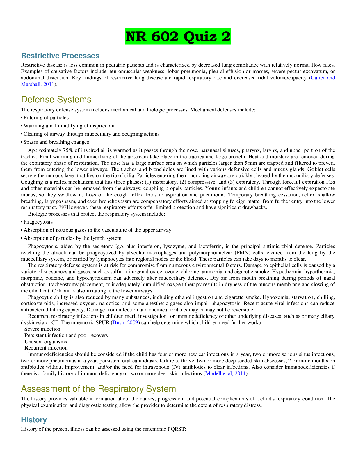





![Preview of WEEK **5 [Chamberlain university]NR 602 IHUMAN Case Week #5 Deborah Arnaudin 54 Year Old](https://browseimages.nyc3.digitaloceanspaces.com/paper-images/2026/03/31/zbQKePwt2026-03-31-11-4669cc328fcad96.png)