OB Exam 2 Study Guide
COMPLICATIONS DURING PREGNANCY:
• Pre-Eclampsia: condition in which HTN develops (>140/90 mmHg) during the last half of pregnancy in a woman who previously had normal blood pressure; renal invol
...
OB Exam 2 Study Guide
COMPLICATIONS DURING PREGNANCY:
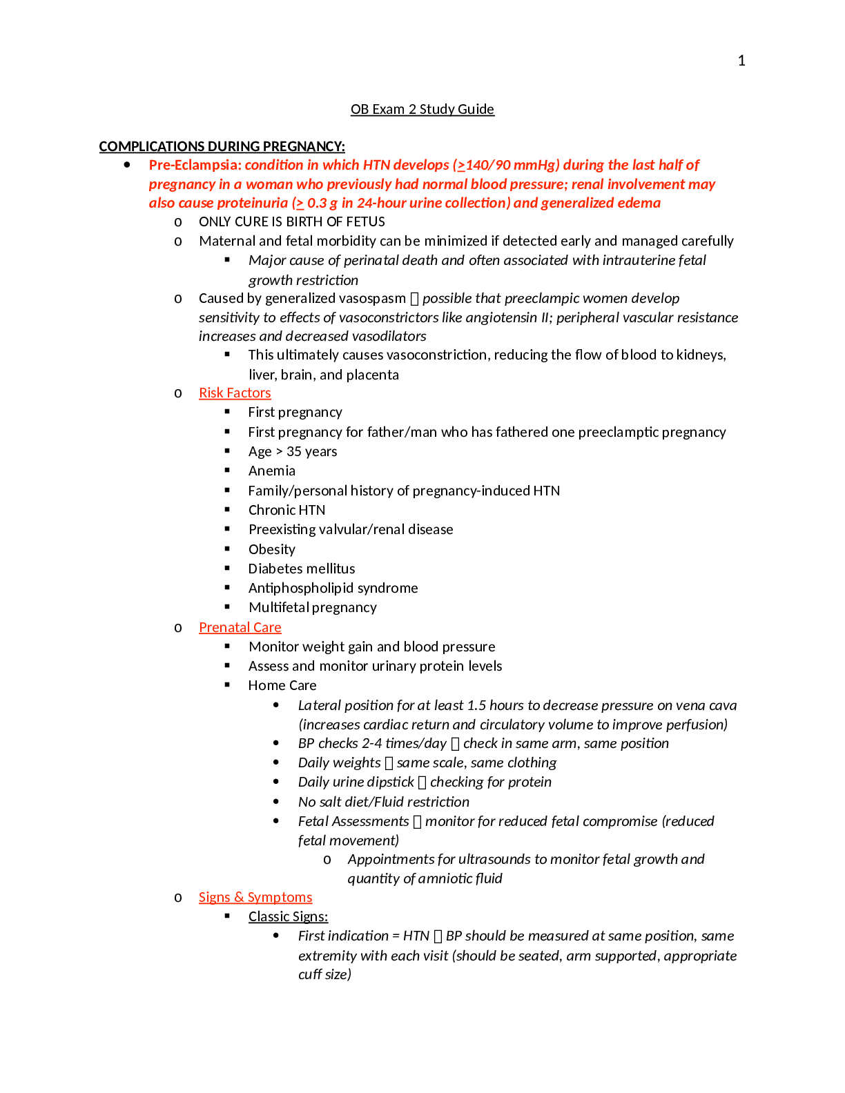
• Pre-Eclampsia: condition in which HTN develops (>140/90 mmHg) during the last half of pregnancy in a woman who previously had normal blood pressure; renal involvement may also cause proteinuria (> 0.3 g in 24-hour urine collection) and generalized edema
o ONLY CURE IS BIRTH OF FETUS
o Maternal and fetal morbidity can be minimized if detected early and managed carefully
▪ Major cause of perinatal death and often associated with intrauterine fetal growth restriction
o Caused by generalized vasospasm possible that preeclampic women develop
sensitivity to effects of vasoconstrictors like angiotensin II; peripheral vascular resistance increases and decreased vasodilators
▪ This ultimately causes vasoconstriction, reducing the flow of blood to kidneys, liver, brain, and placenta
o Risk Factors
▪ First pregnancy
▪ First pregnancy for father/man who has fathered one preeclamptic pregnancy
▪ Age > 35 years
▪ Anemia
▪ Family/personal history of pregnancy-induced HTN
▪ Chronic HTN
▪ Preexisting valvular/renal disease
▪ Obesity
▪ Diabetes mellitus
▪ Antiphospholipid syndrome
▪ Multifetal pregnancy
o Prenatal Care
▪ Monitor weight gain and blood pressure
▪ Assess and monitor urinary protein levels
▪ Home Care
• Lateral position for at least 1.5 hours to decrease pressure on vena cava (increases cardiac return and circulatory volume to improve perfusion)
• BP checks 2-4 times/day check in same arm, same position
• Daily weights same scale, same clothing
• Daily urine dipstick checking for protein
• No salt diet/Fluid restriction
• Fetal Assessments monitor for reduced fetal compromise (reduced fetal movement)
o Appointments for ultrasounds to monitor fetal growth and
quantity of amniotic fluid
o Signs & Symptoms
▪ Classic Signs:
• First indication = HTN BP should be measured at same position, same extremity with each visit (should be seated, arm supported, appropriate cuff size)
• Proteinuria
▪ Additional Signs:
• Vascular constriction and narrowing of small arteries in retina
• Hyperreflexia of DTRs due to decreased brain circulation and edema
• Generalized edema first may manifest as rapid weight gain due to fluid retention, but is usually a nonspecific sign
▪ Symptoms:
• Continuous headache, drowsiness, mental confusion indicates poor cerebral perfusion and are often a precursor to seizures
• Visual disturbances blurred/double vision/spots indicate arterial spasms and retinal edema
• Numbness/tingling of hand/.feet compression of nerves by extra fluid
• Epigastric pain indicate distension of hepatic capsule and may indicate seizure is about to occur
• Decreased urinary output poor perfusion of kidneys and may precede
acute renal failure
▪ Management
• Only cure is delivery MUST OCCUR if any deterioration occurs, either maternal or fetal
o Vaginal birth is preferred
• If < 34 weeks steroids (betamethasone) can be given to accelerate fetal lung maturity and attempt to delay birth for 48 hours
o Severe Preeclampsia >160/110 mmHg and immediate delivery is required
▪ Management
• BR in lateral position
• Quiet, calm environment to reduce incidence of seizures
• Antihypertensives with severe preeclampsia, at higher risk for stroke/CHF
o Hydralazine vasodilator, so increases cardiac output and blood
flow to placenta
o Nifedipine, Labetalol can also be used
o Antihypertensives often contraindication with magnesium sulfate because hypotension can occur, reducing placental perfusion
• Anticonvulsants
o Magnesium sulfate CNS depressant because blocks neuromuscular transmission and decreases amount of acetylcholine
▪ Relaxes smooth muscle, including uterus and reduces vasoconstriction, resulting in moderate BP decrease and promotes circulation
▪ Administered via IV infusion (piggyback) immediate onset
▪ Two RNs to check
▪ May show decreased fetal HR variability but not a cause for concern
▪ Therapeutic level = 4-8 mg/dL magnesium toxicity can occur(respiratory depression)
• Nursing Assessments to monitor for toxicity:
o Serum magnesium levels
o DTRs
▪ Assess for clonus support leg at back of knee and dorsiflex foot to stretch tendon and hold; Clonus will be present if rapid movements are noted
o Respiratory rate/O2 sats
o Urinary output
o Nursing Assessments for Preeclampsia
▪ Weigh on admission/Daily weights
▪ Q4 vitals
▪ Q4 auscultation of lung sounds monitoring for pulmonary edema
▪ Q4 assessment of edema
▪ QHour assessment of urine output Foley if needed
▪ Q4 proteinuria assessment
▪ External fetal monitoring
▪ Assess DTRs report clonus to MD
▪ Assess for headache, visual disturbances, epigastric pain, N&V, or sudden increase in edema
▪ Assess for magnesium toxicity
• Reflexes should be hypotonic but not absent Absent suggests CNS depression and forthcoming respiratory depression
• Respiratory rate and O2 sats
• Monitor for oliguria (<30 mL/hour) Oliguria can cause toxicity, allowing magnesium to build up without adequate voiding
• LOC assessments
▪ Psychosocial assessment may cause anxiety, loss of control, dependent on others, etc.
• HELLP SYNDROME: Hemolysis, Elevated Liver Enzymes, and Low Platelets
o Half of the women affected by HELLP also have severe preeclampsia, but HTN may be absent and may also occur during postpartum phase
o Pathophysiology
▪ Occurs as a result of fragmentation and distortion of erythrocytes during passage through small, damaged blood vessels
▪ Liver enzyme levels increase when hepatic flow is obstructed by fibrin deposits
▪ Hyperbilirubinemia and jaundice can occur due to liver impairment
▪ Low platelets are caused by vascular damage resulting from vasospasm platelets aggregate at sites of damage, resulting in thrombocytopenia, increasing the risk for bleeding, usually in liver
o Symptoms
▪ Pain in RUQ, lower right chest, or mid-epigastric area
▪ Tenderness due to liver distension
▪ Nausea and vomiting
▪ Severe edema
▪ Avoid abdominal palpation and use care in transporting to avoid traumatizing the liver
• Hepatic rupture can lead to fetal and maternal mortality
▪ Sudden increase in intrabdominal pressure leads to rupture of subscapular hematoma, resulting in internal bleeding and hypovolemic shock
o Management
▪ Magnesium sulfate to control seizures
▪ Hydralazine to control BP
▪ IV Fluids to avoid worsening of reduced intravascular volume, but do not give to much which could cause pulmonary edema
▪ Cervical ripening and labor induction if > 34 weeks
• Induction is preferred to avoid bleeding and clotting complications that accompany C-sections and anesthesia is often difficult due to edema; low platelets may reduce safety of epidurals/spinals
• C-section preferred if unfavorable cervix or far from term
▪ Labor is delayed if < 34 weeks if mother is stable
• Can give antenatal steroids (betamethasone) to accelerate fetal lung maturity
• Hypertension Disorders in Pregnancy:
o Gestational Hypertension: > 140/90 mmHg that develops after 20 weeks of pregnancy, but return to normal within 6 weeks postpartum; Proteinuria is not present
o Chronic Hypertension: >140/90 mmHg that existed before pregnancy or develops before 20 weeks gestation; Also diagnosed if HTN does not resolve during postpartum period
▪ Risk Factors
• Older women (> 35 years/late childbearing age)
• Obese
• Diabetes
• African-American women
• Secondary to another disease process (renal disease/autoimmune disorder)
▪ Management
• Monitor BP, proteinuria to due increased risk of development of preeclampsia
• Consult dietician increase protein, reduce salt
• Frequent prenatal visits regular fetal surveillance
• Antihypertensive either continue current meds or Methyldopa
o ACE inhibitors not recommended during pregnancy, but ok during postpartum
o Avoid diuretics because may decrease blood volume, which may already be reduced if preeclamptic
▪ Thiazides are safe during pregnancy
• Rh Incompatibility
o Possible if:
▪ Mother is Rh-negative
▪ Fetus is Rh-positive (thus father is Rh-positive)
o Pathophysiology
▪ When Rh-positive blood enters Rh-negative bloodstream, blood reacts as if it is a foreign substance and develops antibodies against “invading” antigen to destroy the Rh antigen, the whole RBC must be destroyed
▪ Most exposure of maternal blood to fetal blood occurs during third stage of labor, when active exchange of fetal and maternal blood may occur from damaged placental vessels
• First child is not usually affected because antibodies are formed after birth of infant and subsequent fetuses may be affected unless mother receives RhoGAM to prevent antibody formation
▪ Antibodies to Rh factor cross placental and destroy fetal RBCs and thus fetus becomes deficient, which are needed to transport O2 to fetal tissues ERYTHROBLASTOSIS FETALIS
• Fetal bilirubin increases, leading to neurological disease like staining of brain tissue (kernicterus) and bilirubin encephalopathy)
• Results in rapid production of erythroblasts (immature RBCs) which can’t carry O2
• Fetus becomes so anemic that generalized edema results and can end in fetal CHF
o Prenatal Assessment and Management
▪ Initial prenatal visit blood test
• Rh-negative indirect Coombs test to determine whether they are sensitized (have developed antibodies) as a result of previously exposed Rh-positive blood
o If Coombs is negative = test repeated at 28 weeks to detect for subsequent sensitization
o If Coombs is positive = repeated at frequent intervals throughout pregnancy to determine whether the antibody titer is increasing
▪ Increased titer fetal compromise will occur
o RhoGAM
▪ Administered to unsensitized Rh-negative mother at 28 weeks, IM, to prevent sensitization
• Effectively prevents formation of active antibodies against Rh-positive erythrocytes if small amount of Rh-positive fetal blood enters maternal circulation
▪ Repeated IM administration after birth if newborn is determined to be Rh- positive (within 72 hours of delivery)
• Umbilical cord is taken at delivery to determine newborn blood type, Rh
factor, and antibody titer (Coombs)
• Gestational Diabetes
o Risk Factors women with any of these risk factors should be screened for type 2/gestational diabetes at first prenatal visit
▪ Overweight or obese
▪ Maternal age > 25 years
▪ Previous birth outcome associated with GDM (macrosomia, maternal HTN, infant with unexplained congenital abnormalities, previous fetal death)
▪ History of abnormal glucose tolerance test
▪ Family history of diabetes (first-degree relative)
▪ High risk ethnic group (African-American, Hispanic, Asian American, American Indian, Pacific Islander)
o Screening
▪ Glucose Challenge Test administered between 24-28 weeks in both low and high risk women
• Fasting is not necessary
• Women ingests 50g of oral glucose solution and blood sample is taken 1 hour later
o If > 140 mg/dL 3 hour oral glucose tolerance test is recommended
▪ Oral Glucose Tolerance Test gold standard for diagnosis
• Must fast from midnight on day of test
• Should ingest 100g of oral glucose solution and plasma glucose levels are determined at 1, 2, and 2 hours
• Diagnosis is confirmed if fasting blood glucose or if two of the following
values occur:
o Fasting = > 95 mg/dL
o 1 hour = > 180 mg/dL
o 2 hour = > 155 mg/dL
o 3 hours = > 140 mg/dL
o Maternal Risks
▪ In first trimester increased fetal malformations
▪ Increased preeclampsia risk
▪ Increased risk of UTIs
▪ NO increase in spontaneous abortions/ketoacidosis like in normal DM
o Fetal Risks
▪ Major Complications:
• Macrosomia leads to birth injuries/C-section
• Neonatal hypoglycemia
• Congenital malformations
▪ Other:
• Hypocalcemia
• Hyperbilirubinemia
• Respiratory distress
o Management
▪ Diet Consult with dietician, dietary technician, and diabetes educator
• Eliminate concentrated sweets (simple sugars)
• Non-obese = 30 kcal/kg/day
• Obese = 25 kcal/kg/day
▪ Exercise
▪ Blood Glucose Monitoring fasting and postprandial levels should be checked
• If fasting is > 95 mg/dL or postprandial is > 120 mg/dL, insulin therapy is started
▪ Fetal Surveillance may begin as early as 28 weeks if poor glycemic control/ 34
weeks in low-risk
• “Kick counts” fetal movements felt by mother
• Ultrasound for fetal growth and amniotic fluid volume
• Non-Stress Test, Contraction stress test
• Amniocentesis for fetal lung maturity
• Cardiac Disease in Pregnancy affects about 1% of pregnancies
o Alterations in cardiovascular function are necessary in pregnancy to meet additional maternal demands and needs of fetus
▪ Plasma volume, venous return, cardiac output, heart rate, stroke volume all increase
o For women with preexisting conditions/underlying heart disease, these changes can
cause an additional burden on an already compromised heart resulting in cardiac decompensation of CHF
o Rheumatic Heart Disease complication that often occurs after streptococcal pharyngitis, causing scarring of heart valves, resulting in stenosis (narrowing)
▪ Mitral stenosis = most common
• Obstructs blood from left atrium to left ventricle, causing left atrium to become dilated
• Dilation causes pressure in left atrium, pulmonary veins, and pulmonary
capillaries, leading to pulmonary HTN, edema or CHF
▪ Warning signs persistent rales at lung bases, dyspnea on exertion, cough, hemoptysis, progressive edema, tachycardia
o Congenital Heart Disease fetus is more likely to inherit these, as well as affecting the
mother with complications occurring throughout pregnancy
▪ Left-to-Right Shunts (Acyanotic)
• Atrial Septal Defectoften first discovered because symptoms are absent/vague
o Pressure on left side of heart is higher than right side
o Pregnancy is well tolerated and no specific treatment is recommended
o Pulmonary HTN occasionally develops in untreated ASD because additional blood moves to right side of heart and is transported to lungs
• Ventricular Septal Defect more common; usually detected by auscultation and often close with no surgical intervention
o Most women are asymptomatic, but fatigue or symptoms of pulmonary congestion may occur
o Pregnancy is well-tolerated in small to moderate shunts
o Pregnancy occasionally precipitates heart failure/dysrhythmia
o Bacterial endocarditis is common in unrepaired defects and antibacterial prophylaxis is recommended
• Patent Ductus Arteriosus physiological effects are related to size
o Small may be well tolerated during pregnancy, unless complicated by pulmonary HTN
o Tends to become infected antibacterial prophylaxis is recommended before labor
▪ Right-to-Left Shunts (Cyanotic)
• Tetralogy of Fallot combination of 4 defects (VSD, pulm. Valve stenosis, right ventr. Hypertrophy, and displacement of aorta towards right ventricle)
o Women who have undergone repair and have no cyanosis
tolerate pregnancy well
o Untreated high maternal and fetal mortality
• Eisenmenger’s Syndrome develops when pulmonary resistance equals or exceeds systemic resistance to blood flow and a right to left shunt develops
o Late surgical correction often results in woman’s death
o If survive, pregnancy may carry 50% maternal risk, usually from ventricular failure
o Most newborns are preterm and small for gestational age
o Mitral Valve Prolapse may be inherited, or accompany other conditions, such as atrial septal defects and Marfan syndrome – leaflets of mitral valve prolapse into left atrium during ventricular contraction
▪ Considered benign and most women are asymptomatic and tolerate pregnancy well
▪ Some may experience chest pain and dysrhythmias give beta-blockers, like atenolol and metoprolol
▪ Some MDs may consider it a significant risk for bacterial endocarditis and administer prophylactic antibiotics before and during labor
o Peripartum/Postpartum Cardiomyopathy rare condition exclusively associated with pregnancy after exclusion of other causes; have no underlying heart disease, but symptoms are cardiac decompensation appear during last weeks of pregnancy/2-20 weeks postpartum
▪ Symptoms of CHF dyspnea, edema, weakness, chest pain, palpitations
▪ Anticoagulation with low-molecular weight heparins is usually therapy to prevent clot formation during pregnancy when coagulations factors are higher
▪ Fluid restriction to reduce pulmonary edema and treatment of CHF are usually required
▪ Sudden appearance/Abrupt downhill course = occurs in 20% of women and only transplant can save the woman
▪ Often occurs with subsequent pregnancies, often in women who do not have complete recovery of left ventricle
o Therapeutic Management
▪ Mild to Moderate Heart Disease:
• Limit physical activity to remain free of dyspnea, chest pain, tachycardia
• Avoid excessive weight gain to decrease demands on heart; low
sodium diet may be advised
• Prevent anemia anemia will decrease O2-carrying capacity of blood and results in decompensation; most is treated by taking folic acid
• Prevent infection immunization required; administration of
prophylactic antibiotics
• Undergo careful assessment for CHF, pulmonary edema, and cardiac dysrhythmias monitor for rales at lung bases, cough, dyspnea, hemoptysis, increasing edema, tachycardia
▪ Severe Heart Disease:
• Primary goal = prevent decompensation and development of CHF
• Protect fetus from hypoxia and intrauterine growth restriction
• Same as above PLUS bed rest, especially during last trimester, prophylactic anticoagulation therapy/TEDs, SCDs
o Medications
▪ Anticoagulants
• Warfarin (Coumadin) CONTRAINDICATED DURING PREGNANCY – associated with fetal malformations
o OK to use for postpartum anticoagulation
• Subcutaneous/IV heparin does NOT cross placental barrier
o Monitor aPTT, PTT, and platelets throughout therapy
o Withheld during labor and resumed 6 hours after birth/18-24 hours after C-section
• Subcutaneous Enoxaparin (Lovenox) – LMWH – may be used instead of standard heparin because it requires less frequent monitoring for complications
▪ Antidysrhythmics
• Beta-blockers may be used to control maternal HTN
o Have been associated with neonatal respiratory depression, sustained bradycardia, hypoglycemia when administered late in pregnancy or just before delivery
o Atenolol and metoprolol may be preferred because do not cause uterine stimulation that other beta-blockers may cause
• Digoxin, adenosine, and calcium channel blockers also appear to be safe
▪ Anti-infectives amoxicillin, penicillin, gentamycin at delivery; ceftriaxone, vancomycin can also be given
▪ Heart Failure Drugs
• Diuretics may be needed when CHF remains uncontrolled in spite of restriction of activity/sodium intake
o Must continue to monitor electrolytes and water balance
o Thiazide diuretics are commonly used
• Beta-blockers, ACE inhibitors, angiotensin receptor blockers may also be used for pregnancy-associated HF
o Intrapartum Management
▪ With every contraction, 300-500 mL of blood is shifted from uterus and placenta into central circulation
• Extra fluid causes sharp rise in cardiac workload
▪ Careful management of IV fluid administration is essential to prevent fluid overload
▪ Position woman on side, with head and shoulders elevated
▪ O2 can be administered to increase O2 saturation and is monitored with POX
▪ Keep a calm, quiet environment to decrease anxiety and tachycardia
▪ Epidural block may be contraindicated because of potential hemodynamic effects
▪ Fetal compromise/maternal decompensation should be reported immediately
▪ Vaginal birth is often recommended
• Vacuum extraction/forceps are often used to minimize maternal pushing, avoid use of Valsalva maneuver, and to limit prolonged labor
▪ C-section may be chosen for obstetric indications
• Must discuss added stress of major surgery on heart
• Expected blood loss is higher than in vaginal birth
• General anesthesia may be required over epidural, leading to airway management by anesthesiologist
▪ Fourth stage of labor proposes special risks
• After placental delivery, about 500 mL of blood is returned to intravascular volume
• To minimize overloading the heart, positional changes should be avoided
• Uterus should not be massaged to expedite separation of placenta
• Careful assessment of signs of fluid overload, such as bounding pulse, distended neck and peripheral veins, and moist rales in lungs should be done throughout labor and postpartum periods
POSTPARTUM:
• Postpartum Hemorrhage major cause of maternal death and morbidity in the US and world; in US, complicates 1-5% of births
o Blood loss > 500 mL after vaginal birth or 1000 mL after C-section, a decrease in hematocrit > 10% since admission, or the need for a blood transfusion
o Early Postpartum Hemorrhage occurs in first 24 hours after delivery
▪ Usually occurs during 1st hour after delivery and most often caused by uterine atony
▪ Risk Factors/Causes
• Overdistension of uterus (multiple gestation, large infant, hydramnios)
• Multiparity (five or more)
• Precipitate labor/delivery
• Prolonged labor
• Use of forceps/vacuum extraction
• C-section
• Manual removal of placenta
• Uterine Inversion
• Placenta Previa, placenta accrete (adherence to uterine wall), low implantation
• General anesthesia
• Chorioamnionitis
• Clotting disorders
• Previous postpartum hemorrhage or uterine surgery
• Disseminated intravascular coagulation
• Uterine leiomyomas (fibroids)
▪ Uterine Atony refers to lack of muscle tone that results in failure of uterine muscle fibers to contract firmly around blood vessels when the placenta separates
• Relaxed muscles allow rapid bleeding from endometrial arteries at the placental site and bleeding continues until uterine muscle fibers stop flow of blood
• Risk Factors
o Overdistension
o Multiparity
o Obesity
o Ineffective contractions/prolonged labor
o Vigorous contractions/precipitate labor
o Labor augmented/induced with oxytocin
o Retention of part of placenta
• Manifestations
o Fundus that is difficult to locate
o Soft/” boggy” fundus
o Uterus that becomes firm as its massaged but loses tone when massage is stopped
o Fundus that is located above expected level
o Excessive lochia, especially if bright red (soaking pad in 15 mins)
o Excessive clots in lochia with/without uterine massage
• Management
o 1st = massage the fundus! to contract uterine muscle fibers/express clots
▪ DO NOT MASSAGE IS FUNDUS IS FIRM AND CONTINUED BLEEDING
o If fundus is displacement encourage mother to
void/catheterize if necessary and note urine output
o Pharmacologic Measures:
▪ IV Pitocin rapid infusion will increase uterine tone and control bleeding
▪ Methylergonovine (Methergine) can be given IM but elevates BP and should not be given if woman is hypertensive
▪ Prostaglandin 2a analogs effective when given IM or into uterine muscle if oxytocin is ineffective
▪ Prostin (dinoprostone) or misoprostol (Cytotec) can be
given rectally to control bleeding
o If massage/pharmacologic measures are ineffective = MD/midwife will use bimanual compression of uterus
▪ Uterine packing can also be done
▪ Possible need to return to OR to remove placental fragments
▪ Laparotomy used to identify source of bleeding – uterine compression sutures may be placed to stop severe bleeding or ligation of arteries
▪ Hysterectomy is last resort
o Prompt replacement of fluids is required Lactated Ringer’s solution, whole blood/packed RBCs, normal saline, or other plasma extenders can be used
▪ Should be given to maintain urine flow of 30-60 mL/hour
▪ Trauma
• Lacerations should always be suspected if excessive uterine bleeding continues when fundus is contracted firmly and is at expected location
o DO NOT MASSAGE
o Bleeding is often bright red and may be heavy or appear to be a steady trickle
• Hematomas maybe be visible (valvular) or invisible (vaginal/perineum)
o Produce deep, severe, unrelieved pain and feelings of pressure that are not relieved by usual pain measures
o Should be suspected if mother demonstrates systemic signs of concealed blood loss, such as tachycardia, decreasing BP, and when fundus is firm and lochia is within normal limits
• Management Surgical repair is often necessary
o Late Postpartum Hemorrhage occurs after 24 hours after delivery or up to 6-12 weeks after birth
▪ Most common cause = subinvolution delayed return of uterus to its nonpregnant size and consistency and placental fragments remain attached to myometrium when placenta is delivered
• Clots form around retained fragments and excessive bleeding can occur when clots slough away several days after delivery
• Infection can also be a cause
▪ Retaining of placental fragments may be preventable
• Proper inspection to determine if placenta is intact
• If portions mission, HCP should manually explore uterus to locate and remove mission fragments
▪ Secondary Postpartum Hemorrhage hemorrhage occurring between 24 hours and 6 weeks after birth
• Usually occurs after discharge should be taught how to assess fundus and normal characteristics and duration of lochia flow
▪ Risk Factors
• Attempts to deliver placenta before its separation from uterine wall
• Manual removal of placenta
• Placenta accrete
• Previous C-section
• Uterine leiomyomas (fibroids)
▪ Management
• Initial treatment CONTROL BLEEDING
• Oxytocin, Methylergonovine, and prostaglandins are most commonly used
• Ultrasound can be used to identify location of retained placental fragments
• Dilation and Curettage – stretching of cervical os to permit suctioning/scraping of wall of uterus may be necessary to remove fragments
• Broad-spectrum antibiotics can be given if postpartum infection is suspected because of uterine tenderness, foul-smelling lochia, or fever
▪ Hypovolemic Shock
• Normally, woman can tolerate blood loss that is similar to amount of blood added during pregnancy (1500-2000 mL)
o If anemic before birth = less reserve
• Amount of blood lost can be estimated by comparing pre and post hematocrit
o If lower after delivery, woman lost amount of blood added during pregnancy and an additional 500 mL for each 3% drop in hematocrit value
• Signs and Symptoms
o Early sign tachycardia
o Late signs hypotension – usually does not occur until 20-25% of blood volume has been lost; CNS changes, confusion, lethargic, decreased urine output
▪ Narrowing pulse pressure should also be noted
▪ Increased respirations due to anxiety and increased oxygen demands
▪ Vasoconstriction causes pallor, cold, clammy
• Management
o CONTROL BLEEDING AND PREVENT FROM BECOMING IRREVERSIBLE
o Insert secondary large bore IV if need to transfuse and monitor I/Os
o Vasopressors for low BP
o Hemorrhage Medications
▪ Oxytocin (Pitocin) stimulates uterine contractions; given IV
• Action: stimulates uterine smooth muscle, resulting in increased strength, duration, and frequency of contractions
o Sensitivity will increase with continued use makes it less
effective!
• Indications: tachysystole; controls postpartum bleeding
• Contraindications: Placenta Previa, vasa Previa, nonreassuring FHR, abnormal fetal presentation, prolapsed cord, presentation above pelvis,
previous classic/fundal incision, active genital herpes, pelvic deformities, cervical carcinoma
• Side Effects: tachysystole (fetal bradycardia/tachycardia, reduced variability, late decelerations), impaired uterine blood flow, uterine rupture, abruption placentas
• Assess uterus for firmness and height
• Assess lochia
• Assess VS Q15 minutes
▪ Hemabate (carboprost tromethamine) stimulates uterine contractions and causes arterial vasoconstriction; given IV or oral
• Action: stimulates contraction of uterus vasoconstrictor
• Indication: treatment of postpartum hemorrhage caused by uterine atony
• Contraindications: PID; cardiac, pulmonary, renal, or hepatic disease; Caution in history of asthma (causes vasoconstriction), hypo/hypertension, anemia, jaundice, diabetes, epilepsy, previous uterine surgery
▪ Methergine (methylergonovine)
• Action: stimulates sustained contraction of uterus and causes arterial vasoconstriction
• Indication: used for the prevention and treatment of
postpartum/postabortion hemorrhage caused by uterine atony/subinvolution
• Contraindications: HTN, severe hepatic/renal disease, thrombophlebitis, CAD, PVD, hypocalcemia, sepsis, before 4th stage of labor
• Side Effects: N&V, uterine cramping, hypertension, dizziness, headache, chest pain
• Assess BP before administration
▪ Cytotec
• Action: stimulates uterine contractions; given vaginally or rectally
• Subinvolution slower-than-expected return of uterus to its nonpregnant size after childbirth
o Causes
▪ Retained placental fragments
▪ Pelvic Infection
o Signs and Symptoms
▪ Prolonged discharge of lochia
▪ Irregular/excessive uterine bleeding
▪ Profuse Hemorrhage
▪ Pelvic pain/feelings of heaviness
▪ Backache
▪ Fatigue/Persistent malaise
▪ Uterus feels larger and softer than normal than expected
o Management correct cause of subinvolution
▪ Methergine may be given rally to provide long, sustained contractions of uterus
▪ Infection responds to antimicrobial therapy
▪ EDUCATION usually does not occur until other has been discharged
• How to assess fundus (firm vs. boggy/height should decrease by 1 fingerbreadth each day)
• Lochia should decrease in color and amount (no odors or excessive
clots)
• Know signs and symptoms that need to be reported immediately
• Puerperal Infections bacterial infections that occur after childbirth
o Most Common:
▪ Endometritis – infection of inner lining of uterus occur within 36 hours after birth
• Fever (> 100.4), chills, malaise, anorexia, abd. Pain/cramping, uterine
tenderness and purulent, foul-smelling lochia, tachycardia, subinvolution
• Management
o Q2 vitals when febrile; Q4 when afebrile
o IV antibiotics for 24 hours broad spectrum (cephalosporins, clindamycin/gentamycin, ampicillin)
o Monitor lochia
o Antipyretics
o Methylergonovine to increase lochia drainage and promote involution
▪ Wound Infections incisions, lacerations, episiotomy
• Risk factors obesity, diabetes, hemorrhage, anemia, chorioamnioitis, corticosteroids, multiple vaginal exams
• REEDA redness, edema, ecchymosis, discharge, approximation
• After cultured, antibiotics should be given
• Antipyretics/analgesics as necessary
• Surgical debridement is sometimes necessary
▪ UTIs bladder and urethra are hypotonic, with urinary stasis and retention as common problems, along with bacteria in tract
• S/S of burning, frequency, odor, urgency, dysuria, suprapubic pain,
possible hematuria, low-grade, chills; may escalate to pyelonephritis/cystitis
• Treat at home with antibiotics and analgesics like pyridium
• Encourage fluids to prevent
▪ Mastitis – infection of breast often occurs 2-4 weeks postpartum and can developing any time during breastfeeding; usually only affects one breast
• Often caused by Staph. Aureus, E. coli or strep bacteria usually carried on skin of mother or in mouth/nose of newborn
• May enter nipple through cracks/blisters
• Engorgement and stasis of milk may precede mastitis
o Can occur when feeding is skipped, when infant begins to sleep through the night, when breastfeeding is suddenly stopped, too- tight bra, mother is fatigued/stressed
• S/S fatigue, aching muscle, temp > 102.2, chills, malaise, headache, localized lump/wedge-shaped area of pain, redness, heat, inflammation, and enlarged axillary lymph nodes (hard, tender area can be palpated)
o Can progress to abscess
• Management
o Antibiotic therapy
o Continued emptying of breast through breastfeeding/pumping
Q2 hours
o Moist heat
o Breast support (nursing bra)
o Bed rest
o Fluids
o Analgesics
▪ Septic Pelvic Thrombophlebitis often occurs 2-4 days postpartum occurs when infection spreads along venous system and thrombophlebitis develops
• Develops more often in women with wound infection and usually
involves one or both ovarian veins
• Primary symptom pain in groin, abdomen, flank
o Fever, tachycardia, N&V, bloating, GI distress, decreased bowel sounds
o Spiking fever that does not respond to antibiotics and tachycardia in a woman who was treated for endometritis previously
• CBC with differential, blood chemistries, coagulation studies, and cultured can be used to confirm diagnosis
o CT, MRI, pelvic ultrasound also helpful
• Management
o Readmission is usually necessary
o Primary treatment anticoagulation with IV heparin and IV antibiotics
o Warfarin can be given when heparin is discontinued
o Improvement usually occurs within 2-3days of therapy
• Postpartum Mood Disorders:
o Postpartum Depression period of depression with onset during pregnancy or within 4 weeks after childbirth that lasts at least 2 weeks; affects 10-15% of postpartum women
▪ Includes persistent depressed mood/loss of interest in almost all activities, changes in appetite/weight, sleep, and psychomotor activity; decreased energy; feeling of worthlessness/guilt; difficulty thinking, concentrating, or making decisions; recurrent thoughts of death/suicide; death/suicide plans/attempts
• Symptoms last for at least a 2-week period and tend to become worse over time
• Creates strain on family and support system communication is impaired and gradually withdraws
• Appear tense, irritable, feel less competent mothers, less likely to provide health sleep and eating practices
▪ Management
• Combination of psychotherapy, social support, and medication
• If combination is not solely effective medication should be considered (SSRIs, tricyclic antidepressant)
o May take up to 4 weeks to become effective and may be continued for 9-12 months
o Must considered if woman is pregnant/breastfeeding
o Baby Blues transient, self-limit mood disorder; mild depression
▪ Affects 60-80% of postpartum women
▪ Begins in 1st week, peaks around day 5, and ends within 2 weeks
▪ Characterized by insomnia, fatigue, tearfulness, mood instability, and anxiety and do not affect mother’s ability to care for infant
▪ Benefit greatly when empathy and support are freely given by family and health acre team
• Encouraged to rest, take time for herself, and discuss feelings
o Postpartum Psychosis mental state in which person’s ability to recognize reality, communication, and relationship to others is impaired
▪ Can either be depressed psychosis or manic psychosis
▪ Can occur as early as 2 days after delivery and is a psychiatric emergency, requiring hospitalization
▪ S/S agitation, irritability, rapidly shifting moods, disorientations, disorganized
behavior, delusions/hallucinations
▪ Women who have had one episode of psychosis are at increased risk for having another episode
▪ Management requires hospitalization, pharmacologic treatment, and psychiatric
care
• Breastfeeding
o Composition
▪ Colostrum – forms during pregnancy (can occur at 16 weeks) and for use immediately after birth
• Low quantity, high quality
• Baby only needs 2-3 teaspoons
▪ Transitional Milk – occurs 2-3 days after birth and can continue for up to 10 days; like 2% milk; pale yellow
▪ Mature milk – replaces transitional milk – bluish in color, thinner; 20 kcal/oz.; sufficient to meet infant’s nutritional needs
o Benefits
▪ Infant
• Less likely to develop allergies
• Infection prevention/decreased incidence
• Lower incidence of obesity, diabetes, SIDS
• Composition meets specific nutritional needs and will change accordingly
• Easily digested
• Protein, fat, and carbs in appropriate proportions
• Unlikely to be contaminated
• Less likely to overfeeding
• Constipation less likely
• Decreases incidence of jaundice
▪ Mother
• Oxytocin enhances uterine involution
• Less blood loss
• Resuming of ovulation delayed (but not good birth control method)
• Decreased cancer risk
• Burns calories faster weight loss
• Skin-to-skin contact regulates baby’s temperature, heart rate, and respirations
• Convenient and free
• Infant less likely to be ill
o How Often? 1.5 -3 hours; 8-12 feedings in 24 hours
o How Long? Infant may establish; 10-15 minutes on each side; up to 20 mins
o How DO I know if Baby is getting anything? Swallowing, sucking, “Ka/ah” sounds when swallowing
o Positioning
▪ Hands should be C-shaped – breast should fall forward
▪ Erect nipples are best for breastfeeding; Other types require lactation consultation
▪ Latch – fish lips, wide open, lips puckered, whole areola should be in mouth, nipple should be in back of mouth
• Breaking latch place pinky in side of mouth
▪ Cradle Hold use blankets/pillows to support arm, which may tire from holding
▪ Football/Clutch Hold allows mom to see position of infant’s mouth on breast, helps control head and helpful in mothers with heavy breasts; avoids pressure against abdominal incision
▪ Cross-Cradle/Modified Cradle Hold good for preterm infants/shoulder dystocia
– guides infant’s head to breast and see mouth on breast during feedings
▪ Side-Lying avoids pressure on episiotomy or abdominal incisions and allows mother to rest while feeding; Pillows behind back/between legs provide comfort
o How Do You Know Baby is Getting Enough? how many wet diapers? - should be 6-
8/day
o Engorgement
▪ Prevention if breastfeeding breastfeed frequently, minimum Q2 hours
• Treatment = heat packs, ump/breastfeed, pain meds, nursing bra
▪ Treatment if bottle-feeding ice packs, avoid stimulation (no hot showers), pain meds, tight fitting bra
• Postpartum Lab Values SEE HANDOUT
NEWBORN/NEWBORN COMPLICATIONS:
• APGAR method for rapid evaluation of infant’s cardiorespiratory adaptation after birth; the nurse scores the infant at 1 minute and 5 minute in each of the 5 areas
o Each assessment is arranged from most important (heart rate) to least important (color)
and scores are totaled in each area
o Resuscitation should not be delayed until the 1-minute score is obtained
o General guidelines for infant’s care are based on 3 ranges of 1-minute score: 0-2, 3-6; 7- 10
o Heart Rate
▪ 0 = absent
▪ 1 = <100 bpm
▪ 2 = > 100 bpm or higher
o Respiratory Effort
▪ 0 = no spontaneous respirations
▪ 1 = Slow respirations/weak cry
▪ 2 = Spontaneous respirations/Strong, lusty cry
o Muscle Tone
▪ 0 = Limp
▪ 1 = Minimal flexion of extremities; Sluggish movement
▪ 2 = Flexed body posture; Spontaneous and vigorous movement
o Reflex Response
▪ 0 = No response to suction/gentle slap on soles
▪ 1 = Minimal response (grimace) to suction or gentle slap on soles
▪ 2 = Responds promptly to suction or gentle slap to sole with cry/active movement
o Color
▪ 0 = Pallor/cyanosis
▪ 1 = Bluish hands and feet only
▪ 2 = Pink (light skinned) or absence of cyanosis (dark skinned); Pink mucous membranes
• Initiation of Infant Breathing
o Development of Lungs
▪ Fetal life fetal lung fluid expands alveoli and is essential for normal development of lungs and is continuously produced until fetus nears term
• When nears term, fetal lung fluid decreases in preparation for birth,
when fluid must be cleared for infant to take in air
• Absorption of this fluid begins in early labor (moves into interstitial spaces) and is accelerated by secretion of fetal epinephrine and corticosteroids
▪ Surfactant combination of lipoproteins – is detectable around 24-25 weeks
• Lines inside of alveoli and reduces surface tension within alveoli, allowing alveoli to remain partially open when infant begins to breathe in air at birth
• 34-36 weeks enough surfactant is present to prevent respiratory distress/lung collapse
• Secretion increases during labor and immediately after birth to allow spontaneous respirations to occur
▪ Can give steroids, like betamethasone/dexamethasone to help speed up fetal lung maturation/surfactant production
• Example in cases such as intrauterine growth restriction, maternal HTN, heroin addiction, preeclampsia, infection, placental insufficiency, premature rupture of membranes
o Causes of Respirations
▪ First breath must force remaining fluid out of alveoli and into interstitial spaces around alveoli so air can enter lungs why suctioning is often done at birth, since higher pressure is required
▪ Chemical Factors
• Chemoreceptors in carotid arteries and aorta respond to changes in blood caused by hypoxia that occurs with birth
• Decreased PO2 and pH/Increased PCO2 in blood causes receptors to
stimulate respiratory center in medulla
• Occlusion of cord vessels halts respirations and causes diaphragm to contract to bring air into lungs
▪ Mechanical Factors
• Fetal chest is compressed during delivery
• 1/3 of lung fluid is forced out into upper passages and is suctioned out during birth
• When pressure from delivery is released, chest recoils to draw air into
lungs and helps removes viscous fluid
▪ Thermal Factors
• Infant moves from warm, fluid-filled environment into cooler environment
• Sensors on skin respond to sudden change in temperature by sending impulses to medulla that stimulate respiratory center and breathing
▪ Sensory Factors drying infants and swaddling t increase thermal factors; skin to skin contact
o Continuation of Respirations
▪ As alveoli expand, surfactant allows them to stay partially open between respirations
• Because alveoli remain open, subsequent breaths require much less effort
▪ As infant cries, pressure in lungs increases, causing remaining fluid to move into interstitial spaces, where it is absorbed by the cardiopulmonary/lymphatic systems
• Often why lungs sound moist when first auscultated but become clear short time afterward
o Order of Events in Cardiopulmonary Adaptation:
1. Increased blood CO2 level
2. Respirations started
3. Surfactant keeps alveoli open
4. Increased blood oxygen level
5. Ductus arteriosus constricts
6. Increased pressure on left side of heart
7. Foramen ovale closes
8. Fibrosis of ductus venosus
• Methods of Heat Loss
o Evaporation air-drying of skin that resulting in cooling
▪ Drying the infant, especially head, ASAP helps prevent heat loss this way
▪ Insensible water loss from skin and respiratory tract increases heat loss from evaporation
o Conduction movement of heat away from body occurs when newborns have direct contact with objects that are cooler than their skin
▪ Placing infants on cold surfaces or touching them with cool objects causes this type of heat loss
▪ Contact with warm objects increases body heat warming objects before toughing infant or skin-to-skin contact helps prevent conductive heat loss
o Convection transfer of heat from the infant to cooler surrounding air occurs during convection
▪ When infants are in incubators, the circulating warm air helps keep them warm
▪ Providing a warm, draft-free environment avoids convective heat loss
o Radiation transfer of heat to cooler objects that are NOT in direct contact with the infant
▪ Infants in incubators transfer heat to the walls of the incubator
▪ If the walls of the incubator are cold (near a draft, placing something cold on top), the infant is cooled even when the inside temperature of the incubator is warm
▪ Incubators have double walls to prevent this and they should be placed away from windows and outside walls
▪ Newborns can gain heat by radiation through radiant warmers
• Cold Stress
o Nonshivering thermogenesis metabolism of brown fat to produce heat and blood that is passed through brown fat is warmed and carried throughout the body
▪ Begins when thermal receptors in skin detect 35-36 (95-96.8)
▪ Norepinephrine is released in brown fat, stimulating its metabolism
▪ Often starts before change in core temperature occurs (rectal temperature reading)
▪ Brown fat is primarily accumulated during third trimester
• Preterm infants may not have adequate stores
• Hypoxia, hypoglycemia, and acidosis may interfere with infant’s ability to use brown fat to generate heat and are not able to raise their body temperature, causing cold stress
o Increased metabolic rate and metabolism of brown fat that result from cold stress can cause a significant rise in the need for oxygen
▪ Also causes a diminished production of surfactant, obstructing lung expansion and leading to more respiratory distress
▪ Glucose is necessary in larger amounts when metabolic rate rises to produce
heat
• Metabolism of brown fat releases fatty acids, resulting in metabolic acidosis
• Can also interfere with transport of bilirubin, increasing the risk of
jaundice
▪ When infant is trying to conserve heat, vasoconstriction of peripheral vessels occurs to reduce heat loss from skin surface
• Decreased O2 in blood may also cause vasoconstriction of pulmonary
vessels, leading to further respiratory distress
• Normal NB Vitals and Lab Values
o Vitals
▪ HR = 110/120 – 160 bpm
▪ RR = 30-60 breaths/min
▪ Temperature = 36.5 – 37.5 (97.7-99.5), axillary
o Labs
▪ RBCs = 4.8-7.1 mil/mcL
▪ HgB = 15-24 g/dL
▪ HCT = 44-70%
▪ WBCs = 9.1 – 34
▪ Platelets = 84-478 thousand/mm3
▪ Serum glucose:
• Cord = 45-96 mg/dL
• 1-day old = 40-60
• > 1-day old = 50-90
▪ Bilirubin:
• Cord = < 2 mg/dL
• 0-1 day = 1.4-8.7
• 1-2 days = 3.4 – 11.5
• 3-5 days = 1.5-12
• > 5 days = 0.3
• Newborn Screenings/Medications
o PKU phenylketonuria –genetic condition in which infant cannot metabolize the amino acid phenylalanine, which is common in protein foods such as milk
▪ Accumulation of PKU can result in severe intellectual disability and treatment should be started by 3rd week of life
▪ Screening is usually done 24-48 hours after birth blood sample taken from heel
with other 24 hour screenings
o Vitamin K
▪ Given to neonates because they can’t synthesize it in intestines, since they are sterile at birth
▪ Without the synthesis of vitamin K, newborns are at risk for hemorrhagic disease
▪ Given IM within 1st hour of birth (vastus lateralis)
o Erythromycin prophylactic eye ointment to prevent ophthalmia neonatorum in the case that the mother is infected with gonorrhea
▪ Erythromycin 0.5% is most commonly used; tetracycline 1% ointment can also be used
▪ May temporarily blur infant’s vision – parents may wish to delay treatment for short time during initial bonding
▪ Usually given with 1st hour of birth
• Skin-to-Skin uses conduction via mother to warm the infant; enhances initial bonding
• Newborn Reflexes
o Babinski
▪ How to test: stroke lateral sole of foot from heel to across base of toes
▪ Expected Response: toes flare with dorsiflexion of big toe
▪ Abnormal Response: No response bilaterally = CNS deficit; No response unilaterally = local nerve injury
o Gallant (Trunk Incurvature)
▪ How to test: with infant on belly, lightly stroke along side of spine
▪ Expected Response: entire trunk will flex towards side stimulated
▪ Abnormal Response: No response = CNS deficit
o Grasp Reflex (Palmar and Plantar)
▪ How to test: press finger against base of infant’s finger/toe
▪ Expected Response: fingers/toe curl tightly around stimulant
▪ Abnormal Response: Weak/absent = neurologic deficit/muscle injury
o Moro
▪ How to test: let infant’s head drop back approximately 30 degrees
▪ Expected Response: sharp extension and abduction of arms, followed by flexion and adduction to “embrace” position
▪ Abnormal Response: Absent = CNS dysfunction; Asymmetrical = brachial plexus injury, paralysis, fracture; Exaggerated = maternal drug use
o Rooting
▪ How to test: Touch/stroke from side of mouth towards cheek
▪ Expected Response: Infant turns head to side touched; Difficult to test if sleeping or recently fed
▪ Abnormal Response: Weak/absent = neurological deficit; depression from
maternal drug use
o Stepping
▪ How to test: hold infant so feet touch solid surface
▪ Expected Response: Infant lifts alternate feet as if walking
▪ Abnormal Response: Asymmetrical = fracture, neurological deficit
o Sucking
▪ How to test: place nipple/gloved finger in mouth, rub against palate
▪ Expected Response: Infant begins to suck; May be weak if recently fed
▪ Abnormal Response: Weak/absent = prematurity, neurological deficit, maternal drug use
o Swallowing
▪ How to test: Place fluid on back of tongue
▪ Expected Response: Infant swallow; Should be coordinated to sucking
▪ Abnormal Response: Coughing, gagging, choking, cyanosis = prematurity, tracheoesophageal fistula, esophageal atresia, neurologic deficit
o Tonic Neck Reflex
▪ How to test: Gently turn head to one side while infant is on back
▪ Expected Response: Infant extends extremities on side to which head is turned, with flexion on opposite side
▪ Abnormal Response: Prolonged = neurological deficit
• SIDS: Sudden Infant Death Syndrome abrupt death of an infant < 1 year old that is unexplained by history, autopsy, or examination of scene of death
o 3rd leading cause of death of infants from birth to 1-year-old; Most common cause of death from 1 month to 1-year-old
o Occurs in apparently healthy infants during sleep; Affects more male infants; Peaks at 2- 3 months, with 90% occurring before 6 months; African-American, American Indian, Alaskan native infants are 2-3 times more likely to have SIDS
o Sleeping in prone position; sleeping on soft surface; overheating; maternal smoking or drug use during/after pregnancy; young maternal age; low socioeconomic status; Late/no prenatal care; prematurity; low birth weight; male gender; prenatal exposure to nicotine, alcohol, and illicit drugs are all associated with SIDS
o EDUCATION:
▪ Back to sleep
▪ Sleep on firm surface NO couch, armchair, soft mattress, or waterbed
▪ NO bed sharing
▪ Place infant’s bed in parent’s room
▪ NO loose bedding, blankets, bumpers, stuffed animals, soft toys in bed
▪ Dress baby in PJs, sleep, swaddle, or sleep sack
▪ If blankets are used, place them no higher than baby’s waist and tuck edges under mattress
▪ Do not let infant get overheated
▪ Consider pacifier for sleep if breastfeeding, wait until1 month old and breastfeeding is well established
▪ Do not smoke during or after birth/do not let anyone smoke around baby
▪ Avoid alcohol and illicit drug use
▪ Breastfeeding has been found to protect infants from SIDS
▪ Tummy time when baby is awake, with someone watching helps prevent flattening of head and develops muscles of upper body
▪ Do not let infants sleep in car seats, strollers, swings, because may slump down
and obstruct airway
[Show More]



.png)
.png)
.png)