ADULT & ELDER III TEST #2 REVIEW
PRESSURE ULCERS & WOUND CARE
▪ Wound Types:
o Acute
• Surgical
• Trauma
o Chronic
• Vascular ulcers
• Pressure ulcers
• Diabetic foot ulcers
▪ Pressure Ulcer Staging:
o Stage
...
ADULT & ELDER III TEST #2 REVIEW
PRESSURE ULCERS & WOUND CARE
▪ Wound Types:
o Acute
• Surgical
• Trauma
o Chronic
• Vascular ulcers
• Pressure ulcers
• Diabetic foot ulcers
▪ Pressure Ulcer Staging:
o Stage 1non-blanchable redness, skin intact
o Stage 2blisters, partial thickness
o Stage 3full thickness, subcutaneous fat may be visible
o Stage 4full thickness, bone, tendon or muscle exposed, slough or eschar present
o Unstageablefull thickness, depth unknown, completed obscured by slough and/or eschar
o Deep Tissue Injurypurple or maroon localized area of discolored intact skin due to damage of underlying soft tissue from pressure and/or shear
o Location:
• Where is it anatomically located?
o Measurements:
• Length x width x depth
• Greatest length (head to toe)
• Greatest width (side to side)
• Depthuse cotton tipped applicator, note/mark depth & hold against ruler
o Wound Characteristics:
• Describe by % of type of tissue
• Granulation
➢ Red
➢ Cobblestone appearance
➢ Filing-in appearance
• Necrotic
➢ Slough
o Eschar
• Undermining
➢ Separation of tissue from the surface under the edge of the wound
➢ Describe by clock face
• Tunneling
o Wound Drainage & Odor:
• Exudate
➢ Scant, light, moderate, heavy, copious
➢ Clear
➢ Sanguineous (red, thin, watery)
➢ Serosanguineous (thin, watery, clear-pink)
➢ Purulent (sign of infection, green/yellow)
• Odor
➢ Most wounds have an odor
➢ Clean well prior to assessing odor
o Periwound
• Protection is important!
• Assess color, texture, temperature, & skin integrity
o Infection
• All wounds are contaminated, but not necessarily infected
• Contamination on wound surface
• Colonization
• Infection invades soft tissueleads to systemic response
• Look for inflammation, pus, increased/different exudate, fever, pain, & delirium
• Sterile technique during dressing changes
▪ Other factors that contribute to wound healing
o Nutrition/hydration (especially protein)
o Circulation (pressure relief, oxygenation, no smoking)
o Edema
o Glucose control
o Delayed wound healingadvanced age, type-2 diabetes, smoking, etc.
▪ Nursing management:
o Prevention!
• Minimize friction, sheer, & pressure
• Incontinence
• Nutrition
• Education
o Relieve pain
• Use appropriate pain scale
• Try to alleviate pressure where the pressure ulcer is
• Administer pain before debridement or dressing change
• Encourage communication when uncomfortable
o Proper positioning
• Turn & reposition (regardless of support surface)
• Avoid positioning on bony prominences with existing non- blanchable erythema
• Don’t keep on bedpan too long
• 30 degree titled side-lying position
• Moving and shifting in chair
• Do not use donut-shaped devices
• Should not use these devices to elevate heels:
➢ Synthetics sheepskin pads
➢ Cutout, ring, or donut-type devices
➢ IV fluid bags
➢ Water-filled gloves
➢ Use specialized cushioned boots
o Proper nutrition
• Assess ability to swallow
• Ask patient food preferences
• Ensure a high protein diet (supplements like Ensure or Boost)
• Education of patient and family/caregiver
• Hydration
• Consult with registered dietitian
• 30-35 kcalories/kg body weight with 1.25-1.5 grams protein/kg of body weight
• Consider enteral or parenteral nutrition when oral intake is
inadequate
• Older adults we really want BMI above 21 even though normal is 18.5-25…older adults are very frail
o Relieve pressure
• Support surfaces
o Enhanced pressure redistribution, shear reduction, & microclimate control such as air-loss or air fluidized mattress for those with Stage 3, 4, or unstageable pressure ulcers
o Foam mattressesstage 1 or 2
o Fill in dead space if wound is deep
o Protect skin from incontinence
o Protect periwound skin
o DO NOT USE WET to DRY Dressings
o Clean wound with saline or “wound cleaner”
o Debridement
• Wound will not heal with presence of necrotic tissue
• Necrotic tissue increases bioburden
• Firm, dry, stable eschar should not be debrided from heels
• Autolytic, enzymatic, sharp, & biological
▪ Dressing Selection:
o Manage drainage while maintaining a moist wound healing environment
o Dressing Types:
1) Firms
2) Hydrogel
• Doesn’t have a lot of drainage, but has lots of granulation tissue that you want to protect while healing
3) Hydrocolloids
• Shallow stage 3, non-infected, most common
4) Alginates
• Heavy exudate
5) Foams
• Lots of exudate, stage 2/shallow stage 3
6) Gauze
• Pressure ulcers that have been cleaned/debrided
7) Silicone
• To prevent periwound tissue injury when area is fragile
8) Silver-Impregnanted
• Very infected…silver helps kill
9) Honey-Impregnanted
• Stage2/3…promote wound healing
10) Cadexomer Iodine
• Those that have lots of exudate
• Be careful…relates to thyroid
• A lot of people are allergic to iodine
• Be cautious in those with impaired renal function
11) Collagen Matrix
• Stage 3 or 4 to help heal
o Specialty Dressings
• Antimicrobial dressings
o Vacuum-assisted wound treatments (NWPT)
o No recommended in:
▪ Inadequately debrided
▪ Necrotic, or malignant wounds
▪ Where vital organs are exposed
▪ Wounds with no exudate
▪ Individuals with untreated coagulopathy, osteomyelitis or local/systemic clinical infection
▪ Actively bleeding wounds
▪ Wounds in close proximity to major blood vessels
o Hyperbaric oxygen treatment
RESPIRATORY ALTERATIONS
▪ Decrease in # of alveoli as we age
▪ Alteration in shape of alveoliincreases AP diameter
▪ Decreased elastic recoil due to stiffening of elastin & collagen connective tissue
▪ Chest wall stiffness
▪ Arteriosclerosis
▪ Loss of muscle tone
▪ Increase in thoracic rigidity
▪ Change to spine (ie: kyphosis)compresses thoracic cage, can’t expand to breathe as well
▪ Factors that lead to lung problems:
o Age
o Exposure to pollutants
o Cigarette smoking4-5 years of not smoking will bring lungs back to almost normal
o Comorbidities
ASTHMA:
▪ Reversible airflow obstruction & wheezing
▪ Want to consider older adults change in sensitivity of beta-adrenergic receptors
o Bronchoconstriction/dilation
o Beta 2 receptor agonists may not be beneficial
▪ More common in women
▪ Type 1 hypersensitivity reaction
o IgE triggers histamine release from mast cells
▪ Onset in early life (often childhood)
▪ Symptoms may vary day to day
▪ Symptoms may worsen at night/early morning
▪ Family history of asthma
▪ Allergy, rhinitis and/or eczema also present
▪ Clinical manifestations:
o Audible wheezeinitially on expiration…if getting worse will hear on inspiration
o Increased RR
o Increased coughif getting worse sputum would come up (white/thick)… possibly blood if coughing hard enough
o Use of accessory musclesto produce more space for oxygen or to expel air
o Retractionstrying to breathe air in…making negative space
o “Barrel chest” from air trapping
o Long breathing cyclequick intake, but long to get air out
o Cyanosis
o Hypoxemia
▪ Diagnostic tests
o ABGs
• Decreased O2
• Decreased CO2 level early & increase laterpoor gas exchange
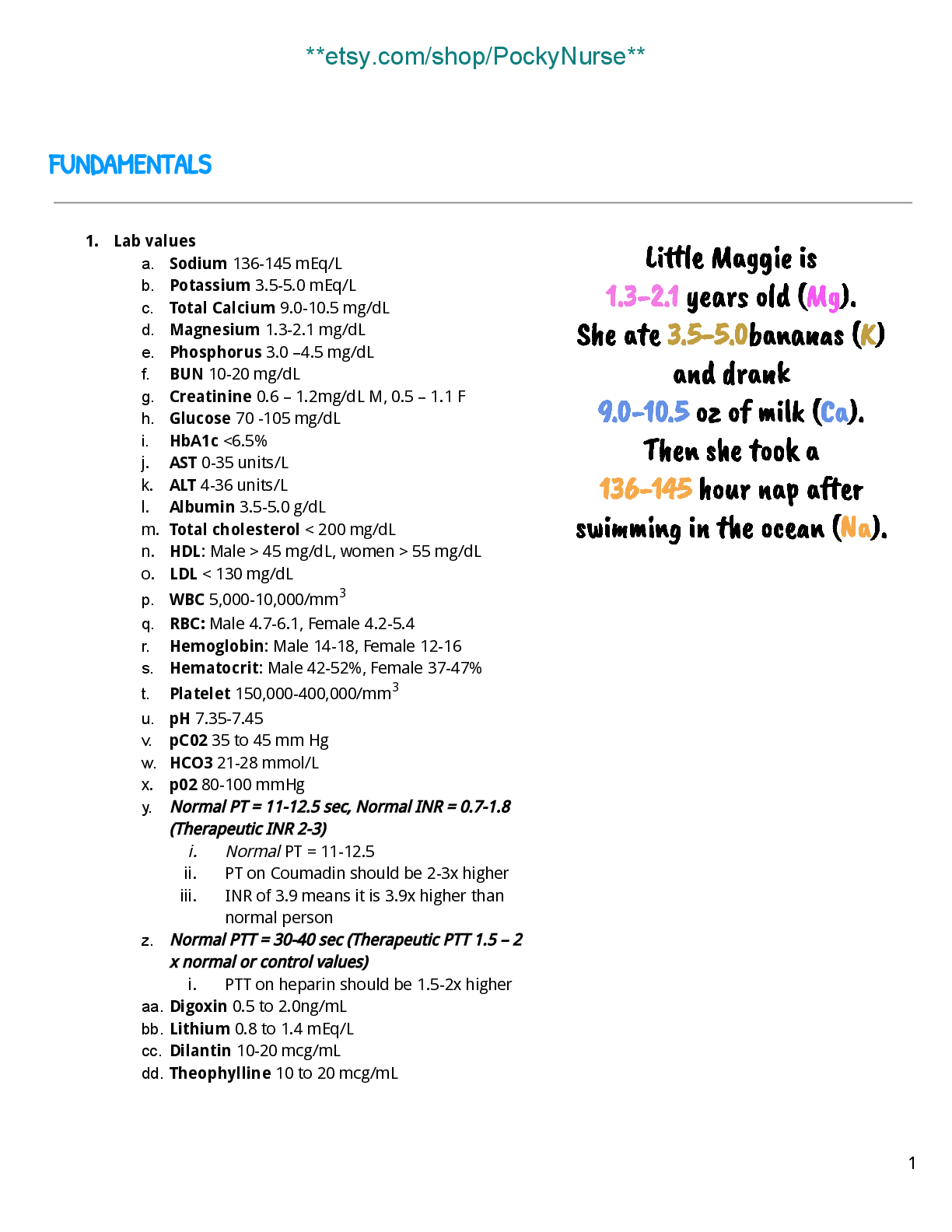
• pH: 7.35-7.45
• CO2: 35-45
• HCO3: 22-26
• O2: 80-100
• Respiratory Acidosis: elevated pCO2 due to ventilation depression
• Respiratory Alkalosis: depression of pCO2 due to alveolar hyperventilation
• Metabolic Acidosis: depression of HCO3 or an increase in non-
carbonic acids
• Metabolic Alkalosis: elevated HCO3 due to excessive loss of metabolic acids
o Allergic asthma will have elevated serum eosinophil count & IgE levels
o Sputum with eosinophils & mucus plugs with shed epithelial cellsfrom coughing
o Pulmonary Function Tests
• Forced vital capacity (FVC)
• Forced expiratory volume in the first second (FEV1)
• Peak expiratory flow rate (PEFR)
▪ Management
o Patient education important!
o Peak flow meter should be used twice a day daily
o Want to rinse mouth after steroidsprone to thrush
▪ Treatment
o Anti-inflammatory agents:
• Corticosteroids
• Leukotriene antagonists
• Immunodulators
o Rescue
• Short-acting beta 2 agonists
➢ Salbutamol
• Anti-cholinergic
➢ Atrovent
o Preventive
• Inhaled corticosteroids
➢ Fluticasone
• Long-acting beta 2 agonists (LABA)give first…this will open up airway so steroids are more effective
➢ Salmeterol
• Leukotriene receptor antagonist (LTRA)
➢ Singulair (pill)
o Other
• Theophylline
➢ Less effective & less well tolerated than long acting bronchodilators
➢ Can reduces exacerbations
• Cromolyn
• Omalimuzab
• Stabilize mast cells, which stop release of histamines
o Administration
• Metered dose inhaler (MDI)
• Dry powder inhaler (DPI)
• Autohalergoes in with breath
o Exercise (ie: swimming) & activitypromotes ventilation & perfusion
o Oxygen therapy
▪ Status Asthmaticus
o Severe, life-threatening, acute episode of airway obstruction
o If not reversed patient may develop pneumothorax and cardiac or respiratory arrest
o Clinical manifestations (that asthma attack worsening)
• Cyanosis
• Inspiratory wheezing
• Altered LOC
• Use of accessory muscles
• Check ABGs or O2 saturation
o Treatment
• IV fluids
• Potent systemic bronchodilator
• Steroids
• Epinephrine
• Oxygen
COPD:
▪ Bronchospasm & dyspnea
▪ Tissue damage is not reversible & increases in severity…eventually respiratory failure
▪ Leading cause of morbidity & mortality worldwide
▪ Associated with significant economic burden
▪ Onset in mid-life
▪ Symptoms slowly progress
▪ Long smoking history
▪ Risk factors:
o Cigarette smoking
• Smoking cessation!
➢ ASK
➢ ADVISE
➢ ASSESS
➢ ASSIST
➢ ARRANGE
o Alpha 1 antitrypsin (AAT) deficiency
• Helps breakdown enzymes around alveoli
• If not working they will breakdown alveoli = emphysema at younger age with no PMH of smoking
o Air pollution
o Gender
o Age
o Respiratory infections
o Socioeconomic status
o Asthma/bronchial hyperreactivity
o Chronic bronchitis
▪ Emphysema
o Loss of lung elasticity
o Hyperinflation of the lung
o Dyspnea
o Increased RR
o Decreased expiratory phase = can’t get as much air in = decreased expiratory even more…and so on
o Air trappingdue to loss of elastic recoil in alveolar walls, overstretching, & enlargement of the alveoli into bullae, & collapse of small airways (bronchioles)
o Clinical manifestations
• “Pink Puffer”
• Pursed lip breathingexhale slowly, help expand to get air out that’s “deep down”
• Dyspnea
• Hyperessonance on chest percussiondue to air trapping
• Orthopnea
• Barrel chest
• Prolonged expiratory time
• Speaks in short, jerky sentences
• Anxious
• Use of accessory muscles
• Thin appearance
• Tripod positioning
▪ Chronic Bronchitis
o Inflammation of the bronchi & bronchioles caused by chronic exposure to irritants, especially tobacco smoke
o Inflammation, vasodilation, congestion, mucosal edema, & bronchospasm
o Affects only the airways NOT the alveoli
o Production of large amount of thick mucus
o Hard to get rid of air on way out due to blockage
o Clinical manifestations
• “Blue Bloater”
• Cyanotic
• Recurrent cough & sputum production
• Hypoxia
• Hypercapnea
• Respiratory acidosis
• Increased hemoglobin (secondary polycythemia)
• Increased RR
• Dyspnea or exertion
• Increased incidence in heavy cigarette smokers
• Digital clubbing
• Cardiac enlargement
• Use of accessory muscles
• Leads to right-sided heart failure
▪ Complications
o Hypoxemia
o Acidosis
o Respiratory infections
o Lung cancer
o Diabetes
o Osteoporosis
o Cardiac failure, especially cor pulmonale (hypertrophy/right-sided heart failure)
• Air trapping = taking up more space = more resistance = more
pressure = right ventricle working harder to pump blood
o Cardiac dysrhythmias
• Hypertrophy of heartincreases distance electrical conduction has to travel
o Ventilation failure (not getting air in/out)
o Oxygenation failure (not getting O2 to blood)
o Or Combination
o Anxiety & depression
▪ Diagnostic tests
o Dyspnea assessment tool
o ABGs
o Sputum samples (culture & sensitivity, infection?)
o CBC (hemoglobin/hematocrit)
o Serum electrolyte levels (dilutional hyponatremia)
o Serum AAT levels (Alpha 1 Antitrypsin deficiency?)
o Chest x-ray
o Pulmonary function test
o Spirometry: required to establish diagnosis
• Should be performed after administration of dose of short-acting inhaled bronchodilator to minimize variability
• FEV1/FVC < 0.70 confirms airflow limitation
• 2 or more incidences that FEV1 < 50% OR 1 + hospitalizations for COPD exacerbations = HIGH RISK
▪ Treatment
o Pulmonary rehab
• Incentive spirometer 10x/hour
• Chest physiotherapy
• Hydrationif not fluid overloaded
• Exercise
o Beta-adrenergic agents
o Cholinergic antagonists
o Methyxanthines
o Corticosteroids
o Mucolyticsto get mucus out
o Want to stop Robitussin or any other cough suppressants…stop cough, which will prevent them from removing secretions
o Lung transplantationfor end-stage
• Large midline incision or a transverse anterior thoracotomy
▪ Management
o Ineffective breathing
• Breathing techniques
• Positioning to help alleviate dyspnea or drainage
• Energy conservationnot doing too much at one time
➢ Encourage patient to pace activities
➢ Do not rush through morning activities
➢ Gradually increase activities
o Ineffective airway clearance
• Possible suctioning
• Controlled coughing
• Chest physiotherapy with postural drainage
• Hydration via beverage & humidifier
• Flutter-valve mucus clearance devices
• Tracheostomy
o Risk for imbalance nutrition
• Often in a hyper-metabolic stateneed more calories
• Prevent protein-calorie malnutrition through dietary consultation
• Monitor weight, skin condition, & serum pre-albumin levels
• Dyspnea management
• Food selection to prevent weight loss
o Prevent anxietythis can worsen symptoms
o Risk for pneumonia & other respiratory infections
• Avoid large crowds
• Pneumonia vaccine
• Yearly influenza vaccine
LUNG CANCER:
▪ Leading cause of cancer deaths
▪ Poor long-term survival because of late-stage diagnosis
▪ Bronchogenic carcinomas
▪ Staged to assess size & extent of disease
▪ Warning signs
o Hoarseness
o Change in respiratory pattern
o Persistent cough
o Blood in sputum
o Chest pain/tightness
o Shoulder pain
o Recurrent pneumonias
o Dyspnea
o Wheezing
o Weight loss
o Possible JVD & edema in upper extremities
▪ Management
o Can be curative (if caught early) to increase survival time, and/or palliative
o Chemotherapy
o Targeted therapy (to de-bulk, then do in & remove it)
o Radiation therapy (to de-bulk, then do in & remove it)
o Photodynamic therapy
o Wedge resection
o Lobectomy
o Pneumonectomy
o Postoperative care
• Chest tube placement (except for pneumonectomy)
➢ Sterile technique
➢ Place drainage system below level of patient
➢ Look for kinks
➢ Tidaling
➢ Vaseline dressing
• Pain management
• Respiratory management (mechanical ventilation)
▪ Interventionsto maintain quality of life, ADLs, IADLs
o Oxygen therapy
o Drug therapy
o Radiation therapy
o Thoracentesis (remove fluid from pleural space)
o Pleurodesis (injection to “close off” pleural space so fluid can’t get it)
o Dyspnea management
o Pain management
o Hospice care (~6 months)
SLEEP APNEA:
▪ Breathing disruption during sleep that lasts at least 10 seconds & occurs a
minimum of 5 times an hour
▪ Results in upper airway obstruction by the soft palate or tongue
▪ Risk factors
o Obesity
o Large uvulasometimes they will laser off the uvula
o Short neck
o Smoking
o Enlarged tonsils/adenoids
o Oropharyngeal edema
• Can be from ACE inhibitorsstop drug
▪ Clinical manifestations
o Heavy snoring
o Excessive daytime sleepiness
o Inability to concentrate
o Irritability
o Headaches
o Personality changes
MECHANICAL VENTILATION:
▪ Modes of ventilation
o BiPAP
• 2 levels of positive airway pressure via nasal or oral mask, nasal pillow, or mouthpiece
• Higher level of pressure during inspiration augments spontaneous breathskeep soft palate & tongue away from back of throat
• Lower level of pressure during exhalation
o CPAP
• Delivers a set positive airway pressure continuously during each cycle of inhalation & exhalation
• Keeps alveoli open during inhalation & prevent collapse during
exhalation
o Assist-control (A/C)
• Set tidal volume & rate (how fast they will breathe, but can initiate breaths)
• All breaths (assisted or controlled) are delivered with the same set
pressure/flow rate
• Used in initiating mechanical ventilation and/or when patient at high risk for respiratory arrest
• If start breathing on own, probably not the best because they can
be overventilated
o Synchronized intermittent mandatory ventilation (SIMV)
• Preset tidal volume & rate
• Allows spontaneous breaths
• Starting to “wean” patient off ventilator
• Breaths synchronized with patient’s effort
o Pressure support (PS)
• Positive pressure to augment inspiratory effort
• Patient controls RR, flow rate, & tidal volume
• Used for weaning off or in combination with other modes
• Can have with A/C or SIMV (as an extra boost)
• All breaths on own, but assists with breathing in
• “Last step”
▪ Reasons for Ventilation
o Improve gas exchange & decrease work of breathing
o Doesn’t cure anything
o Support patient until natural breathing can resume
▪ Ventilator Settings
o RRwhen weaning off, will set lower so they breathe on their own
o Tidal volume
o Oxygen concentration
o Flow ratehow fast in
o Positive end-expiratory pressure (PEEP)to keep alveolar open & prevent lungs from collapsing
o Pressure supportextra push when they breathe in
▪ Ventilator Alarms
o High pressure
• Secretions or mucus plug
• Coughing, gagging, biting tube
• Obstruction
• Asynchrony (“fighting the ventilator”)may need to sedate, or it may indicate they are ready to be weaned off
• Airway displaced or movedwant to auscultate
• Pulmonary complications (ie: bronchospasm, pneumothorax, pneumonia)
o Low pressure
• Leak in circuit
• Cuff leak
• Loss of spontaneous breathing or decreased respiratory effort
• If patient can talk to you with ET tubeLEAK
▪ Complications
o Cardiac problems
• May need to address fluid overload, electrolytes)
o Lung problems (ie: barotrauma)
o GI & nutritional issues (ie: ulcersmay need Nexium)
o Infections
o Ventilator dependence
MUSCULOSKELETAL ALTERATIONS
OSETOPOROSIS:
▪ Chronic metabolic disease in which bone loss causes decreased density & possible fractures
▪ Osteoblasts put stuff into bone, osteoclast take stuff out…more osteoclastic
activity than osteoblastic activity
▪ Primary
o Most common in postmenopausal women & men in 60-70 (especially those thin & frail)
o Bone mineral density (BMD) decreases rapidly post-menopausal
▪ Secondary
o From an associated medical condition
• Hyperparathyroidismpulling Ca+ out of bone = bone weaker
• Long-term drug therapy
• Long-term immobility (may have MS or Parkinson’s)
• Chronic drug use (ie: corticosteroids, heparin, anticonvulsants)
▪ Regional
o When limb is immobilized (ie: related to fracture)
o Can build bone back up usually, but we worry about age
▪ Risk factors:
o Older age
o Family history
o Low body weight/thin build
o Chronic low calcium & vitamin D intakeactivate Ca+ to move around body to get into bone
o Estrogen/androgen deficiencyaffects bone density
o Early menopause
o Smokingaffects bone density
o High alcohol intake (1 drink for women, 2 for men)
o Lack of physical exercise/immobility
o White, thin women are likely to develop osteoporosis at an earlier age
▪ Health Promotion/Illness Prevention!!!
o Teaching should start with young women who begin to lose bone density after 30 years of age
o Focus of osteoporosis prevention is to decrease modifiable risk factors
o Ensure adequate nutrition
• Vitamin D
• Calcium
• Make sure not getting too frailkeep good BMI
o Avoid sedentary lifestyle
o Continue program of weight-bearing exercises
▪ Physical assessment:
o “Dowager’s hump”due to bone density loss, kyphosis
o Loss of height
o Complains of back painif spine is losing a lot of bone density, affect cartilage, possible compression of nerves
o Fallophobiamay be sedentary due to fear of falling
▪ Diagnostic tests:
o No definitive lab tests
o Serum calcium, vitamin D, & phosphorus to rule out secondary osteoporosis & other metabolic bone diseases
o Dual X-ray Absorptiometry*
• To measure bone mineral density
• Look at hip and spine
• T Score
➢ More commonly used
➢ To help determine risk of fracture (fragility fracture)
❖ Might just be walking & fracture a hip
➢ < -2.5 = osteoporosis
❖ Times # by 10 to get % of bone loss (ie: -2.5 x 10 = 25% loss of bone density compared to that of a 30- year-old female)
➢ -1 to -2.5 = osteopenia
❖ Want to start treating in this stage to prevent osteoporosis
➢ < -1.5 & has risk factors = should start some kind of
treatment
➢ < -2 should start treatment right away even without risk factors
➢ Compares score to that of a normal 30-year-old female
• Z Score
➢ This compares the patient to someone of the same relative age & gender
➢ Not really used in diagnostic testing
▪ Interventions:
o Nutritional therapy
• Calcium & vitamin D
• Protein, magnesium, vitamin K, & other minerals
o Lifestyle changes
• Muscle strengthening & weight-bearing exercises
• Water aerobics are good too
• Walking for 30 minutes 3-5 times/week
• Avoid smoking
o Drug therapy
• Calcium & vitamin D (Os-Cal, Citracal)
➢ 1-1.5 grams in divided doses
➢ Vitamin D is in units
• Estrogen Agonist/Antagonist (Raloxifine)
➢ Hormone therapy
➢ Normal for post-menopausal therapygreat risk for DVTs, especially if also smoking
➢ Watch for DVT & monitor LFTs
• Bisphosphonates [Alendronate (Fosamax), Risedronate (Actonel), Ibandronate (Boniva)]
➢ Can affect lower esophageal sphicter
➢ Side effect: Esophagitis
➢ Take on empty stomach first thing in morning with full glass of water & stay upright/sitting for 30 minutes-1 hr.
• Other agents
➢ Parathyroid hormone
➢ Calcitonin
➢ Fortero (Pen)subQ daily
❖ Possibly helps increase bone density, but definitely helps with further loss
OSTEOARTHRITIS:
❖ Lots of education!
❖ Rotate site (deltoid, abdomen, thigh)
❖ Clean site
❖ Proper disposal
▪ Most common type of arthritis
▪ Joint pain & loss of function characterized by deterioration & loss of cartilage in the joints
▪ Development of osteophytes
▪ Less we use it, less mobility & function
▪ Most common in hips, knees, & ankles
▪ Bone hypertrophybony outgrowths
▪ Bone rubbing against bone = pain & inflammation
▪ Primary
o Triggered by aging or genetics
o Weight-bearing joints mostly affected (ie: knees)
▪ Secondary
o Results from other musculoskeletal conditions such as rheumatoid arthritis (can lead to osteoarthritis), congenital anomalies, joint sepsis, obesity, smoking, & other metabolic conditions (ie: diabetes)
▪ Clinical manifestations:
o Chronic joint pain & stiffness
o Pain may be present at rest (getting worse)
o Interruption of sleep patterns
o Tenderness on palpation/ROM
o Enlarged joint
o Heberden’s (DISTAL) & Bouchard’s nodes (PROXIMAL)
o Joint effusion
o Atrophy
o Loss of function
▪ Diagnostic tests:
o Elevated ESR (Normal: 0-22 men/ 0-29 women)
o Elevated C-reactive protein (Normal: < 1)lot of inflammation throughout body
• The more joints affected, the higher the ESR & CRP will be
o Better diagnostic tests:
• X-raychanges in bone structure, bone spurs, hypertrophy
• MRIwhat’s going on in joint, joint space, & blood flow to area
• CT
▪ Nursing management:
o Manage pain
o Get them moving
o Address secondary causes
o Analgesics
• Tylenol is the drug of choice* (keep under 4g/day to prevent overdose)
➢ Remember that other drugs may have Tylenol in them
• Lidoderm 5%patch
➢ Follow instructions on how long to have it on
➢ Don’t use bare hands/be careful applying it
➢ Change location (don’t want to wear skin out)
• NSAIDsworry about GI problems
o Other
• Cortisone injectionsinto joint space to decrease inflammation
• Hyaluronate injectionssimilar to a compound in our joint spaces, help lubricate & move properly
• Muscle relaxantshelpful especially with back spasms
o Rest & positioning
• Positioning depends on wear the pain is
o Weight control
o Hot or cold application
• Heat may decrease muscle tension
• Hot shower/baths
• Cold to reduce swelling/inflammation
• Depends on the patient & what works for them
o Complementary/alternative therapies
• Worry about drug interactions!
• Chondroitin & glucosamine (for osteoarthritis), & turmeric (strong anti-inflammatory)
• Acupuncture
• Massage therapy
• TENS (Transcutaneous electric nerve stimulation)get blood flowing & muscle moving
o No therapy will halt progression
o Current therapy directed at relief of pain & minimizing functional disabilityaddress ADLs, IADLs, pain, & mobility
o Agents for pain relief
• Topical agents
• Systemic oral agents (acetaminophen, NSAIDs)
• Intra-articular agents
➢ Corticosteroids for synovial inflammation
➢ Hyaluronic acid
▪ Surgical management:
o Total Joint Arthroplasty (most commonhip or knee replacement)
• Pre-Op
➢ Teaching what to expect post-op
➢ Medications (insulin , BP meds, Coumadin, Lovenox, aspirin, etc.)
➢ Pain management
• Operative
• Post-Op
➢ Prevention of discoloration
➢ Make sure placement stays
➢ Prevention of infection
❖ Prophylactic antibiotics
➢ Thromboembolic complications
➢ Assess for bleeding
➢ Management of anemia
HIP FRACTURE:
▪ Most common injury in older adults
▪ High mortality rate
▪ Especially concerned with femoral neck fracturecould lead to avascular necrosis of the femoral head
▪ Clinical manifestations:
o Injured leg shortened
o Externally rotated
o Extreme pain prevents movement
▪ Treatment:
o Immobilize immediately to prevent further damage
o Buck’s traction may be used before surgery
• Little boot around lower leg (traction & weight)
• Make sure weight is NOT on floor
• DO NOT turn side to side
• Trapeze can be used to help the patient move (sit upright more)
• No pins
• Assess skin
• Look at circulation
• May need to reposition
o Surgery is treatment of choice
• Open Reduction Internal Fixation (ORIF)
➢ Femoral neck fracture repairs = pins
➢ Intertrochanteric fracture repairs = bidirectional
o Surgical procedure depends on
• Type of injury
• Condition of the person
• Preexisting orthopedic conditions
o With acute or chronic diseases the risk of surgery may be too greatmedical management may be preferred
▪ Nursing management:
o ABCs!
o Pain assessment
o Skin assessment
o Body alignmentabduction pillow
o Ambulation
• Provide analgesic before first time out of bed
o Education to return home
o Monitor for complications
• Dislocation of the device
• Avascular necrosis
• Infection
• Delayed healing
• PEfrom DVT or fracture surgery, fat embolism or something else from rupturing the tissue, etc.
• DVTdue to immobility
• Compartment Syndrome
➢ Due to lack of perfusion
➢ Reduced circulation in an area due to edema
➢ Pain, pressure, paralysis, parathesia, pallor, pulselessness (use Doppler)
➢ Notify HCP!
• Poor alignmentdislocation of the prosthesis
• Infection
• Skin alterations
• Hazards of immobility
*Side note: For NCLEX questions, pain is considered psychosocial, BUT chest pain is ABCs!
HIP REPLACEMENT SURGERY:
▪ Precautions
o DO NOT stand or sit for long periods
o DO NOT flex hips more than 90 degrees
o Abduction pillow
o Avoid internal rotation
▪ Prevention of complications
o Continuous Passive Movement (CPM)
• Give pain medication before
o Assess for neurovascular compromise
o Manage pain
o Progression of activityup & moving post-op day 2
o Promotion of self-careOT
ADVANCED CARE PLANNING & END OF LIFE
▪ Barriers to Quality EOL Care:
o Limited evidence base
o Fear & discomfort about death
o Lack of experience with death
o Death denying culture
o Uncertainty about communication
▪ Advanced Directives
o An order to let the provider know what they want ahead of time in case a decision needs to be made
o Progressive, ongoing discussion
o Patient status & wishes often change with time
o Should be about values, not documents
o Durable Power of Attorney (Health Care Proxy)
• A person who can speak for you when you cannot speak for yourself
o Living Will
• DNR (Do Not Resuscitate)
• DNI (Do Not Intubate)
• AND (Allow Natural Death)
• DNH (Do Not Hospitalize)
• Can decide about other things we may not think about such as artificial hydration & nutrition, dialysis, antibiotics, re- hospitalization, etc.
▪ MOLST (Medical Orders for Life Sustaining Treatment)
▪ FIVE WISHES (person you want to speak for you, who you want around you, etc.)
▪ Where people want to die, differs from where people actually do die
o 50% Hospitals
o 25% Nursing Homes
o 25% Home
▪ Communication Skills:
o Assessment first
o Open-ended questions
o Reflecting/Validating
o Therapeutic use of silence
▪ Moral Uncertainty (just doesn’t feel right)
o Uncertain if a problem exists, unsure about its nature, & unclear which values conflict
▪ Moral Dilemma (discomfort feeling that can be identified)
o 2 or more ethical principles clash
▪ Moral distress (we can identify, but feel like we can’t do anything about it)
o Emotion resulting from inability to act upon the right course of action
▪ Our values may not match the patient’s & that’s okay!
▪ Autonomy: the right to make your own choices
▪ Beneficence: doing good
▪ Nonmaleficence: do no harm
▪ Veracity: truthfulness
▪ Confidentiality
▪ Fidelity: doing what you promised the patient
▪ Justice: equality across all boundaries
CAPACITY COMPETENCY
Task specific Global
Clinical determination Legal determination
Waxes & wanes Global
Patient must understand:
1. Nature of intervention
2. Risk/benefits
3. Alternatives
▪ Anytime you think patient doesn’t understandknow their capacity
▪ Ethics Committees
o Advisory services designed to assist patients, families, & health professional in identifying, analyzing, & resolving ethical dilemmas
o Interdisciplinary
o Confidential
o In most institutions anyone can call an ethics consultation
▪ Euthanasiaillegal everywhere, patient is “killed”
▪ Assisted Suicidehelp patient to kill themselves, legal in 5 states
o Nurses should promote comfort & relieve suffering even if death hastened
o Withholding/withdrawing can be ethically acceptable
o Nurses should seek to understand request for assisted suicide
o Nurses should create environment where patient can feel comfortable expressing their thoughts
o Get help for pain & symptom management
▪ Many patients have an issue of control: fear of pain, uncontrolled symptoms, loss of dignity, being a burden
▪ Palliative Care
o Focus on Quality of Life
o Pain & symptom management (throughout treatment)
o Aligning goals with treatment
o Interdisciplinary team
o Given at any point in illness along with active treatment
o Almost anyone with a chronic illness can benefit from palliative care
o It works...when people feel better they do more for themselves
▪ Hospice Care
o Medicare benefit
o Care provided in the last 6 months of life
▪ Care of an Actively Dying Patient:
o Change in Rhythm of Care
• More intensive care
o Change in Goals of Care
o Shift in Focus of Care
• More about quality of life
• Focus on existing or new symptoms
• Initiation of new measures for comfort
• Discontinuing non-essential treatment
• Family needs may become more pronounced
o Change in priorities
• Life prolonging treatments
• Vital signs
• Diagnostic tests
• Fluid & nutrition
• Medications (re-evaluate)
• Increased focus on family!
o Nursing response
• Validate
• Normalize
• Intervene
o Signs of Pending Death
• Cold, mottled, clammy skinbody shunting blood from extremities to brain & heart
• Cyanosis
• Changes in breathing pattern (Cheyne-Stokes)
➢ Periods of rapid respirations followed by apnea
➢ Oxygen
➢ Opioids
➢ Fan
➢ Family education
➢ Morphine (for respiratory distress or dyspnea)
• Decreased LOC
• “Death Rattle”
➢ Noisy secretionssaliva on voice box, sounds worse than it feels
➢ Raise HOB
➢ Positioning
➢ No deep suction
➢ Anticholinergic
➢ Glycopyrrolateto dry up secretions
• Agitation
➢ Address causes (ie: pain, urine retention)
➢ Palliative sedation
➢ Anti-psychotics (Haldol or Ativan)
• Some may die with eyes openloss of subcutaneous fat or blink reflex
➢ Eye drops to prevent dryness
• Decreased PO intake
➢ Oral care at end of life is essential!
➢ Avoid IVs/hydration3rd spacing can occur where fluid in vessels leak into tissue = increased secretions, increased incontinence, edema, etc.-->substitute with other “caring” habits (ie: do hair, massage, oral care)
o Emotional-Spiritual Symptoms
• Withdrawal
➢ Some family members experience loss of intimacytell them its okay to touch them
➢ We believe hearing is the last to go…encourage family to
speak to the patient even if he/she cannot respond
• Letting go: Restlessness
• Visions
• Saying Goodbye
o Cultural Competence
➢ Self-reflection…how do you view this issue?
➢ Identify & integrate knowledge of most common cultural groups in your practice
o Cultural Humility
➢ Commitment to self-reflection & self-critique
➢ Developing mutually beneficial, respectful partnerships
➢ Is there anything about your culture/religion that would help me take care of the patient?
o The Death Vigil
• Family presence
• Common fears
➢ Being alone with patient
➢ Time of death: how do you know death has occurred
➢ Missing the moment
o When Death Occurs…
• Cessation of integrated tissue & organ function manifested by lack of heartbeat, absence of respirations, or irreversible brain dysfunction
• No pulse
• No respirations
• Fixed & dilated pupils
• Relaxation of muscles & sphincters
• Eyes may remain open, jaw may drop
[Show More]