*NURSING > QUESTIONS & ANSWERS > CHAPTER 13 JARVIS WORKBOOK QUESTIONS AND ANSWERS 2022 LATEST (All)
CHAPTER 13 JARVIS WORKBOOK QUESTIONS AND ANSWERS 2022 LATEST
Document Content and Description Below
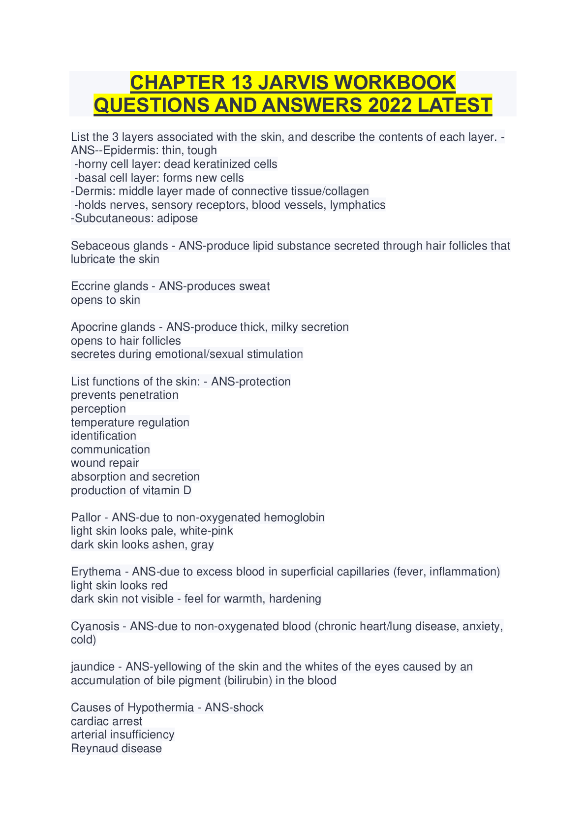
CHAPTER 13 JARVIS WORKBOOK QUESTIONS AND ANSWERS 2022 LATEST List the 3 layers associated with the skin, and describe the contents of each layer. - ANS--Epidermis: thin, tough -horny cell layer: d... ead keratinized cells -basal cell layer: forms new cells -Dermis: middle layer made of connective tissue/collagen -holds nerves, sensory receptors, blood vessels, lymphatics -Subcutaneous: adipose Sebaceous glands - ANS-produce lipid substance secreted through hair follicles that lubricate the skin Eccrine glands - ANS-produces sweat opens to skin Apocrine glands - ANS-produce thick, milky secretion opens to hair follicles secretes during emotional/sexual stimulation List functions of the skin: - ANS-protection prevents penetration perception temperature regulation identification communication wound repair absorption and secretion production of vitamin D Pallor - ANS-due to non-oxygenated hemoglobin light skin looks pale, white-pink dark skin looks ashen, gray Erythema - ANS-due to excess blood in superficial capillaries (fever, inflammation) light skin looks red dark skin not visible - feel for warmth, hardening Cyanosis - ANS-due to non-oxygenated blood (chronic heart/lung disease, anxiety, cold) jaundice - ANS-yellowing of the skin and the whites of the eyes caused by an accumulation of bile pigment (bilirubin) in the blood Causes of Hypothermia - ANS-shock cardiac arrest arterial insufficiency Reynaud disease Causes of Hyperthermia - ANS-increased metabolic rate trauma infection sunburn hyperthyroidism Causes of Diaphoresis - ANS-thyrotoxis, MI, anxiety, pain Mobility - ANS-the ease of skin to rise (decreased with edema, Scleroderma) Turgor - ANS-the skin's ability to return to place promptly when released (decreased with dehydration, extreme weight loss) Lunula - ANS-the white linear markings that are normally visible through the nail and on the pink nail bed Mongolian spot - ANS-common variation of hyperpigmentation in Black, Asian, American Indian, and Hispanic newborns Cafe au lait spot - ANS-large round or oval patch of light brown pigmentation - usually normal erythemia toxicum - ANS-rash, small pustules, normal, will go away Cutis marmorata - ANS-transient mottling in the trunk and extremities in response to cooler room temperatures - forms reticulated red or blue pattern over skin Physiologic jaundice - ANS-yellowing of the skin, sclera, and mucous membranes that develops after the 3rd or 4th day of life due to increased numbers of RBCs that hemolyze after birth Milia - ANS-tiny white papules on cheeks and forehead and across the nose and chin caused by sebum that occludes the opening of the follicles Lentigines - ANS-common variations of hyperpigmentation; "liver spots" Seborrheic keratosis - ANS-raised, thickened areas of pigmentation that look crusted, scaly, warty, dark, greasy, and "stuck on" Actinic keratosis - ANS-red-tan plaques that increase over the years to become raised and roughened Acrochordons (skin tags) - ANS-overgrowths; form stalk; are polyp-like Sebaceous hyperplasia - ANS-raised yellow papules with a central depression Petechiae - ANS-a small red or purple spot caused by bleeding into the skin. hematoma - ANS-a solid swelling of clotted blood within the tissues. bruise - ANS-an injury appearing as an area of discolored skin on the body from ruptured blood vessels. Measles (Rubeola) rash - ANS-red-purple maculopapular blotchy rash first appears behind ears looks coppery and does not blanch Koplik spots in mouth (red-based elevations of 1-3mm) German Measles (Rubella) rash - ANS-pink, papular rash (similar to Measles, but paler) Distinguished from measles by: neck lymphadenopathy no Koplik spots Chickenpox (Varicella) - ANS-shiny vesicles on erythmatous base - become pustules, then crusts very pruritic Stage I Pressure Ulcer - ANS--intact skin, red, does not blanch Stage II Pressure Ulcer - ANS-partial-thickness skin erosion - loss of epidermis and possibly dermis -looks like shallow abrasion or open blister Stage III Pressure Ulcer - ANS-full-thickness- extends into subcutaneous tissue -may see subcutaneous fat, but not muscle, bone, or tendon Stage IV Pressure Ulcer - ANS-full-thickness - involves all skin layers and extends into supporting tissue -exposes muscle, tendon, bone, slough (stringy matter attached to wound bed), eschar (black or brown necrotic tissue) lesion - ANS-tissue destruction macule - - ANS-flat skin lesion papule - ANS-small palpable skin lesion, plaque - - ANS-collection of papules, nodule - - ANS-larger elevated skin lesion tumor - - ANS-abnormal growth of tissue, wheal - - ANS-raised skin lesion with interstitial fluid, vesicle - - ANS-elevated cavity containing free fluid, pustule - ANS-elevated cavity with thick, cloudy fluid. crust - - ANS-thick, dried-out exudate from burst vesicles or pustules like in chickenpox scale - - ANS-shedded dead skin cell fissure - - ANS-crack in skin going into dermis erosion - ANS-shallow depression in skin such as stage 2 of pressure ulcers. ulcer - ANS-sloughing of necrotic, inflamed tissue such as a stage 3 pressure ulcer. primary skin lesions: - ANS-macule papule plaque nodule tumor wheal vesicle pustule secondary skin lesions - ANS-crust scale fissure erosion ulcer [Show More]
Last updated: 2 years ago
Preview 1 out of 6 pages
Buy this document to get the full access instantly
Instant Download Access after purchase
Buy NowInstant download
We Accept:

Reviews( 0 )
$7.50
Can't find what you want? Try our AI powered Search
Document information
Connected school, study & course
About the document
Uploaded On
Sep 23, 2022
Number of pages
6
Written in
Additional information
This document has been written for:
Uploaded
Sep 23, 2022
Downloads
0
Views
34







.png)




 (1).png)
