Paramedic Cardiology Questions and
Answers Already Passed
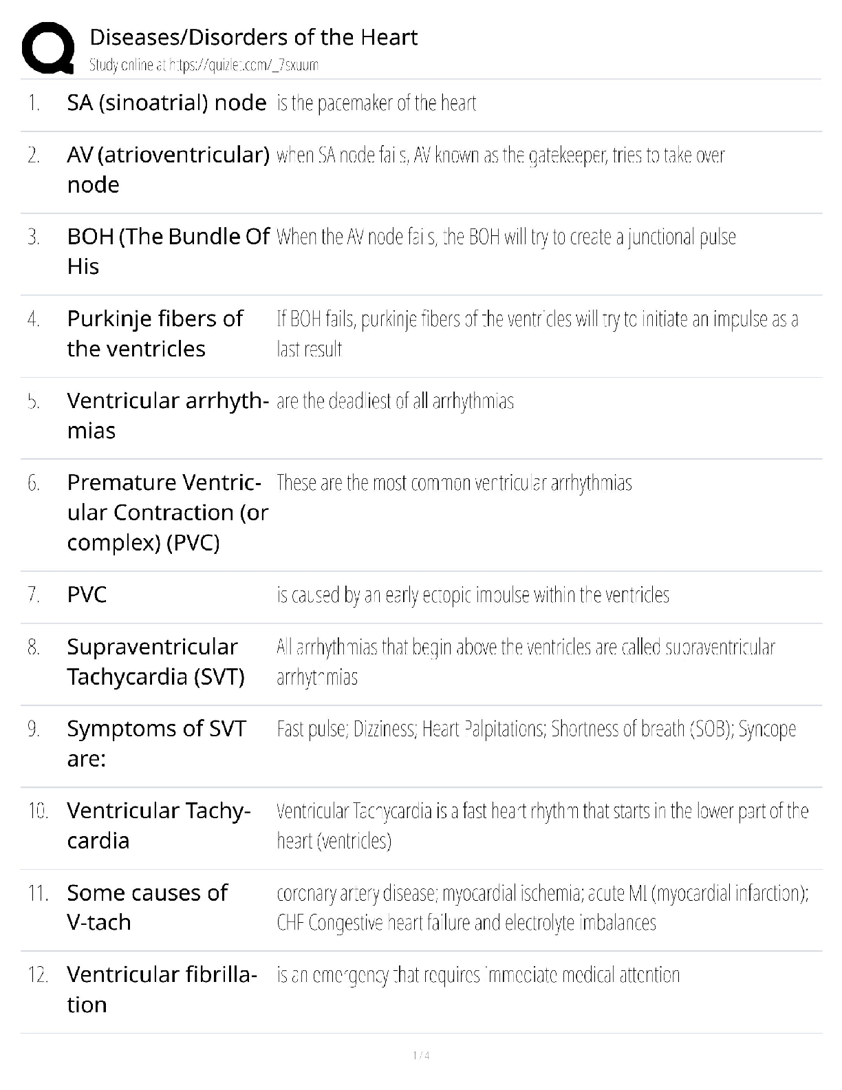
SA Node Rate ✔✔60-100 BPM
AV Node Rate ✔✔40-60 BPM
Bundle of His/Perkinjie Rate ✔✔>40 BPM
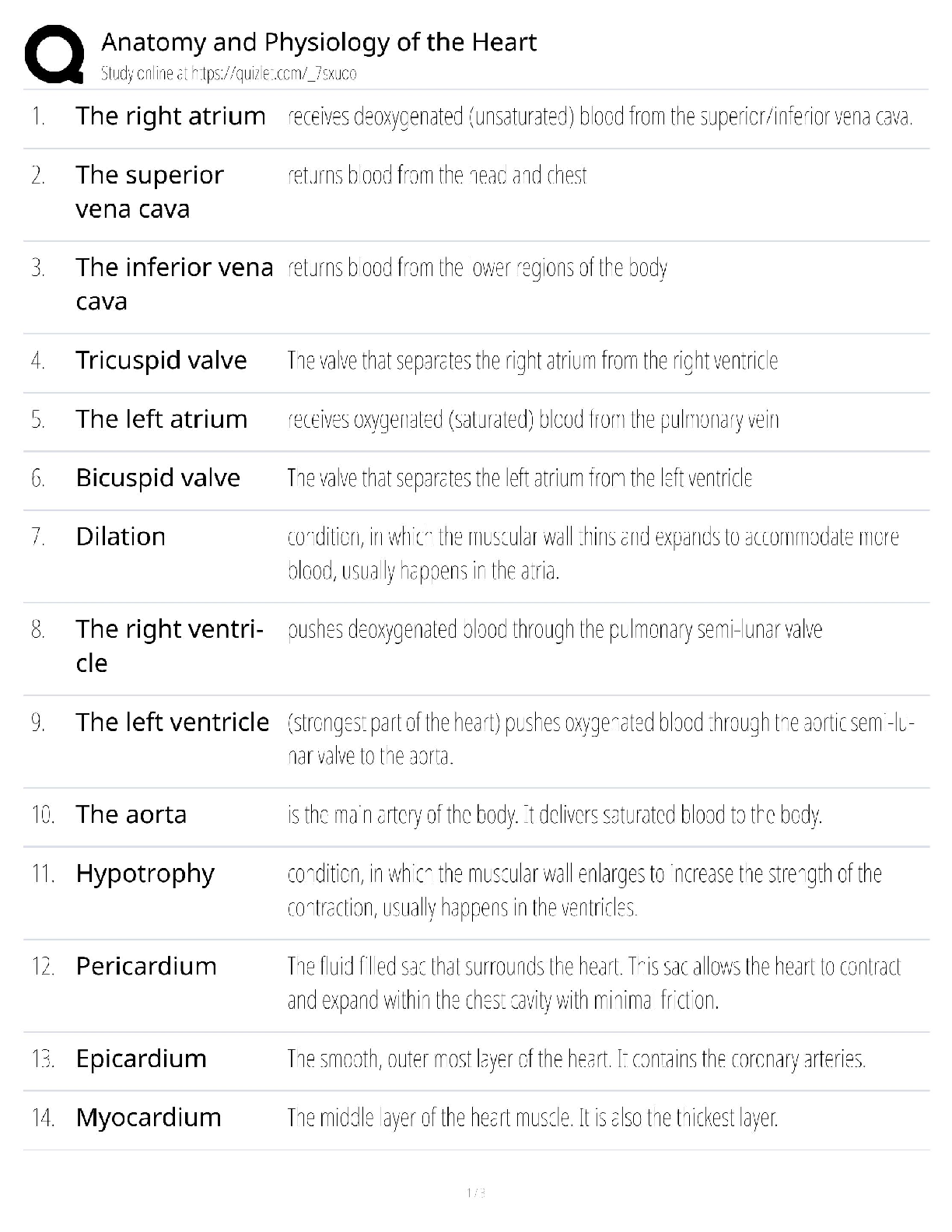
Flow of blood through the heart ✔✔from the body
superior and infer
...
Paramedic Cardiology Questions and
Answers Already Passed
SA Node Rate ✔✔60-100 BPM
AV Node Rate ✔✔40-60 BPM
Bundle of His/Perkinjie Rate ✔✔>40 BPM
Flow of blood through the heart ✔✔from the body
superior and inferior vena cavas
right atrium
tricuspid valve
right ventricle
semilunar valve
pulmonary artery
lungs
pulmonary veins
left atrium
bicuspid valve
left ventricle
semilunar valve
aorta
to the body
Inotropic ✔✔Beats Harder
Chronotropic ✔✔Beats Faster
Dromotropic ✔✔Improved contractility
"P" Wave ✔✔the P wave represents the electrical activity associated with the contraction of the
atria, or atrial depolarization
"P-R" Interval ✔✔Time required for conduction from the SA node to AV node. The time
between atrial and ventricular depolarization. This is normally 0.12 to 0.2 seconds.
"Q" Wave ✔✔The first portion of the QRS complex that is seen with a downward deflection and
is usually not obvious on the EKG of the normal heart. Often the first portion of the deflection is
positive, signifying no Q wave. This is the beginning of ventricular depolarization.
"QRS" Complex ✔✔Represents ventricle depolarization. The QRS Interval is measured from the
start of the Q wave or from the beginning of the R wave if no Q wave is present. If no Q wave is
present, a normal QRS interval should be less than 0.12 seconds in all leads.
"ST" Segment ✔✔Indicates that ventricle depolarization is complete and that repolarization is
about to begin. Also called early ventricle repolarization. The ST Segment is measured from the
end of the QRS to the beginning of the T wave, and represents a portion of ventricular
repolarization. The normal segment is usually flat, or isoelectric. The absolute measurement of
this segment is not as clinically important as is the configuration, whether depressed or elevated.
"T" Wave ✔✔the T wave represents the electrical recovery of the ventricles, or ventricle
repolarization. the muscle cells are recovering in preparation for another impulse
"R-R" Interval ✔✔duration of ventricular cardiac cycle (an indicator of ventricular rate)
Cardiac Output ✔✔heart rate x stroke volume, Total blood volume pumped from the left
ventricle per minute. Can be increased by increasing heart rate or stroke volume. SV X HR= CO
Stroke Volume ✔✔the volume of blood pumped out by a ventricle with each heartbeat
Pre-load ✔✔Volume of blood in the ventricles at the end of diastole, immediately before
ventricular contraction.
After-load ✔✔the pressure that must be exceeded before ejection of blood from the ventricles
can occur
Diastole ✔✔relaxation phase of the cardiac cycle, the widening of the chambers of the heart
between two contractions when the chambers fill with blood
Systole ✔✔the contraction of the chambers of the heart (especially the ventricles) to drive blood
into the aorta and pulmonary artery
Beta 1 ✔✔located in the heart + ionotrphic and chronotrophic, also in kidneys renin release in
blood helping regulation of bp causing vasoconstriction
Beta 2 ✔✔bronchodilation, uterine relaxation, glycogenolysis, vasodialation, skeletal muscle
contraction
Alpha 1 ✔✔vasoconstriction, mydriasis, contraction of bladder neck and prostate capsule
Alpha 2 ✔✔Decrease Sympathetic outflow thus Vasodilation and decreased BP.
Epinephrine ✔✔sympathomimetic; elevates heart rate, blood pressure
Sympathomimetic ✔✔denotes mimicking of the actions of the sympathetic nervous system (EPI
& Nor-EPI)
Sympatholytic ✔✔drug or other substance that blocks the actions of the sympathetic nervous
system (also call antiadrenergic).
(Clonidine)
Cholinergic ✔✔DRUGS USED TO DECREASE HEART RATE AND ACT ON SMOOTH
AND SKELETAL MUSCLES (Parasympathetic System)
Anticholinergic ✔✔Block the action of acetylcholine in the parasympathetic nervous system;
also known as cholinergic blocking agents, antispasmodics, and parasympatholytic agents
(Atropine)
Sympathetic Nervous System ✔✔the division of the autonomic nervous system that arouses the
body, mobilizing its energy in stressful situations (Fight or Flight) EPI and NorEPI
Parasympathetic Nervous System ✔✔the division of the autonomic nervous system that calms
the body, conserving its energy (Feed or Breed)
Norepinephrine ✔✔affects arousal and memory; raises blood pressure by causing blood vessels
to become constricted, but also carried by bloodstream to the anterior pituitary which relaxes
ACTH thus prolonging stress response
(levophed)
Atropine ✔✔anti-cholinergic drug that acts on muscarinic receptors. anti-parasympathetic drug,
blocks the Parasympathetic NS not stimulating the Sympathetic NS
Bundle Branch Block ✔✔heart block; occurs when electrical impulse is blocked from traveling
down the bundle branches; results in the ventricles beating at a different rate than the atria
Right Bundle Branch Block ✔✔widened QRS (>0.12s); double peaked QRS in V1 (best to look
in V1 and V2); T wave can look abnormal - inverted (does not indicate ischemia)
Right Bundle Branch ✔✔carry electricity through right ventricle 20 - 40 bpm
Left Bundle Branch ✔✔carries electricity through left ventricle 20 - 40 bpm
Right Coronary Artery ✔✔supplies blood to right atrium, portions of both ventricles and cells of
AV nodes and SA node.
Left Coronary Artery ✔✔Branches into two arteries: 1. Anterior interventricular artery - also
called the left anterior descending artery, supplies the anterior surface of both ventricles and
most of the interventricular septum 2. Circumflex artery - supplies the left atrium and ventricle
SA Node ✔✔the pace-maker of the heart; where the impulse conduction of the heart usually
starts; located in the top of the right atrium
AV Node ✔✔an area of specialized tissue between the atria and the ventricles of the heart,
which conducts the normal electrical impulse from the atria to the ventricles.
Perkinje Fibers ✔✔End of the conduction system; sends impulses through the ventricles, causing
them to contract
Bundle of HIS ✔✔neurological fibers, extending from the AV node to the right and left bundle
branches, that fire the impulse from the AV node to the Purkinje fibers
AV Junction ✔✔The atrioventricular junction; the portion of the electric conduction system of
the heart located in the upper part of the interventricular septum that conducts the excitation
impulse from the atria to the bundle of His
Atria ✔✔the upper chambers of the heart
Ventricles ✔✔the two lower chambers of the heart, and they pump blood out to the lungs and
body.
Sinus Bradycardia ✔✔When the SA node fires at a rate of less than 60 beats per minute. The P
wave and the QRS complex are normal.
Sinus Rhythm ✔✔a heart rhythm established by impulses from the SA node. Irregularities
include sinus bradycardia, sinus tachycardia, sinus arrest, and sinus arrhythmia
Sinus Tachycardia ✔✔P Waves present, constant, each constant P Wave is followed by a
constant QRS complex (normal width - slim), constant intervals between QRS Complexes are
equal, heart rate is 100-150 BPM
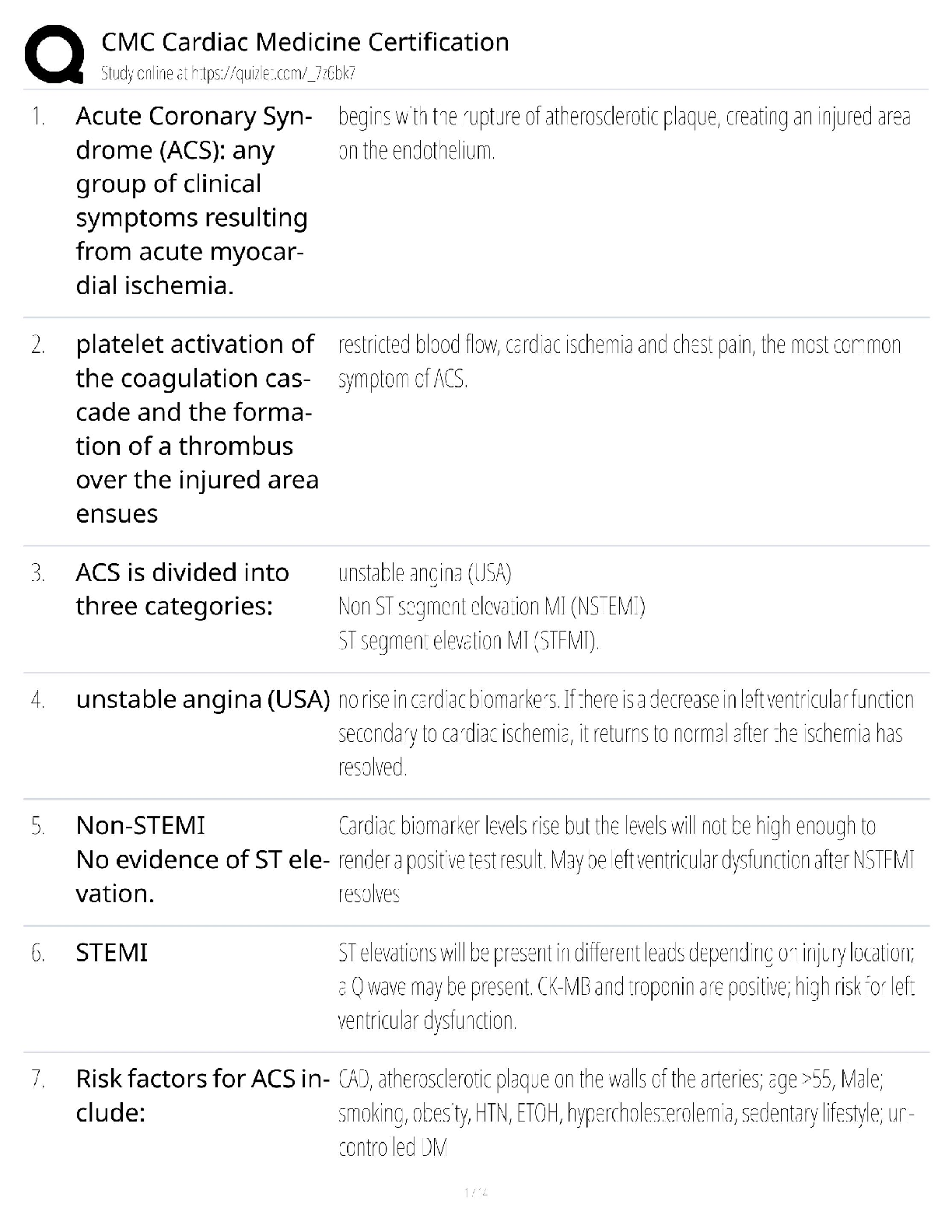
Atrial Fibrilation ✔✔irregularly irregular. rate varies and depends on the conduction rate. no
identifiable P waves. Baseline shows fibrillartory waves.
Atrial Flutter ✔✔Characterized by a "saw-toothed" wave formation with a slower ventricular
rate. The atrial rate generally ranges form 220 to 350 beats/min. Cardioversion may be used.
Junctional Rhythm ✔✔the SA node is nonfunctional, P waves are absent, and heart is paced by
the AV node at 40-60 beats/min
Accelerated Junctional Rhythm ✔✔regular rhythm, rate 60-100, p waves inverted or occur
before during or after QRS, PRI measured if p before QRS <0.12
Supraventricular Tachycardia ✔✔A heart rate of over 150 bpm caused above the ventricle, either
the atria, SA node, or AV junction.
Wolff-Parkinson-White syndrome ✔✔is a syndrome of pre-excitation of the ventricles of the
heart due to an accessory pathway known as the bundle of Kent. This accessory pathway is an
abnormal electrical communication from the atria to the ventricles. WPW is a type of
atrioventricular reentrant tachycardia. Has a "Delta Wave" on the EKG
Ventricular Tachycardia ✔✔Rhythm: Regular
Rate: 100-250 bpm
P-waves: Absent
PR-Interval: Absent
QRS complex: Wide and bizarre
*rhythm is unstable, compromises BP and CO
Ventricular Fibrilation ✔✔parts of the myocardium contract random, no blood is pumped,
treated with direct-current shock
Sinus Arrest ✔✔The SA node will fail momentarily and will not initiate an impulse
Multi-focal Atrial Tachycardia ✔✔Rate 100-250/bpm P wave two or more ectopic P waves with
different morphologies QRS normal Rhythm irregular
Lead I ✔✔records the heart's voltage difference between the right arm and the left arm LA+ RALead II ✔✔records the dfference between the right arm and left leg; shows the heart's rhythm
more clearly than the other two
RA- LL+
Lead III ✔✔records the difference between the left arm and the left leg
LA- LL+
V1 Placement ✔✔fourth intercostal space just right of sternum
V2 Placement ✔✔fourth intercostal space just left of sternum
V3 Placement ✔✔halfway between v2 and v4
V4 Placement ✔✔fifth intercostal space midclavicular
V5 Placement ✔✔5th intercostal space, midway between V4 & V6
V6 Placement ✔✔midaxillary line at the same level as v4 (5th intercostal space)
Inferior Leads ✔✔Leads II,III, aVF Look at electrical activity from the vantage point of the
inferior surface
Lateral Leads ✔✔Leads I, aVL, V5 and V6 Look at the electrical activity from the vantage point
of the lateral wall of left ventricle.
Septal Leads ✔✔V1 and V2 Look at electrical activity from the vantage point of the septal wall
of the ventricles
Anterior Leads ✔✔leads V3 and V4, which are positioned over the front (anterior) of the left
ventricle
[Show More]