BioLab3
Microscope and the Cell Lab Report
Name, date, course and section required for password:
I. Purpose of the Microscope
1. Define resolution.
2. Microscopes magnify the image of a spec
...
BioLab3
Microscope and the Cell Lab Report
Name, date, course and section required for password:
I. Purpose of the Microscope
1. Define resolution.
2. Microscopes magnify the image of a specimen. How is this accomplished?
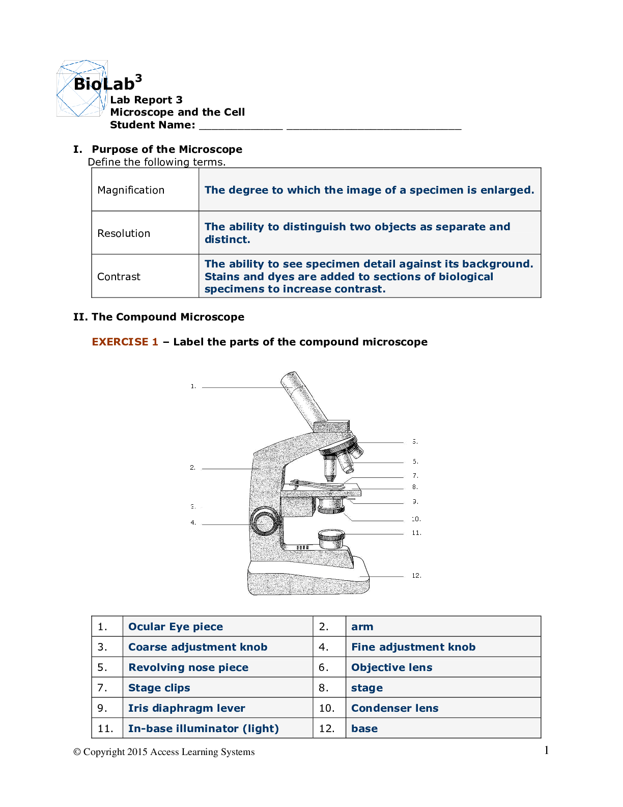
II. Compound Microscope
1. Describe the location and function of the following parts of the microscope:
a. revolving nosepiece -
b. iris diaphragm lever –
c. base
d. fine adjustment knob –
2. Describe how total magnification is calculated.
III. Viewing Microscope Slides
1. Describe in at least five steps, how to focus a microscope in order to view root cells on high magnification.
2. Go to lab, Section III, Exercise 4 to study specimen orientation and answer the following questions:
a. With the scanning lens in place, the prepared slide is moved to the right. In which direction does the image appear to move?
b. The slide is moved away from you. In which direction does the image move?
IV. Depth of Field
1. What diaphragm adjustment should be made to more easily determine the order of the colored threads?
2. Explain how depth of field can be used to reconstruct structure.
V. Microscopic Measurement
1. What is meant by the “field of view”?
2. Go to lab, Section V, Exercise 6 to discover how to measure the diameter of the field of view.
a. What is the diameter of the field of view under scanning power (40X) in micrometers (µm)?
b. What is the diameter of the field of view under low power (100X) using the following equation?
Diameter of field of view of 100X (µm) = (4400µm) x (40X) =
100X
VI. Cells
1. Identify and describe the two major cell types.
2. What three parts do all cells have in common?
VII. Prokaryotic Cells
1. Go to lab, Section VII, Exercise 8 to observe bacteria cells. Answer the following questions:
a. Which magnification is used to view individual bacteria?
b. Can you see organelles within the cytoplasm?
c. Measure the approximate size (μm) of a bacterium.
2. Draw and label a typical bacterial cell, then provide functions for at least five of the labeled structures. Sign, date and prepare an image of your drawing and include it with this lab report.
VIII. Eukaryotic Cells
1. Explain how to make a wet mount of cheek cells. What are two ways to increase contrast?
Summary Questions
1. Explain how the microscope is used as an instrument of measurement.
2. Determine the total magnification of a microscope which has an eyepiece lens (10X) and an objective lens (100X).
3. Name three differences between prokaryotic and eukaryotic cells.
Eukaryotic Cells
4. You are observing an onion epidermal cell under the microscope. Describe what you could do to enlarge the image.
5. Compare the relative size of a bacteria cell, human cheek cell, and Elodea cell.
6. Describe three differences between plant and animal cells.
7. Do all plant cells contain chloroplasts?
8. What is the purpose of a coverslip?
[Show More]


.png)