NR 602 Week 6 Quiz
Anovulation
Anovulation is the failure of the ovary to release ova over a period of time generally exceeding 3 months. The
normal functioning ovary releases one ovum every 25–28 days. This average t
...
NR 602 Week 6 Quiz
Anovulation
Anovulation is the failure of the ovary to release ova over a period of time generally exceeding 3 months. The
normal functioning ovary releases one ovum every 25–28 days. This average time between ovulation events is
variable, especially during puberty and the perimenopause period.1 For nonpregnant women aged 16–40
anovulation is considered abnormal and a cause of infertility in 30% of fertility patients.2
One of the cardinal signs of anovulation is irregular or absent menstrual periods. Oligomenorrhea is defined as
more than 36 days between menstrual cycles or fewer than eight cycles per year.3 In the absence of
pregnancy, menstruation follows ovulation by approximately 14 days. Because menstruation is linked to
ovulation, the clinical finding of oligomenorrhea correlates with oligoovulation. This predictable pattern of
ovulation and menstruation is regulated by a cyclic change in hormones. Consequently, the diagnosis of
ovulation dysfunction includes the assessment of the hormones and systems involved in ovulation and not just
the symptom of amenorrhea.
The most important consideration in the workup of anovulation is to determine the patient’s goals. Treatment
of the patient who wants to get pregnant differs from that of the patient who is concerned about the risks of
early menopause. In patients who desire pregnancy, the clinician needs to determine if they are actively trying
for pregnancy, or are planning for pregnancy several years in the future. The approach outlined below begins
with the patient who is actively trying for pregnancy.
History
Many patients with anovulation will present with amenorrhea. Primary amenorrhea is failure to menstruate
and no secondary sexual characteristics by age 14 or no menstruation by age 16 with normal sexual
development.129 Secondary amenorrhea is the cessation of menstruation for more than 3 months.130 The
etiologies of primary and secondary amenorrhea differ. Primary amenorrhea is often seen in congenital
disorders.131 The most common cause of secondary amenorrhea in women of childbearing age is pregnancy,
consequently the workup for anovulation should begin with a pregnancy test.
Details of a patient’s previous medical history can direct the evaluation of anovulation. Chronic disease can
affect ovulation and may increase risks during pregnancy.132 Psychiatric problems are also often associated
with ovulation dysfunction. The use of any antipsychotic medications should be noted.40 Details of previous
pregnancies are likewise important in the evaluation of ovulation and can help distinguish genetic disorders
from later onset anovulation.
Physical
The physical exam should include an evaluation of vital signs, height, weight, BMI, and appearance. Obesity is
commonly associated with anovulation and PCOS.77 Very thin patients may have anorexia or nutritional
deficits. Hirsutism may suggest PCOS, CAH or an androgen secreting tumor. Visual field testing is useful in
patients who report visual changes suggesting a pituitary tumor. Palpation of the thyroid and abdomen should
also be performed to evaluate for masses. Evaluation of the patient with primary amenorrhea should include a
bimanual exam to determine the presence of a patent outflow tract and uterus.
Laboratory tests
In patients with amenorrhea, pregnancy should be considered and a pregnancy test performed early in theworkup. Evaluation of the HPO axis should be performed in a stepwise fashion. Serum estradiol and
gonadotropins determine ovarian function. FSH measurements have been standardized for day 3 of the
menstrual cycle. However, in patients with amenorrhea a random FSH is appropriate. Measurement of LH has
limited clinical use. The ratio of LH to FSH has been studied for PCOS but is not included in the definition of the
syndrome and is therefore not necessary.77
Elevated FSH indicates an ovarian problem. In patients under 30 years old with an elevated FSH, a karyotype
should be performed. An increased risk of ovarian cancer is seen in XY females with gonadal
dysgenesis.133 Turner syndrome (45,XO) is associated with increased risk for cardiovascular, thyroid, and renal
disease.134 For these patients, a karyotype is very useful in the workup particularly relating to future
pregnancy and health. In patients with elevated FSH and a normal karyotype, ovarian resistance and POI are
considered. A trial of ovulation induction may be performed using clomiphene citrate as described later. If
there is no benefit of clomiphene, exogenous gonadotropins may be effective.
Normal or decreased FSH values suggest dysfunction of the HPO axis. Subsequent testing includes prolactin,
TSH, and T4. Thyroid abnormalities are very common and may be seen in up to 4% of patients with
infertility.135 Treatment for thyroid disease often restores HPO axis function. Hyperprolactinemia should direct
the clinician to obtain an MRI of the pituitary. Serum prolactin levels greater than 250 μg/L are seen in
prolactin secreting macroadenomas.136 Macroadenomas may require surgery, while many microadenomas can
be successfully treated with medical therapy.137
In patients with signs of hirsutism, serum androgens including testosterone and dehydroepiandrosterone
(DHEAS) can be evaluated. A testosterone level is a useful androgen test in determining the cause of hirsutism
in women.138 Elevated free testosterone is seen in 70% of women with PCOS. Due to technical limitations in
testing for free testosterone, measurement of total testosterone can be used. DHEAS is produced primarily
from the adrenal gland and elevated levels suggest an adrenal tumor. Many androgen-secreting tumors,
however, cause severe signs of hyperandrogenism including virilization and clitoromegally.139 A normal DHEAS
level should direct attention to the ovary as the origin of excess androgens. Another useful hormone test in
hirsutism is 17-hydroxyprogesterone. This is produced in the adrenal gland and the ovary, and is elevated in
CAH. Most patients with hirsutism and PCOS will have elevated testosterone levels, while only 25–35% will
have elevated DHEAS.138
An additional laboratory test for patients with PCOS is a 2-hour GTT.85 This test involves examining insulin and
glucose levels following administration of a 75 g glucose bolus.140 The glucose tolerance test is useful for
determining insulin resistance. Additionally, obese PCOS patients are at increased risk for dyslipidemia and
metabolic syndrome and a serum lipid profile is appropriate.141 Elevated lipid levels, particularly in young
patients, may increase the risk of cardiovascular disease later in life. Diet, weight loss, and lifestyle
modifications should be recommended to patients with metabolic syndrome risks.
Imaging tests
Ultrasound is an invaluable tool for the evaluation of gynecologic problems including the assessment of
ovarian architecture, which is a criterion for the diagnosis of PCOS. Transvaginal ultrasound provides a reliable
measurement of the thickness of the endometrial lining.142 A thickened endometrial lining suggesys the
presence and effect of estrogen. Long term anovulation leads to chronic estrogen stimulation of the uterus
and increases the risk of uterine cancer.143 Since there is not good correlation between thickness and absence
of endometrial hyperlasia or cancer,144 it is justified to sample the endometrial lining in chronic anovulatory
patients independent of the endometrial thickness. Ultrasound can also be used to evaluate the ovaries and
measure the number antral follicles. Antral follicle count is a sensitive test for determining ovarian reserve and
response to ovarian stimulation.145 A low number of antral follicles during the follicular phase of the menstrualcycle is an indication of poor ovarian reserve. Ultrasound evaluation of the ovary is useful in the diagnosis of
PCOS. To satisfy the definition of polycystic ovaries, each ovary must contain more than 12 follicles 2–9 mm in
size or a calculated ovarian volume more than 10 mL.146
Abnormal/Dysfunctional Uterine Bleeding
Essentials of Diagnosis
Abnormal uterine bleeding includes abnormal menstrual bleeding and bleeding due to other causes such as
pregnancy, systemic disease, or cancer.
In childbearing women, a complication of pregnancy must always be considered.
Exclusion of all possible pathologic causes of abnormal bleeding establishes the diagnosis of dysfunctional
uterine bleeding (nearly 60% of cases).
General Considerations
Abnormal uterine bleeding includes abnormal menstrual bleeding and bleeding due to other causes such as
pregnancy, systemic disease, or cancer. The diagnosis and management of abnormal uterine bleeding present
some of the most difficult problems in gynecology. Patients may not be able to localize the source of the
bleeding from the vagina, urethra, or rectum. In childbearing women, a complication of pregnancy must
always be considered, and one must always remember that more than 1 entity may be present, such as
uterine myomas and cervical cancer.
Patterns of Abnormal Uterine Bleeding
The standard classification for patterns of abnormal bleeding recognizes 7 different patterns.
Menorrhagia (hypermenorrhea) is heavy or prolonged menstrual flow. The presence of clots may not be
abnormal but may signify excessive bleeding. “Gushing” or “open-faucet” bleeding is always abnormal.
Submucous myomas, complications of pregnancy, adenomyosis, IUDs, endometrial hyperplasias, malignant
tumors, and dysfunctional bleeding are causes of menorrhagia.
Hypomenorrhea (cryptomenorrhea) is unusually light menstrual flow, sometimes only spotting. An obstruction
such as hymenal or cervical stenosis may be the cause. Uterine synechiae (Asherman's syndrome) can be
causative and are diagnosed by a hysterogram or hysteroscopy. Patients receiving oral contraceptives
occasionally complain of light flow and can be reassured that this is not significant.
Metrorrhagia (intermenstrual bleeding) is bleeding that occurs at any time between menstrual periods.
Ovulatory bleeding occurs midcycle as spotting and can be documented with basal body temperatures.
Endometrial polyps and endometrial and cervical carcinomas are pathologic causes. In recent years,
exogenous estrogen administration has become a common cause of this type of bleeding.
Polymenorrhea describes periods that occur too frequently. This usually is associated with anovulation and
rarely with a shortened luteal phase in the menstrual cycle.
Menometrorrhagia is bleeding that occurs at irregular intervals. The amount and duration of bleeding also
vary. Any condition that causes intermenstrual bleeding can eventually lead to menometrorrhagia. Sudden
onset of irregular bleeding episodes may be an indication of malignant tumors or complications of pregnancy.
Oligomenorrhea describes menstrual periods that occur more than 35 days apart. Amenorrhea is diagnosed if
no menstrual period occurs for more than 6 months. Bleeding usually is decreased in amount and associated
with anovulation, either from endocrine causes (eg, pregnancy, pituitary-hypothalamic causes, menopause) or
systemic causes (eg, excessive weight loss). Estrogen-secreting tumors produce oligomenorrhea prior to other
patterns of abnormal bleeding.Contact bleeding (postcoital bleeding) is self-explanatory but must be considered a sign of cervical cancer until
proved otherwise. Other causes of contact bleeding are much more common, including cervical eversion,
cervical polyps, cervical or vaginal infection (eg, Trichomonas), or atrophic vaginitis. A negative cytologic smear
does not rule out invasive cervical cancer, and colposcopy, biopsy, or both may be necessary.
Evaluation of Abnormal Uterine Bleeding
Detailed history, physical examination, cytologic examination, pelvic ultrasound, and blood tests are the first
steps in the evaluation of abnormal uterine bleeding. The main aim of the blood tests is to exclude a systemic
disease, pregnancy, or a trophoblastic disease. The blood tests usually include complete blood count, assay of
the β subunit of human chorionic gonadotropin (hCG), and thyroid-stimulating hormone (TSH).
History
Many causes of bleeding are strongly suggested by the history alone. Note the amount of menstrual flow, the
length of the menstrual cycle and menstrual period, the length and amount of episodes of intermenstrual
bleeding, and any episodes of contact bleeding. Note also the last menstrual period, the last normal menstrual
period, age at menarche and menopause, and any changes in general health. The patient must keep a record
of bleeding patterns to determine whether bleeding is abnormal or only a variation of normal. However, most
women have an occasional menstrual cycle that is not in their usual pattern. Depending on the patient's age
and the pattern of the bleeding, observation may be all that is necessary.
Physical Examination
Abdominal masses and an enlarged, irregular uterus suggest myoma. A symmetrically enlarged uterus is more
typical of adenomyosis or endometrial carcinoma. Atrophic and inflammatory vulvar and vaginal lesions can
be visualized, and cervical polyps and invasive lesions of cervical carcinoma can be seen. Rectovaginal
examination is especially important to identify lateral and posterior spread or the presence of a barrel-shaped
cervix. In pregnancy, a decidual reaction of the cervix may be the source of bleeding. The appearance is a
velvety, friable erythematous lesion on the ectocervix.
Cytologic Examination
Although most useful in diagnosing asymptomatic intraepithelial lesions of the cervix, cytologic smears can
help screen for invasive cervical (particularly endocervical) lesions. Although cytology is not reliable for the
diagnosis of endometrial abnormalities, the presence of endometrial cells in a postmenopausal woman is
abnormal unless she is receiving exogenous estrogens. Likewise, women in the secretory phase of the
menstrual cycle should not shed endometrial cells. Of course, a cytologic examination that is positive or
suspicious for endometrial cancer demands further evaluation.
Tubal or ovarian cancer can be suspected based on a cervical smear. The technique of obtaining a smear is
important, because a tumor may be present only in the endocervical canal and may not shed cells to the
ectocervix or vagina. Laboratories should report the presence or absence of endocervical cells. The current use
of a spatula and endocervical brush has significantly increased the adequacy of cytologic smears from the
cervix. Any abnormal smear requires further evaluation (see Chapter 48).
Pelvic Ultrasound Scan
Pelvic ultrasonography has become an integral part of the gynecologic pelvic examination. The scan can be
performed either transvaginally or transabdominally. The transvaginal examination is performed with an
empty bladder and enables a closer look with greater details at the pelvic organs. The transabdominal
examination is performed with a full bladder and enables a wider, but less discriminative, examination of the
pelvis. The ultrasound scan can add many details to the physical examination, such as a description of theuterine lining and its width and regularity (Fig. 38–1) and the presence of intramural or submucous fibroids
(Fig.38–1), intrauterine polyps, and adnexal masses. Persistent thick and irregular endometrium is one of the
preoperative predictors of endometrial pathology and demands further evaluation and tissue biopsy.
All Menstrual Cycle Phases
Uterine Cycle
The events that occur in the uterus during the menstrual cycle terminate with the menstrual flow. By the end
of each menstrual period, all but the deep layer of the endometrium has sloughed. Under the influence of
estrogen secreted from the developing follicles, the endometrium regenerates from the deep layer and
increases rapidly in thickness during the period from the fifth to 16th days of the menstrual cycle. As the
thickness increases, the uterine glands are drawn out so that they lengthen (Fig. 4–6), but they do not become
convoluted or secrete to any degree. These endometrial changes are called proliferative, and this part of the
menstrual cycle is sometimes called the proliferative phase. It is also called the preovulatory or follicular phase
of the cycle. After ovulation, the endometrium becomes more highly vascularized and slightly edematous
under the influence of estrogen and progesterone from the corpus luteum. The glands become coiled and
tortuous (Fig. 4–6), and they begin to secrete clear fluid. Consequently, this phase of the cycle is called the
secretory or luteal phase. Late in the luteal phase, the endometrium, like the anterior pituitary, produces
prolactin. The function of this endometrial prolactin has yet to be determined, though it has been suggested
that prolactin may play a role in implantation.
The endometrium is supplied by 2 types of arteries. The superficial two-thirds of the endometrium, the
stratum functionale, is shed during menstruation and is supplied by the long, coiled spiral arteries. The deep
layer, which is not shed, is called the stratum basale and is supplied by short, straight basilar arteries.
When the corpus luteum regresses, hormonal support for the endometrium is withdrawn, causing vascular
spasms in the spiral artery, ultimately leading to endometrial ischemia. The endometrium becomes thinner,
which adds to the coiling of the spiral arteries. Leukocyte infiltration into the endometrial stroma initiates the
breakdown of the extracellular matrix in the functionalis layer. Foci of necrosis appear in the endometrium
and walls of the spiral arteries, which coalesce and lead to spotty hemorrhages that become confluent and
ultimately produce menstrual flow.
Spiral artery vasospasm serves to limit blood loss during menstruation and probably is produced by locally
released prostaglandins. There are large quantities of prostaglandins in the secretory endometrium and in
menstrual blood. Infusions of prostaglandin F2a (PGF2a) produce endometrial necrosis and bleeding. One
theory of the onset of menstruation holds that in necrotic endometrial cells, lysosomal membranes break
down and release proteolytic enzymes that foster the formation of prostaglandins from cellular phospholipids
while promoting further local tissue destruction.
From the point of view of endometrial function, the proliferative phase of the menstrual cycle represents the
restoration of the epithelium from the preceding menstruation, while the secretory phase represents the
preparation of the uterus for implantation of the fertilized ovum. The length of the secretory phase is
remarkably constant, about 14 days. The variations seen in the length of the menstrual cycle are mostly due to
variations in the length of the proliferative phase. When fertilization fails to occur during the secretory phase,
the endometrium is shed, and a new cycle begins.
Premenstrual Syndrome/ Premenstrual dysphoric disorder (PMDD)
Essentials of DiagnosisSymptoms include mood symptoms (irritability, mood swings, depression, anxiety), physical symptoms
(bloating, breast tenderness, insomnia, fatigue, hot flushes, appetite changes), and cognitive changes
(confusion and poor concentration).
Symptoms must occur in the second half of the menstrual cycle (luteal phase).
There must be a symptom-free period of at least 7 days in the first half of the cycle.
Symptoms must occur in at least 2 consecutive cycles.
Symptoms must be severe enough to require medical advice or treatment.
General Considerations
Premenstrual syndrome (PMS) has been defined as “the cyclic occurrence of symptoms that are of sufficient
severity to interfere with some aspects of life and that appear with consistent and predictable relationship to
the menses.” Although the symptoms themselves are not unique, the restriction of the symptoms to the luteal
phase of the menstrual cycle is pathognomonic of PMS. It is a psychoneuroendocrine disorder with biologic,
psychological, and social parameters that is both difficult to define adequately and quite controversial. One
major difficulty in detailing whether PMS is a disease or a description of physiologic changes is its
extraordinary prevalence. Up to 75% of women experience some recurrent PMS symptoms; 20–40% are
mentally or physically incapacitated to some degree, and 5% experience severe distress. The highest incidence
occurs in women in their late 20s to early 30s. PMS is rarely encountered in adolescents and resolves after
menopause. Evidence suggests that women who have suffered with PMS and premenstrual dysphoric disorder
are more likely to suffer from perimenopausal symptoms.
The symptoms of PMS may include headache, breast tenderness, pelvic pain, bloating, and premenstrual
tension. More severe symptoms include irritability, dysphoria, and mood lability. When these symptoms
disrupt daily functioning, they are clustered under the name premenstrual dysphoric disorder (PMDD).
Other symptoms commonly included in PMS are abdominal discomfort, clumsiness, lack of energy, sleep
changes, and mood swings. Behavioral changes include social withdrawal, altered daily activities, marked
change in appetite, increased crying, and changes in sexual desire. In all, more than 150 symptoms have been
related to PMS. Thus the symptom complex of PMS has not been clearly defined.
Pathogenesis
The etiology of the symptom complex of PMS is not known, although several theories have been proposed,
including estrogen–progesterone imbalance, excess aldosterone, hypoglycemia, hyperprolactinemia, and
psychogenic factors. A hormonal imbalance previously was thought to be related to the clinical manifestations
of PMS/PMDD, but in the most recent consensus, physiologic ovarian function is believed to be the trigger.
This is supported by the efficacy of ovarian cyclicity suppression, either medically or surgically, in eliminating
premenstrual complaints.
Further research has shown that serotonin (5-hydroxytryptamine [5-HT]), a neurotransmitter, is important in
the pathogenesis of PMS/PMDD. Both estrogen and progesterone have been shown to influence the activity
of serotonin and gamma-aminobutyric acid (GABA) centrally. Many of the symptoms of other mood disorders
resembling the features of PMS/PMDD have been associated with serotonergic dysfunction. Disturbances in
cortical GABA neuronal function and modulation by neuroactive steroids have been implicated as potentially
important contributors to the pathogenesis of PMS/PMDD. GABA levels are decreased in women with
PMS/PMDD during the late luteal phase compared with normal women.
DiagnosisNo objective screening or diagnostic tests for PMS and PMDD are available; thus special attention must be
paid to the patient's medical history. Certain medical conditions (eg, thyroid disease and anemia) with
symptoms that can mimic those of PMS/PMDD must be ruled out.
The patient is instructed to chart her symptoms during the month. According to the American College of
Obstetricians and Gynecologists criteria, PMS can be diagnosed if the patient reports at least 1 affective
symptom (depression, angry outbursts, irritability, anxiety, confusion, or social withdrawal) and somatic
symptom (breast tenderness, abdominal bloating, headache, or swelling of extremities) during the 5 days
before menses in each of the 3 prior menstrual cycles. These symptoms should be relieved within 4 days of
the onset of menses, without recurrence until at least cycle day 13, and should be reproducible during 2 cycles
of prospective recording.
Clinical Findings
A careful history and physical examination are most important to exclude organic causes of PMS localized to
the reproductive, urinary, or gastrointestinal tracts. Most patients readily describe their symptoms, but careful
questioning may be needed with some patients who may be reluctant to do so. Although it is important not to
lead a patient to exaggerate her concerns, it is equally important not to minimize them.
Symptoms of PMS may be specific, well localized, and recurrent. They may be exacerbated by emotional
stress. Migraine-like headaches may occur, often preceded by visual scotomas and vomiting. Symptomatology
varies among patients but often is consistent in the same patient.
One of the most common symptoms of PMS is mastodynia, or mastalgia (pain, and usually swelling, of the
breasts caused by edema and engorgement of the vascular and ductal systems). A positive correlation
between degree of ductal dilatation and degree of breast pain has been documented. Mastodynia specifically
refers to a cyclical occurrence of severe breast pain, usually in the luteal phase of the menstrual cycle, and it
may be the primary symptom of this syndrome in some. It has been shown to be related to high gonadotropin
levels. Estrogen stimulates the ductal elements, whereas progesterone stimulates the stroma. An augmented
response to prolactin has also been suggested. Examination is always necessary to rule out neoplasm,
although most malignant tumors are painless. The presence of solitary or multiple cystic areas suggests
fibrocystic change. The diagnosis usually can be confirmed by aspiration, but excisional biopsy occasionally is
necessary. Serial mammograms or ultrasound examinations can be used to help monitor these patients (see
Chapter 5).
A psychiatric history should be obtained, with special attention paid to a personal history of psychiatric
problems or a family history of affective disorders. A mental status evaluation of affect, thinking, and behavior
should be performed and recorded. A prospective diary correlating symptoms, daily activities, and menstrual
flow can be useful to document changes and to encourage patient participation in her care.
If underlying psychiatric illness is suspected, a psychiatric evaluation is indicated. The most common
associated psychiatric illness is depression, which generally responds to antidepressant drugs and
psychotherapy. Recall that psychiatric illnesses have premenstrual exacerbations, so medications should be
altered accordingly.
Treatment
Treatment of PMS/PMDD depends on the severity of the symptoms. For some women, changes in eating
habits—limiting caffeine, alcohol, tobacco, and chocolate intake, and eating small, frequent meals high in
complex carbohydrates—may be sufficient. Decreasing sodium intake may alleviate edema. Stress
management, cognitive behavioral therapy, and aerobic exercise have all been shown to improve symptoms.Low-risk pharmacologic interventions that may be effective include calcium carbonate (1000–1200 mg/d) for
bloating, food cravings, and pain; magnesium (200–360 mg/d) for water retention; vitamin B6 (note that
prolonged use of 200 mg/d may cause peripheral neurotoxicity) and vitamin E; nonsteroidal anti-inflammatory
drugs (NSAIDs); spironolactone for cyclic edema; and bromocriptine for mastalgia. Traditional Chinese herbal
medicines are frequently used to treat PMS in China. A recent well-designed study has reported on the
effectiveness of Jingqianping in the treatment of PMS. However, currently there is insufficient evidence to
support the use of Chinese herbal medicine for PMS.
For symptoms of severe PMS and PMDD, further pharmacologic intervention may be necessary. Large, welldesigned, randomized, placebo-controlled trials of fluoxetine and sertraline and smaller trials of several other
serotonin reuptake inhibitors have shown clear benefit compared with placebo for women diagnosed with
PMDD, with at least moderately beneficial response rates in 50–60% of women taking active drug. This is not a
generic antidepressant effect, because agents with different mechanisms are not effective. Treatment should
be given 14 days prior to the onset of menstruation and continued through the end of the cycle. Anxiolytics
such as alprazolam and buspirone also have been shown to be efficacious, but their side effects and potential
for dependence must be seriously considered.
Hormonal interventions have been shown to be effective. Use of selected oral contraceptives and transdermal
estradiol patch has been suggested because they suppress ovulation. Recently, the US Food and Drug
Administration approved the use of a pill containing drospirenone, which is a progestin derived from
spironolactone rather than 19-nortestosterone, for treating PMDD. This approval permits the manufacturer to
make claims about the drug's effectiveness in treating PMDD. However, the product label notes that
effectiveness after 3 cycles is unknown.
Use of gonadotropin-releasing hormone (GnRH) agonists leads to a temporary “medical menopause” and an
improvement in symptoms. Their limitations lie in a hypoestrogenic state and a risk for osteoporosis, although
“add-back” therapy with estrogen and progesterone may obviate these problems. There is no conclusive
evidence that progesterone can help women with PMS. Danazol may improve mastalgia. Finally, bilateral
oophorectomy is a definitive surgical treatment option; again, estrogen replacement would be recommended.
Cervical Cancer
Essentials of Diagnosis
Early disease is frequently asymptomatic, underscoring the importance of cervical cytology screening.
Abnormal uterine bleeding and vaginal discharge are the most common symptoms.
A cervical lesion may be visible on inspection as a tumor or ulceration; cancer within the cervical canal may be
occult.
Diagnosis must be confirmed by biopsy.
General Considerations
In the United States, an estimated 12,710 new cases of invasive cervical cancer are diagnosed annually, and
there are 4290 deaths from the disease. In contrast, with more than 529,000 new cases diagnosed annually
and a >50% mortality rate, cervical cancer is the second most common cause of cancer-related morbidity and
mortality among women in developing countries. This dichotomy is largely the result of a 75% decrease in the
incidence of cervical cancer in developed countries following the implementation of population-based
screening programs and treatment of preinvasive disease. The average age at diagnosis of patients with
cervical cancer is 51 years. However, the disease can occur in the second decade of life and during pregnancy,and nearly 20% of cervical cancers are diagnosed in women age 65 or older. More than 95% of patients with
early cancer of the cervix can be cured.
Pathogenesis
The major epidemiologic risk factors for cervical cancer are the same as those for CIN and were discussed
earlier. HPV is central to the development of cervical neoplasia. HPV DNA is found in 99.7% of all cervical
carcinomas. HPV-16 is the most prevalent HPV type in squamous cell carcinoma and adenocarcinoma,
followed by HPV-18 and HPV-45. Other associated risk factors are tobacco use, immunosuppression, infection
with HIV or a history of other STIs, high parity, and oral contraceptive use.
HPV is epitheliotropic. Once the epithelium is acutely infected with HPV, 1 of 3 clinical scenarios ensues:
Asymptomatic latent infection;
Active infection in which HPV undergoes vegetative replication but not integration into the genome (eg,
leading to condyloma or CIN I); or
Neoplastic transformation following integration of oncogenic HPV DNA into the human genome.
The virus infects a subset of primitive basal cells in the epithelium where it will undergo replication. The
infected cell then enters the proliferating epithelial component. Active infection, in which HPV undergoes
replication but remains episomal, is characterized by minimal viral oncogene expression. However, integration
of HPV into the human genome is associated with cell immortalization, allowing for malignant transformation.
This involves an upregulation of the viral oncogenes E6 and E7. These oncoproteins interfere with cell-cycle
control in the human host cell. E6 and E7 have the ability to complex with the tumor suppressor genes p53
and Rb, respectively. The disabling of these 2 major tumor suppressor genes is thought to be central to host
cell immortalization and transformation induced by HPV and the observed increased genetic instability.
Incipient cancer of the cervix is generally a slowly developing process. Most cervical cancers probably begin as
a high-grade dysplastic change (see previous section) or CIS with gradual progression over a period of several
years. At least 90% of carcinomas of the cervix develop from the intraepithelial layers, almost always within 1
cm of the squamocolumnar junction of the cervix either on the portio vaginalis of the cervix or slightly higher
in the endocervical canal.
Early stromal invasion (stage IA1) up to a depth of 3 mm below the basement membrane is a localized process,
provided there is no pathologic evidence of lymphovascular space involvement. Penetration of the stroma
beyond this point carries an increased risk of lymphatic metastasis (Table 48–2). When the lymphatics are
involved, tumor cells are carried to the regional pelvic lymph nodes (parametrial, hypogastric, obturator,
external iliac, and sacral) (Fig. 48–6). The more pleomorphic or extensive the local disease, the greater is the
likelihood of lymph node involvement. As the tumor grows, it also spreads by direct extension to the
parametria.
Prevention
Until now, prevention of morbidity and death from cervical cancer largely involved recognition and treatment
of preinvasive and early invasive disease. Over 60% of women who develop cervical cancer in developed
countries either never have been screened or have not been screened in the preceding 5 years. Risk factors
must be recognized, and screening, treatment intervention, and patient education must be modified.Universal cytologic screening of all women age 21 years or older must be continued on a regular basis until
better, more sensitive and specific means of screening are found and outreach into underserved areas is
improved. Women with preinvasive cervical neoplasia should be treated and followed up closely (Fig. 48–5). It
is important to remember that cervical cytology smears are of limited value in detecting frankly invasive
disease, with some studies finding false-negative rates up to 50%. Sexual abstinence is an effective but
impractical prophylactic measure. Education of young women and men about risk factors and the necessity for
regular screening, as well as information about the association of HIV infection and smoking with the
development of cervical cancers, is crucial.
The role of HPV vaccination has been discussed earlier.
Clinical Findings
Symptoms & Signs
Abnormal vaginal bleeding is the most common symptom of invasive cancer and may take the form of a
blood-stained leukorrheal discharge, scant spotting, or frank bleeding. Leukorrhea, usually sanguineous or
purulent, odorous, and nonpruritic, is frequently present. A history of postcoital bleeding may be elicited on
specific questioning.
Pelvic pain, often unilateral and radiating to the hip or thigh, is a manifestation of advanced disease, as is the
involuntary loss of urine or feces through the vagina, a sign of fistula formation. Weakness, weight loss, and
anemia are characteristic of the late stages of the disease, although acute blood loss and anemia may occur in
a bulky or ulcerating stage I lesion.
Physical examination findings include a grossly normal-appearing cervix with preclinical disease. As the local
disease progresses, physical signs appear. Infiltrative cancer produces enlargement, irregularity, and a firm
consistency of the cervix and eventually of the adjacent parametria. The growth pattern can be endophytic,
leading to a barrel-shaped enlargement of the cervix, or exophytic, where the lesion generally appears as a
friable, bleeding, cauliflowerlike lesion of the portio vaginalis. Ulceration may be the primary manifestation of
invasive carcinoma; in the early stages, the change often is superficial, so that it may resemble an ectropion or
chronic cervicitis. With further progression of the disease, the ulcer becomes deeper and necrotic, with
indurated edges and a friable, bleeding surface. The adjacent vaginal fornices may become involved next.
Eventually, extensive parametrial involvement by the infiltrative process may produce a nodular thickening of
the uterosacral and cardinal ligaments with resultant loss of mobility and fixation of the cervix.
Biopsy
Because of the failure of malignant cells to desquamate and the obscuring effect of inflammatory cells, it is not
uncommon for an invasive carcinoma of the cervix to exist despite a negative cytologic smear. Any suspicious
lesion of the cervix should be sampled by adequate biopsy, regardless of cytologic examination result. Biopsy
of any Schiller-positive areas or of any ulcerative, granular, nodular, or papillary lesion provides the diagnosis
in most cases. Colposcopically directed biopsies with endocervical sampling or conization of the cervix may be
required when reports of suspicious or probable exfoliated carcinoma cells are made by the pathologist and a
visible or palpable lesion of the cervix is not evident. Colposcopic warning signs of early invasive cancer in a
field of CIN include capillaries that are markedly irregular, appearing as commas, corkscrews, and spaghettishaped vessels with great variation in caliber and abrupt changes in direction, often causing acute angles.
Ulcerations or a markedly irregular appearance of the cervix with a waxy, yellowish surface and numerous
bizarre, atypical blood vessels are common. Bleeding may occur also after slight irritation.
Differential DiagnosisA variety of lesions of the cervix can be confused with cancer. Entities that must sometimes be ruled out
include cervical ectropion, acute or chronic cervicitis, condyloma acuminata, cervical tuberculosis, ulceration
secondary to STD (syphilis, granuloma inguinale, lymphogranuloma venereum, chancroid), abortion of a
cervical pregnancy, metastatic choriocarcinoma or other cancers, and rare lesions such as those of
actinomycosis or schistosomiasis. Histopathologic examination is usually definitive.
Complications
The complications of cervical cancer, for the most part, are those related to tumor size or invasion, necrosis of
the tumor, infection, and metastatic disease. The natural history of the disease was outlined earlier. There are
also problems pertaining to treatment of the disease (eg, radical surgery or radiation therapy; see next
section, Treatment).
Treatment
Invasive carcinoma of the cervix spreads primarily by direct extension and lymphatic dissemination. The
therapy of patients with cervical cancer needs to address not only the primary tumor site, but also the
adjacent tissues and lymph nodes. This is generally accomplished by either radical hysterectomy and pelvic
lymphadenectomy, radiation with concomitant chemotherapy, or a combination thereof.
Endometrial Cancer
Endometrial Carcinoma
Endometrial cancer is characterized by obvious hyperplasia and anaplasia of the glandular elements, with
invasion of underlying stroma, myometrium, and vascular spaces. Although atypical complex hyperplasia is
thought to be a precursor lesion, only approximately 25% of patients with endometrial carcinoma have a
history of hyperplasia.
Important prognostic factors include stage, histologic grade and cell type, depth of myometrial invasion,
presence of lymphovascular space involvement (LVSI), lymph node status, involvement of the lower uterine
segment, and size of tumor.
Endometrial cancers of endometrioid histology of any grade with no myometrial invasion are almost never
associated with lymph node metastases. The depth of myometrial invasion and histologic grade are correlated
with the incidence of pelvic and aortic lymph node metastases. In the surgical pathology study GOG-33, nodal
disease was more frequent with increasing grade (3% grade 1, 9% grade 2, 18% grade 3), depth of invasion
(1% endometrium only, 5% inner one-third, 6% middle one-third, 25% outer one-third myometrial invasion),
and LVSI (27% with LVSI, 7% without LVSI). Patients with poorly differentiated deeply invasive cancers have
about a 35% incidence of involved pelvic nodes and a 10–20% incidence of aortic node metastases. Because
patients with lymph node metastases are at very high risk for recurrence, these pathologic features have
serious implications for treatment planning.
Endometrial cancer can spread by 4 possible routes: direct extension, lymphatic metastases, peritoneal
implants after transtubal spread, and hematogenous spread. Undifferentiated lesions (grade 3) may spread to
the pelvic and aortic nodes while still confined to the superficial myometrium. In serous and clear cell
subtypes, the spread pattern is similar to that of ovarian cancer, and upper abdominal metastases are
common. Hematogenous metastases to the lungs are uncommon with primary tumors limited to the uterus
but do occur with recurrent or disseminated disease. Endometrial cancer spreads via a dual pathway to the
pelvic and aortic lymph nodes (Fig. 49–1). The aortic nodes are rarely involved when the pelvic nodes are freeof metastases. The lymph nodes most commonly involved in endometrial cancer are found in the obturator
space.
Vaginal metastases occur by submucosal lymphatic or vascular metastases in approximately 3–8% of patients
with clinical stage I disease. The concept that these metastases occur by spillage of tumor through the cervix
at the time of surgery lacks convincing support. However, vaginal metastases are more common with higher
histologic grade and with lower uterine segment or cervical involvement.
Pathologists recognize various histologic types of endometrial carcinoma. Approximately 80% of all
endometrial cancers are of the endometrioid type with several variants: villoglandular, secretory, with
squamous differentiation, and with ciliated cells. These types have similar presenting symptoms and signs,
patterns of spread, and general clinical behavior. For this reason, they can be considered collectively for
purposes of clinical workup, differential diagnosis, and treatment. Endometrial adenocarcinomas of the
nonendometrioid phenotype show mucinous, serous, clear cell, squamous, small cell, mixed, or transitional
cell differentiation.
Adenocarcinoma
The most common type of endometrial carcinoma is adenocarcinoma, composed of malignant glands that
range from well-differentiated (grade 1) to anaplastic carcinoma (grade 3). To determine stage and prognosis,
the tumor is usually graded by the most undifferentiated area visible under the microscope (Fig. 49–2). In the
United States, adenocarcinoma comprises 80% of endometrial carcinomas.
Prevention
A doubling of the incidence of endometrial cancer in the 1970s correlated with unopposed estrogen use in
hormone replacement and sequential oral contraceptives over the previous 10 years. The declining incidence
in the 1980s paralleled progesterone use in hormone replacement regimens and low-dose estrogen
combination birth control pills.
Estrogens are implicated as a causative factor in endometrial carcinoma based mainly on the high incidence of
this disease in patients with presumed alterations in estrogen metabolism and in those who take exogenous
estrogens. Furthermore, patients with anovulatory cycles are at higher risk of developing endometrial cancer
because of prolonged periods of estrogenic stimulation of the endometrium without the opposing effects of
progesterone. Progesterone has an antiproliferative effect on the endometrium and can induce apoptosis of
endometrial cells.
Classically, endometrial carcinoma affects the obese, nulliparous, infertile, hypertensive, and diabetic white
woman, but it can occur in the absence of all these factors. Unlike cervical cancer, it is not related to sexual
history.
Prophylactic hysterectomy and bilateral salpingo-oophorectomy have been shown to be effective strategies
for preventing endometrial and ovarian cancer in these high-risk patients.
Several modifiable risk factors for endometrial carcinoma have been described, including obesity, diabetes,
hypertension, and nulliparity. Prevention of endometrial cancer is primarily based on weight control, physical
exercise, adequate control of diabetes and hypertension, and increased surveillance of women at high risk. In
addition, a careful family history of each patient will help identify patients with a genetic predisposition for
endometrial cancer, for example, as part of the HNPCC syndrome. If appropriate, these patients should
undergo genetic counseling and genetic testing. A hysterectomy after the completion of childbearing is
appropriate for patients with HNPCC syndrome given the lifetime risk for endometrial cancer of up to 70%.
Hormone therapy in postmenopausal patients without hysterectomy should always include a progestationalagent to oppose the action of estrogens on the endometrium. Estrogens should be administered either
continuously or cyclically using the lowest dose that controls symptoms. Progesterone (10 mg of
medroxyprogesterone acetate or 200 mg of micronized progesterone) should be added for the last 10–14 days
of the cycle to neutralize the risk of endometrial carcinoma. Alternatively, if estrogen and progesterone are
administered continuously, 2.5 mg of medroxyprogesterone acetate is given daily.
Clinical Findings
The onset of endometrial bleeding facilitates detection in the earlier stages of disease. The most common
presenting symptom is abnormal vaginal bleeding, particularly postmenopausal bleeding. Less frequently,
severe cramps from hematometra or pyometra caused by an obliterated endocervical canal in elderly patients
may be the presenting symptom.
Symptoms & Signs
Abnormal bleeding occurs in approximately 80% of patients and is the most important and early symptom of
endometrial carcinoma. An abnormal vaginal discharge, especially after menopause or intermittent spotting, is
reported by some patients. During the premenopausal years, the bleeding is usually described as excessive
flow at the time of menstruation. However, bleeding may occur as intermenstrual spotting or premenstrual
and postmenstrual bleeding. Approximately 5–10% of patients with postmenopausal bleeding have underlying
cancer, but the probability increases with age and depends on underlying risk factors. Approximately 10% of
patients complain of lower abdominal cramps and pain secondary to uterine contractions caused by detritus
and blood trapped behind a stenotic cervical os (hematometra). If the uterine contents become infected, an
abscess develops and sepsis may occur.
Physical examination is usually unremarkable but may reveal medical problems associated with advanced age.
Speculum examination may confirm the presence of bleeding, but because it may be minimal and
intermittent, blood might not be present. Atrophic vaginitis is frequently identified in these elderly women,
but postmenopausal bleeding should never be ascribed to atrophy without a histologic sampling of the
endometrium to rule out endometrial carcinoma. Bimanual and rectovaginal examination of the uterus in the
early stages of the disease will be normal unless hematometra or pyometra is present. If the cancer is
extensive at the time of presentation, the uterus may be enlarged and may be misdiagnosed as a benign
condition such as leiomyomata. In advanced cases, the uterus may be fixed and immobile from parametrial
extension.
Vaginal, vulvar, or inguinal–femoral lymph node metastases are rarely identified in early disease but are not
uncommon in advanced cases or with recurrence following treatment. Ovarian metastases may cause marked
enlargement of these organs.
Laboratory Findings
Routine laboratory findings are normal in most patients with endometrial carcinoma. If bleeding has been
prolonged or profuse, anemia may be present. Cytologic study of specimens taken from the endocervix and
posterior vaginal fornix can reveal adenocarcinoma in symptomatic patients. More important, endometrial
carcinoma will be missed in 40% of symptomatic patients by routine cytologic examination. Accuracy has been
greatly increased by aspiration cytologic study or biopsy (discussed under Special Examinations). Nevertheless,
the Papanicolaou (Pap) smear is an integral part of the examination of all patients, because it identifies a small
but definite percentage of patients with asymptomatic disease. Furthermore, the presence of benign
endometrial cells in the cervical or vaginal smear of a menopausal or postmenopausal woman is associatedwith occult endometrial carcinoma in 2–6% of cases and in up to 25% with postmenopausal bleeding. Thus,
any postmenopausal woman who shows endometrial cells on a routine cervical Pap smear requires evaluation
for endometrial cancer, including endometrial sampling.
Routine blood counts, urinalysis, endocervical and vaginal pool cytology, chest radiography, stool guaiac, and
sigmoidoscopy are useful ancillary diagnostic tests in patients with endometrial carcinoma. Liver function
tests, blood urea nitrogen, serum creatinine, and a blood glucose test (because of the known relationship to
diabetes) are considered routine. Serum CA-125 (cancer antigen-125), a well-established tumor marker for
epithelial ovarian cancer, might be useful for endometrial cancer. Approximately 20% of patients with clinical
stage I disease (preoperatively, the tumor appears to be confined to the uterus) have an elevated CA-125. In
cases with extensive intraperitoneal spread or enlarged uterus, the tumor marker CA-125 may be markedly
elevated. However, in contrast to patients with ovarian cancer, the value of CA-125 in the management of
patients with endometrial cancer is limited.
Imaging Studies
Chest radiography might reveal metastases in patients with advanced disease but is rarely positive in the early
stages. Colonoscopy is usually unnecessary in a patient with a negative stool guaiac test and normal
sigmoidoscopic examination but should always be performed in the patient with gross or occult
gastrointestinal bleeding or symptoms. In patients from families with HNPCC, a colonoscopy should be
performed preoperatively, particularly if the patient screens positive for the HNPCC-associated DNA mismatch
repair gene mutations.
Hysteroscopy can increase the diagnostic accuracy over office endometrial biopsy or dilatation and curettage.
Hysteroscopy promotes the transtubal spread of tumor cells into the peritoneal cavity. However, the presence
of a positive peritoneal cytology after hysteroscopy does not seem to alter the prognosis. Computed
tomography is useful in assessing pelvic anatomy, visualizing enlarged lymph nodes in the pelvis and periaortic
areas and diagnosing distant metastasis in the liver and lungs. Magnetic resonance imaging (MRI) is
particularly helpful in identifying myometrial invasion and lower uterine segment or cervical involvement.
Treatment
The mainstay of treatment is surgery, including a total hysterectomy with bilateral salpingo-oophorectomy
and staging with pelvic and periaortic lymphadenectomy. Further postoperative therapy depends on the
particular histologic characteristics and the extent of the tumor.
The majority of endometrial cancer cases are diagnosed at an early stage and can be treated with high cure
rates. The most important treatment modality is surgery with total hysterectomy, bilateral salpingooophorectomy, and staging, including pelvic and periaortic lymphadenectomy. Primary radiation therapy is
used only in patients with medical contraindications for surgery or advanced pelvic disease. It has been
repeatedly demonstrated that radiation therapy can cure endometrial carcinoma in some patients. However,
radiation therapy averages about a 20% lower cure rate compared to surgery in stage I disease. Primary
chemotherapy is used infrequently and mostly in patients with metastatic disease. High-dose progesterone
therapy, commonly with medroxyprogesterone acetate or megestrol acetate, may be used for patients who
are inoperable or in younger patients who elect for fertility preservation. The overall response to high-dose
progesterone therapy is up to 75% in grade 1 endometrial cancer cases limited to the endometrium. To verify
that the patient is responding to therapy, regular endometrial sampling needs to be performed.
Adjuvant treatment is dependent on the results of surgical staging and histology. For example, adjuvant
radiation therapy is frequently used in high-risk endometrial cancers of endometrioid histology to prevent
pelvic recurrences. Advanced pelvic disease may be treated with radiation followed by systemicchemotherapy. Serous cancers of the endometrium behave biologically similar to ovarian cancer and are
treated with adjuvant platinum-based chemotherapy possibly in conjunction with radiation.
Atrophic Vaginitis
Clinical Findings
Prepubertal, lactating, and postmenopausal women lack the vaginal effects of estrogen production. The pH of
the vagina is abnormally high, and the normally acidogenic flora of the vagina may be replaced by mixed flora.
The vaginal epithelium is thinned and more susceptible to infection and trauma. Although most patients are
asymptomatic, many postmenopausal women report vaginal dryness, spotting, presence of a serosanguineous
or watery discharge, and/or dyspareunia. Some of the symptoms of irritation are caused by a secondary
infection. On examination, the vaginal mucosa is thin, with few or absent vaginal folds. The pH is 5.0–7.0. The
wet mount shows small, rounded parabasal epithelial cells and an increased number of polymorphonuclear
cells.
Treatment
Treatment includes intravaginal application of estrogen cream. Because approximately one-third of the vaginal
estrogen is systemically absorbed, this treatment may be contraindicated in women with a history of breast or
endometrial cancer. The estradiol vaginal ring, which is changed every 90 days, may provide a preferable route
of administration for some women. Estradiol hemihydrate (Vagifem) 1 tablet intravaginally daily for 2 weeks
and then twice a week for at least 3–6 months may be more convenient. Systemic estrogen therapy should be
considered if there are no contraindications.
Postcoital bleeding
Postcoital bleeding mainly comes from surface lesions of the genital tract to include cervical polyps, cervicitis,
ectropion, cervical intra-epithelial lesion (CIN), or carcinoma [7]. The prevalence of cervical cancer in women
with postcoital bleeding is 3.0 to 5.5% and prevalence of CIN is 6.8% to 17.8% [6, 8–13].
The differential diagnosis for women who present with postcoital bleeding is broad. Most women with
postcoital bleeding have benign disease, which is reassuring given that the initial concern for both patient and
provider is the possibility of underlying malignancy. Table 1outlines some of the most common causes for
postcoital bleeding.
At this time, there are no established guidelines from the American College of Obstetricians and Gynecologists
or the Royal College of Obstetricians and Gynaecologists or evidence from randomized clinical trials to base
recommendations on diagnosis and treatment of postcoital bleeding. The following discussion provides
various considerations to take into account when approaching a patient with postcoital bleeding. Figure
1 presents a diagnostic algorithm for women with postcoital bleeding.
A thorough emphasis on patient history often leads to an accurate diagnosis of postcoital bleeding. With all
gynecologic patients, it is important to obtain an accurate menstrual history. Factors which should be elicited
from the patient include the frequency of the patient's menstrual cycle, days of menstruation, presence of
heavy bleeding, presence of intermenstrual bleeding, and whether cycles are regular or irregular. The duration
of normal menstrual flow is 5 days with cycles typically lasting between 21–35 days [43]. Clinicians should also
evaluate if the patient is postmenopausal which is defined as 12 months of amenorrhea without any other
physiologic or pathologic cause. Moreover, history should focus on whether the patient's postcoital bleeding is
truly bleeding that occurs as a direct result of intercourse or if it is secondary to irregular menstrual bleeding.
History may also help to differentiate between whether bleeding is originating from the uterus or cervix.
Patients with abnormal uterine bleeding often report heavy periods, intermenstrual bleeding not related to
intercourse, and irregular menstrual cycles.There are multiple considerations to take into account for patients past medical history. Screening should be
performed as to whether the patient has been diagnosed or has any symptoms concerning a bleeding
disorder. Regarding surgical history, determine whether there have been surgeries on the genital tract with
focus on timing and indication for the surgery. A detailed sexual history should be obtained with focus on
number of partners, new partners, and history of any sexually transmitted infections for either the patient or
her partners. It is imperative to also screen patients for domestic abuse and/or sexual abuse as genital tract
trauma can lead to postcoital bleeding. Patients may not be willing to volunteer this information for either
embarrassment or fear of retaliation. Providers should attempt to establish rapport with the patient and
create an environment in which patients may be willing to share this information. If the patient's partner is
present, then strategies may be employed to have the partner step outside the exam room during the time of
pelvic exam, at which point one may also evaluate the patient privately for concerns of abuse. Finally,
providers should ensure cervical cancer screening is up-to-date.
There are also multiple factors to ask on review of symptoms that can help establish a diagnosis. For example,
one should inquire about pain with focus on pain during menstruation (dysmenorrhea) or with intercourse
(dyspareunia). Regarding the latter, a detailed history should be obtained as to when the dyspareunia occurs:
at all times, with deep penetration, or in certain positions. Patients should be asked if there has been any
change in discharge, specifically color, consistency, frequency, and odor. Finally, patients should be screened
for symptoms concerning for pelvic organ prolapse such as a feeling of heaviness in the vagina, sensation that
things are dropping, need to splint in order to have bowel movement or urination, and visualization of organs
prolapsing from the vagina.
Every woman presenting with postcoital bleeding requires a thorough examination of the genital tract. A
bivalve speculum exam should be performed to evaluate the vaginal rugae and cervix. Attention should be
focused to determine if there are any lacerations or trauma to the vaginal walls. Upon examining the cervix,
one should evaluate any obvious gross lesions on the cervix or lesions protruding through the cervical canal.
Colposcopy may be considered if there are any suspicious lesions on the cervix to further evaluate the lesion
under high power. In obtaining cultures or clearing mucus from the cervix, one should also determine whether
gentle palpation alone of the cervix with a swab is able to recreate bleeding.
Considerations may then be made to break down the bivalve speculum and perform an inspection of the
vagina with one blade of the speculum. This may allow for a better visualization of the vaginal rugae as there is
less risk of obstruction by the blades of the speculum. This technique may be used to evaluate signs of pelvic
organ prolapse. A blade should be placed along the anterior vaginal wall, while having the patient Valsalva, to
evaluate prolapse of the posterior structures.
A bimanual exam is performed to evaluate the size and contour of the uterus as well as the presence of any
adnexal masses. During this exam, one may delineate whether there is presence of cervical motion tenderness
which may help with diagnosing an underlying infection. If the patient has complained of dyspareunia or pelvic
pain, then it is also important to delineate the location of the pain. Most women will not find a bimanual exam
comfortable, so it is important to specifically ask what on exam reproduces the patient's pain. Finally, if there
is concern for underlying malignancy, then one should also evaluate the inguinal lymph nodes to determine if
there is any lymphadenopathy. A rectovaginal exam should be performed to determine if there are any
masses or nodularities located on the anterior surface of the rectum or extension of disease into the
parametrium.
On speculum exam, there are multiple cultures that may be obtained to further evaluate postcoital bleeding.
Nucleic acid amplification testing (NAAT) for N. gonorrhoeae, C. trachomatis, and T. vaginalis should routinelybe obtained in women presenting with postcoital bleeding. Even though wet mount is the most cost-effective
means of diagnosing Trichomonas, the overall sensitivity is low and is dependent on the inoculum size; thus,
NAAT testing has become popular due to its relatively high sensitivity and specificity. Women who are not
recent on cervical cancer screening may also undergo cervical cytology, with or without testing for high risk
HPV. Nevertheless, it is important to note that the false negative rate for Pap smears in the presence of
invasive cancer is 50%; thus, gynecologists must be cognizant that a normal smear does not rule out
underlying malignancy in women presenting with postcoital bleeding [44].
There are multiple variations based on expert opinion on which patients with postcoital bleeding should be
referred for colposcopy. There is little debate that women with an abnormal pap smear or a grossly visible
lesion that is suspicious for an underlying malignancy should be referred for colposcopy. Nevertheless, there is
controversy on whether colposcopy should be performed on women with no visible lesions and negative
cervical cancer screening results on recently performed testing. One may argue that postcoital bleeding alone
is not an absolute indication for colposcopy [12]. Providers should discuss with their patients that there are no
guidelines or evidence to base recommendations in these scenarios [19]. One retrospective study of 314
women with postcoital bleeding seen by a gynecologic service found that 20% of women diagnosed with
cervical cancer or vaginal cancer on colposcopy had a normal speculum exam with negative cytology prior to
the procedure [11]. In short, there is limited evidence to base recommendations on colposcopy for women
with negative Pap smears and no obvious lesion on exam. However, the Working Group of the Royal
Australian College of General Practice and of Obstetrics and Gynecology [7] recommend that general
practitioners refer women for colposcopy if they have one of the following qualifications; nevertheless, it is
important to realize that these recommendations are not evidence based [15]:persistent postcoital bleeding,
postcoital bleeding associated with a single smear suggestive of LGSIL or worse, postcoital bleeding associated
with repeated smears with minor atypia or wart virus changes.
Directed biopsy with colposcopy remains the standard for disease detection [43]. Recent studies, however,
have compared directed biopsy to blind four-quadrant ectocervical biopsies or loop excision procedure as
diagnostic criteria [45, 46]. These studies found that the presence of CIN 2 and higher was missed on directed
biopsy but detected on random four-quadrant biopsies in 18.6–31.6% of times [46, 47]. Another study,
however, demonstrated that diagnosis of CIN 2 and higher was found in 57.1% of women with colposcopy
directed biopsy versus 37.4% with random biopsy [48]. Based on these studies, the American College of
Obstetricians Gynecologists recommends that biopsies should be performed on all visible lesions [49]. These
recommendations and studies pertain to patients with abnormal cytology. It is hard to interpret these
recommendations in women with postcoital bleeding and no history of abnormal cytology.
There are multiple ways to evaluate the endocervical and endometrial cavity for sources of postcoital
bleeding. One option is to perform an office endometrial biopsy which can evaluate for the presence of
endometrial hyperplasia, malignancy, and endometrial polyps. If the patient is not amenable to this procedure
or if further imaging is indicated, then a saline infused sonohysterogram is another useful diagnostic technique
to evaluate the contours of the uterine cavity. Finally, depending on the presence of other complaints, one
may also consider diagnostic hysteroscopy to evaluate the cervical canal and uterine cavity; although this
procedure should be reserved for patients with complaints of abnormal uterine bleeding which may suggest
an endometrial source for the abnormal bleeding.
The clinical approach to postmenopausal women presenting with postcoital bleeding warrants other
considerations to exclude carcinoma of the endometrium. The American College of Obstetricians and
Gynecologists reports that there are two acceptable methods for evaluating malignancy: endometrial biopsy
or transvaginal ultrasonography. An endometrial thickness of greater than 4 mm in a patient with
postmenopausal bleeding requires further evaluation with sonohysterography, office endometrial biopsy, orhysteroscopy. Alternatively, providers may also decide to initiate the evaluation of postmenopausal bleeding
with performing an endometrial biopsy [50].
The majority of women presenting to their primary care physician with the complaints of postcoital bleeding
will be found to have no obvious underlying cause for their bleeding based on history, exam, or laboratory
investigation [11]. Nevertheless, the reassuring aspect is that 60% of naturally menstruating women with
postcoital bleeding will have spontaneous resolution of symptoms within six months [4]. Half of these women
will maintain resolution for two years [4].
Diagnostic Laboratory Procedures
Vaginal Dryness
VAGINAL DRYNESS OVERVIEW
Vaginal dryness, also known as atrophic vaginitis, is a common condition in postmenopausal women. This
condition is also common in women who have had both ovaries removed at the time of hysterectomy.
Some women have uncomfortable symptoms of vaginal dryness, such as pain with sex, burning vaginal
discomfort or itching, or abnormal vaginal discharge, while others have no symptoms at all.
Fortunately, there are several effective treatments for vaginal dryness. If you think you have vaginal dryness,
talk to your healthcare provider about which treatment is right for you. (See "Clinical manifestations and
diagnosis of genitourinary syndrome of menopause (vulvovaginal atrophy)".)
VAGINAL DRYNESS CAUSES
Estrogen helps to keep the vagina moist and to maintain thickness of the vaginal lining. Vaginal dryness occurs
when the ovaries produce a decreased amount of estrogen. This can occur at certain times in a woman's life,
and may be permanent or temporary. Times when less estrogen is made include:
●At the time of menopause.
●After surgical removal of the ovaries, chemotherapy, or radiation therapy of the pelvis for cancer.
●After having a baby, particularly in women who breastfeed.
●While using certain medications, such as danazol, medroxyprogesterone (brand names: Provera or
DepoProvera), leuprolide (brand name: Lupron), or nafarelin. When these medications are stopped, estrogen
production resumes.
Women who smoke cigarettes have been shown to have an increased risk of an earlier menopause transition
as compared to non-smokers. Therefore, atrophic vaginitis symptoms may appear at a younger age in this
population.
VAGINAL DRYNESS TREATMENT
There are several treatment options for women with vaginal dryness: vaginal moisturizers or lubricants,
vaginal estrogen, a pill called ospemifene (brand name: Osphena), and a vaginal tablet called prasterone, or
dehydroepiandrosterone (DHEA). All vaginal dryness treatments work temporarily. The vaginal dryness will
return when the treatment is stopped unless the ovaries make more estrogen.Vaginal lubricants and moisturizers — Vaginal lubricants and moisturizers can be purchased without a
prescription. These products do not contain any hormones and have virtually no side effects.
Lubricants are designed to reduce friction and discomfort from dryness during sexual intercourse. The
lubricant is applied inside the vagina or on the penis just before having sex. Products designed as vaginal
lubricants (eg, Astroglide) are more effective than lubricants that are not designed for this purpose, such as
petroleum jelly (Vaseline).
Oil-based lubricants, such as petroleum jelly, baby oil, or mineral oil, may damage latex condoms and/or
diaphragms and make them less effective in preventing pregnancy or sexually transmitted infections.
Polyurethane condoms can be used with oil based products. Also, lubricants that are made with water or
silicone can be used with latex condoms and diaphragms.
Natural lubricants, such as olive, avocado or peanut oil, are easily available products that may be used as a
lubricant with sex. Again, natural oils are not recommended for use with latex condoms or diaphragms; the oil
can damage the latex, potentially making it less effective in preventing pregnancy or sexually transmitted
infections.
Vaginal moisturizes (eg, Replens, Moist Again, Vagisil, K-Y Silk-E, and Feminease) are formulated to allow water
to be retained in the vaginal tissues. Moisturizers are applied into the vagina three times weekly to allow a
continued moisturizing effect. These should not be used just before having sex, as they can be irritating.
Hand and body lotions should not be used to relieve vaginal dryness since they can be irritating to the vaginal
tissues.
Vaginal estrogen — Vaginal estrogen is the most effective treatment option for women with vaginal dryness.
Vaginal estrogen must be prescribed by a healthcare provider.
Very low doses of vaginal estrogen can be used when it is put into the vagina to treat vaginal dryness. A small
amount of estrogen is absorbed into the bloodstream, but only about 100 times less than when using estrogen
pills or tablets. As a result, there is a much lower risk of side effects, such as blood clots, breast cancer, and
heart attack, compared with other estrogen-containing products (birth control pills, menopausal hormone
therapy).
Several types of vaginal estrogen products are available:
●Estrogen cream (eg, Premarin, Estrace cream) is inserted into the vagina every day for two to three weeks,
and then one or two times weekly. The cream can be difficult to measure accurately and insert into the vagina.
●The vaginal estrogen insert (Vagifem) looks like a small tablet but is inserted inside the vagina. The vaginal
tablet is packaged in a disposable applicator. Vagifem is usually inserted every day for two weeks and then
twice weekly.
●The vaginal estrogen ring, called Estring, is a flexible plastic ring that is worn inside the vagina all the time. It
is replaced every three months by the woman or her healthcare provider. The ring does not need to be
removed during sex or bathing. It cannot be felt by most women or their sexual partners. In women who have
previously had a hysterectomy, the ring will sometimes fall out.Estring should not be confused with the estrogen replacement vaginal ring (Femring), which releases a much
higher dose of estrogen and is intended to be absorbed into the body to relieve hot flashes. (See "Patient
education: Menopausal hormone therapy (Beyond the Basics)".)
How long can I use vaginal estrogen? — Vaginal estrogen is thought to be safe and can probably be used
indefinitely, although there are no long-term studies confirming its safety.
Is vaginal estrogen safe for women with a history of breast cancer? — The safety of vaginal estrogen in women
who have a past history of breast cancer is unclear. A small amount of estrogen can be absorbed from the
vagina into the bloodstream. If you have a history of breast cancer, talk to your healthcare provider or your
oncologist about the potential risks and benefits of vaginal estrogen.
Ospemifene — Ospemifene is a prescription medication that is similar to estrogen, but is not estrogen. In the
vaginal tissue, it acts similarly to estrogen. In the breast tissue, it acts as an estrogen blocker. It comes in a pill,
and is prescribed for women who want to use an estrogen-like medication for vaginal dryness or painful sex
associated with vaginal dryness, but prefer not to use a vaginal medication. The medication may cause hot
flashes as a side effect. This type of medication may increase the risk of blood clots or uterine cancer. Further
study of ospemifene is needed to evaluate the risk of these complications. This medication has not been
tested in women who have had breast cancer or are at a high risk of developing breast cancer.
Prasterone (dehydroepiandrosterone) — Prasterone, also known as dehydroepiandrosterone (DHEA), is also
an option for women with vaginal dryness due to menopause. It comes in the form of a suppository that is
inserted into the vagina once a day. Vaginal estrogen therapy is more commonly used than prasterone,
because vaginal estrogen has been studied more thoroughly and the twice-weekly dosing may be more
convenient. However, prasterone is a reasonable option for women who can't take estrogen or prefer to avoid
it, but who can use other vaginal hormones.
Sexual activity — Vaginal estrogen improves vaginal dryness quickly, usually within a few weeks. You may
continue to have sex as you treat vaginal dryness because sex itself can help to keep the vaginal tissues
healthy. Vaginal intercourse may help the vaginal tissues by keeping them soft and stretchable and preventing
the tissues from shrinking.
Primary dysmenorrhea
Dysmenorrhea, or painful menstruation, is one of the most common complaints of gynecologic patients. Many
women experience mild discomfort during menstruation, but the term dysmenorrhea is reserved for women
whose pain prevents normal activity and requires medication, whether an over-the-counter or a prescription
drug.
There are 3 types of dysmenorrhea: (1) primary (no organic cause), (2) secondary (pathologic cause), and (3)
membranous (cast of endometrial cavity shed as a single entity). This discussion focuses mainly on primary
dysmenorrhea. Secondary dysmenorrhea is discussed elsewhere in this book in association with specific
diseases and disorders (eg, endometriosis, adenomyosis, pelvic inflammatory disease, cervical stenosis,
fibroids, and endometrial polyps). Membranous dysmenorrhea is rare; it causes intense cramping pain due to
passage of a cast of the endometrium through an undilated cervix. Another cause of dysmenorrhea that
should be considered is cramping due to the presence of an intrauterine device (IUD).
PathogenesisPain during menstruation has long been known to be associated with ovulatory cycles. The mechanism of pain
has been attributed to prostaglandin activity. Advances in the last 3 decades and current understanding
suggest that in primary dysmenorrheal there is abnormal and increased prostanoid and possibly eicosanoid
secretion, which in turn induces abnormal uterine contractions. The contractions reduce uterine blood flow,
leading to uterine hypoxia.
Other studies have confirmed increased leukotriene levels as a contributing factor. Vasopressin was thought
to be an aggravating agent, but atosiban, a vasopressin antagonist, has shown no effect on menstrual pain.
Psychological factors may be involved, including attitudes passed from mother to daughter. Girls should
receive accurate information about menstruation before menarche; this can be provided by parents, teachers,
physicians, or counselors. Emotional anxiety due to academic or social demands may be a cofactor.
Clinical Findings
Reactions to pain are subjective, and questioning by the physician should not lead the patient to exaggerate or
minimize her discomfort. History taking is most important and should include the following questions: When
does the pain occur? What does the patient do about the pain? Are there other symptoms? Do oral
contraceptives relieve or intensify the pain? Does the pain become more severe over time?
Because dysmenorrhea almost always is associated with ovulatory cycles, it does not usually occur at
menarche but rather later in adolescence. As many as 14–26% of adolescents miss school or work as a result
of pain. Typically, pain occurs on the first day of the menses, usually about the time the flow begins, but it may
not be present until the second day. Nausea and vomiting, diarrhea, and headache may occur. The specific
symptoms associated with endometriosis are not present.
The physical examination does not reveal any significant pelvic disease. When the patient is symptomatic, she
has generalized pelvic tenderness, perhaps more so in the area of the uterus than in the adnexa. Occasionally,
ultrasonography or laparoscopy is necessary to rule out pelvic abnormalities such as endometriosis, pelvic
inflammatory disease, or an accident in an ovarian cyst.
Differential Diagnosis
The most common misdiagnosis of primary dysmenorrhea is secondary dysmenorrhea due to endometriosis.
With endometriosis, the pain usually begins 1–2 weeks before the menses, reaches a peak 1–2 days before,
and is relieved at the onset of flow or shortly thereafter. Severe pain during sexual intercourse or findings of
adnexal tenderness or mass or cul-de-sac nodularity, particularly in the premenstrual interval, help to confirm
the diagnosis (see Chapter 56). A similar pain pattern occurs with adenomyosis, although in an older age group
and in the absence of extrauterine clinical findings.
Treatment
NSAIDs or acetaminophen may relieve mild discomfort. Addition of continuous heat to the abdomen in
addition to NSAIDs decreases pain significantly. For severe pain, codeine or other stronger analgesics may be
needed, and bed rest may be desirable. Occasionally, emergency treatment with parenteral medication is
necessary. Analgesics may cause drowsiness at the dosages required.
Antiprostaglandins
Antiprostaglandins are now used for treatment of dysmenorrhea. The newer, stronger, faster-acting drugs
appear to be more useful than aspirin. Ibuprofen and naproxen, NSAIDs that are available over the counter,
have been extremely effective in reducing menstrual prostaglandin and relieving dysmenorrhea. More specific
cyclooxygenase-2 (COX-2) inhibitors are now available, but concerns about their adverse effects have recentlyattracted attention. Rofecoxib, valdecoxib, and lumiracoxib are effective for treating primary dysmenorrhea.
Thus far, COX-2 inhibitors are equally effective but not better than naproxen. Given the above considerations,
concerns about safety of COX-2 inhibitors, the short duration of therapy for relieving primary dysmenorrhea,
and the low costs of NSAIDs, it is prudent to recommend established NSAIDs with track records of long-term
safety as the preferred pharmacologic agent. The drug must be used at the earliest onset of symptoms, usually
at the onset of, and sometimes 1–2 days prior to, bleeding or cramping.
Antiprostaglandins work by blocking prostaglandin synthesis and metabolism. Once the pain has been
established, antiprostaglandins are not nearly as effective as with early use.
Oral Contraceptives
Cyclic administration of oral contraceptives, usually in the lowest dosage but occasionally with increased
estrogen, prevents pain in most patients who do not obtain relief from antiprostaglandins or cannot tolerate
them. The mechanism of pain relief may be related to absence of ovulation or to altered endometrium
resulting in decreased prostaglandin production. In women who do not require contraception, oral
contraceptives are given for 6–12 months. Many women continue to be free of pain after treatment has been
discontinued. NSAIDs act synergistically with oral contraceptive pills to improve dysmenorrhea.
Surgical Treatment
In a few women, no medication controls dysmenorrhea. Cervical dilatation is of little use. Laparoscopic
uterosacral ligament division and presacral neurectomy are infrequently performed, although some physicians
consider these procedures to be important adjuncts to conservative operation for endometriosis.
Adenomyosis, endometriosis, or residual pelvic infection unresponsive to medical therapy or conservative
surgical therapy eventually may require hysterectomy with or without ovarian removal in extreme cases.
Rarely, a patient with no organic source of pain eventually requires hysterectomy to relieve symptoms.
Adjuvant Treatments
Continuous low-level topical heat therapy has been shown to be as effective as ibuprofen in treating
dysmenorrhea, although its practicality in daily life may be questionable. Many studies have indicated that
exercise decreases the prevalence and/or improves the symptomatology of dysmenorrhea, although solid
evidence is lacking.
A recent Cochrane review analyzed 7 randomized controlled trials of transcutaneous electrical nerve
stimulation (TENS) compared with placebo or no treatment. Overall, high-frequency TENS is more effective for
pain relief in primary dysmenorrhea than placebo TENS.
Currently, there is insufficient evidence to recommend the use of herbal and dietary therapies for
dysmenorrhea.
Vaginitis
Vaginitis
Candidiasis
Essentials of Diagnosis
Intense vulvar pruritus
A white vaginal discharge
Vulvar erythemaFilaments and spores in vaginal discharge can be seen in saline (“wet prep”) and KOH preparations
The gold standard for diagnosis is a vaginal culture
Pathogenesis
Approximately 75% of women experience an episode of vulvovaginal candidiasis during their lifetime. Candida
albicans, the most common Candida species, causes symptomatic vulvovaginitis in approximately 90% of the
cases. C albicans frequently inhabits the mouth, throat, large intestine, and vagina. Clinical infection is
dependent on considerable growth and colonization and may be associated with a systemic disorder (diabetes
mellitus, HIV, obesity), pregnancy, medication (antibiotics, corticosteroids, oral contraceptives), and chronic
debilitation.
Prevention
Nonabsorbent undergarments should be avoided. The vulva and vaginal area should be kept dry. Controlling
any underlying metabolic illnesses, especially diabetes, can prevent candidal growth. Even when diabetes is
not present, a low-sugar diet is recommended, as the glucose in a vaginal discharge may promote the growth
of the yeast. Complicating medications, especially antibiotics, estrogen, or oral contraceptive, should be
discontinued if possible. Some experts recommend administering a prophylactic dose of an antifungal
medication simultaneous to every antibiotic administration.
Clinical Findings
SYMPTOMS & SIGNS
Vulvovaginal candidiasis presents with intense vulvar pruritus; a white, cheesy vaginal discharge; and vulvar
erythema. A burning sensation may follow urination, particularly if there is excoriation of the skin from
scratching. Widespread involvement of the skin adjacent to the labia may suggest an underlying systemic
illness. The labia minora may be erythematous and edematous.
WET PREP EVALUATION
Diagnosis is based on a normal vaginal pH ≤4.5 and microscopic evaluation of vaginal secretions both in a
saline preparation (wet prep) and mixed with 10% KOH solution. Identification of C albicans requires
detections of filamentous forms (pseudohyphae) of the organism (Fig. 39–5). Spores may be present as well,
but the presence of spores alone may indicate a Candida glabrata infection. The gold standard for diagnosis is
a vaginal culture.
Differential Diagnosis
Genital herpes and localized provoked vulvodynia should be included in the differential diagnosis. Other
causes of vaginal discharge are discussed later in this chapter.
Treatment
The current medical treatment of candidal infection is by imidazoles, fungistatic agents that interfere with the
production of the sterol of the cell wall (Table 39–11). These are available as topical creams, vaginal
suppositories, and oral agents. Application of a topical steroid may be beneficial to the patient with severe
vulvar itch or edema. In evaluating the patient with complicated candidal vulvovaginitis, underlying
predisposing disease processes should be addressed. Additionally, cultures of the vagina should be taken to
identify resistant strains. C glabrata and Candida tropicalis, which are detected with increasing frequency,
require prolonged periods of treatment.
Treatment regimens for complicated candidal vulvovaginitis include prolonging antifungal therapy for at least
2 weeks, consistent with the life cycle of yeast; self-medication for 3–5 days upon first evidence of symptoms;
and prophylactic treatment for several days before menstruation or during antibiotic therapy. Oral
administration of fluconazole 150 mg weekly for 6 months or itraconazole 100 mg daily for 6 months mayreduce the frequency of recurrence to 10% during maintenance therapy. Liver function should be monitored
during prolonged oral therapy. Treatment of the partner may be considered in cases of symptomatic balanitis.
Gentian violet 1%, an aniline dye, has demonstrated effectiveness against C albicans and C glabrata when
painted over vaginal surfaces once weekly. Boric acid compounded in a 600-mg suppository form,
administered daily for 6 weeks, is also effective treatment for candidiasis and yeast infestation. Polyenes, such
as nystatin, which is not absorbed in the gastrointestinal tract, may be taken orally to reduce intestinal
colonization. Flucytosine may be administered in resistant cases.
[Show More]








![Preview of WEEK **5 [Chamberlain university]NR 602 IHUMAN Case Week #5 Deborah Arnaudin 54 Year Old](https://browseimages.nyc3.digitaloceanspaces.com/paper-images/2026/03/31/zbQKePwt2026-03-31-11-4669cc328fcad96.png)





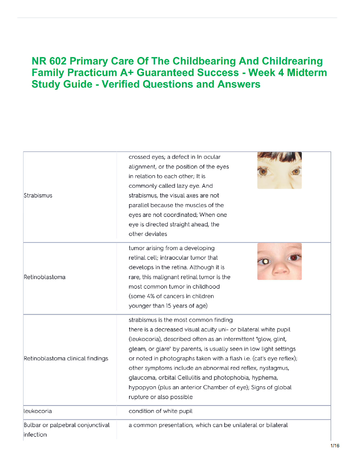



.png)