Biology > QUESTIONS & ANSWERS > Chapter 08: Use of Colony Morphology for the Presumptive Identification of Microorganisms. All Answe (All)
Chapter 08: Use of Colony Morphology for the Presumptive Identification of Microorganisms. All Answers
Document Content and Description Below
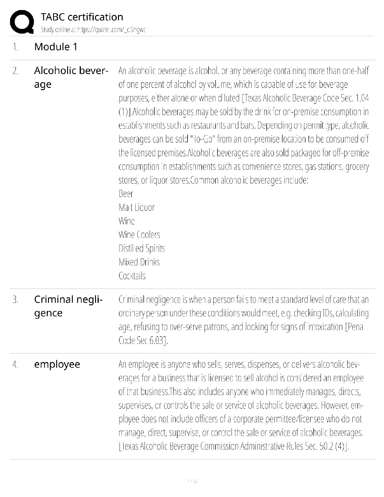
MULTIPLE CHOICE 1. Colony morphology is defined as colony: a. characteristics and form. b. size and color. c. smell and shape. d. constitution and environmental conditions. A Colony cha ... racteristics and forms take into consideration all the different shapes, colors, smells, makeup, texture, and architecture. All the other choices point to a different aspect of a colony’s characteristics and forms. REF: 170 OBJ: Level 1: Recall 2. Which of the following descriptions will directly facilitate identification of bacteria? a. Environmental growth temperature b. Physical characteristics of colony morphology c. Environmental growth atmosphere d. Colony color B Many specific microorganisms have characteristics that distinguish them in a crowd of genera or species. This characterization of colonies on culture media facilitates presumptive identification of commonly isolated organisms. REF: 170 OBJ: Level 1: Recall 3. When microbiologists encounter a sputum specimen, they know that the upper respiratory tract contains many indigenous organisms and to identify every organism in the culture would be a time-consuming, cost-prohibitive, and insurmountable task. So microbiologists must: a. work quickly with commercially available kits to identify all organisms present in a sputum specimen for the best quality patient care available. b. work up only the gram-negative rods present in the culture. c. differentiate the potential pathogens from the “usual” inhabitants of the upper respiratory tract and direct the diagnostic workup only to potential pathogens. d. be able to ascertain whether the results generated by a commercial or automated system correlate with the suspected organism. C The microbiologist must go through a mental catalog of all the organisms on the plate to differentiate between the indigenous organisms and the pathogenic organisms. Then, the pathogenic organisms are the only organisms that need to be identified and worked up for antimicrobial susceptibility testing. REF: 170 OBJ: Level 1: Recall 4. When microbiologists set up a biochemical identification using a commercial system on a suspected pathogen from a culture, they must: a. recognize the colony characteristics of the pathogen. b. observe the culture after 18 to 24 hours of incubation. c. use quality control organisms to ensure the identification of the organism is correct. d. correlate colony characteristics with the suspected identification of the organism. D When a microbiologist suspects a specific organism, a biochemical identification using a commercial system is set up. When the results are available, the microbiologist checks the identification against the colony characteristics on the inoculating plate. If the identification does not coincide with the colony characteristics, a mixed culture should be suspected and the biochemical identification should be redone. REF: 170 OBJ: Level 2: Interpretation 5. Generally, microbiologists observe the colonial morphology of organisms on primary culture after how many hours of incubation? a. 18 to 24 hours b. 6 to 12 hours c. 24 to 48 hours d. 72 hours A The first reading of the primary culture usually occurs from 18 to 24 hours after plating. If done sooner than this, the bacterial colonies may be too small to differentiate. If done later than this, the bacterial colonies may look different from the earlier time. REF: 170 OBJ: Level 1: Recall 6. Plate reading is: a. examining the colonial morphology of bacteria. b. a comparative examination of bacteria growing on a variety of culture media. c. setting up biochemical tests to identify bacteria. d. determining if the inoculum is mixed. B Each type of agar plate that the initial specimen is plated on is examined in relationship to the other. Therefore, as a culture set from a specimen, growth on these three culture media illustrates the comparative colonial examination of plate reading. The colonial morphology and the biochemical reactions with particular media are all important in determining the identification of the bacteria. REF: 170 OBJ: Level 1: Recall 7. A microbiologist is reading plates from a cerebrospinal fluid (CSF) culture. She notices that there is growth on chocolate agar (CHOC), but no growth on sheep blood agar (SBA) or MacConkey (MAC) agar. The Gram stain showed a gram-negative bacillus. What organism should be suspected? a. Streptococcus pneumoniae b. Staphylococcus aureus c. Haemophilus influenzae d. Escherichia coli C H. influenzae is a tiny, gram-negative rod that is very fastidious. It will not grow on MAC and will not grow on SBA. It will grow on CHOC because additional nutritional growth requirements are added to the media. Streptococcus pneumoniae and Staphylococcus aureus are gram-positive cocci that grow well on BAP but do not grow on MAC. E. coli is a gram-negative rod that grows well on SBA, CHOC, and MAC. REF: 171 OBJ: Level 3: Synthesis 8. MacConkey agar is used to: a. differentiate between hemolytic and nonhemolytic gram-negative rods. b. show the clear, pinpoint colonies of Haemophilus influenzae. c. demonstrate the large, pink colonies of Neisseria meningitidis. d. differentiate between lactose fermenters and lactose nonfermenters. D MacConkey (MAC) agar is a differential media used to distinguish between lactose fermenters and lactose nonfermenters. Lactose fermenters produce dark pink to red colonies, and lactose nonfermenters produce colorless colonies. Hemolysis is determined only on sheep blood agar. H. influenzae and N. meningitidis are two fastidious species that do not grow on MAC because they require additional nutrients that are found in chocolate agar. REF: 171 OBJ: Level 1: Recall 9. A microbiologist is reading stool culture plates. She is looking for enteric pathogens on the MacConkey (MAC) plate. What do they look like? a. Clear, colorless colonies b. Large, bright pink colonies c. Small, mucoid, green colonies d. Small, orange colonies A MAC is used to differentiate lactose fermenters (bright pink) from lactose nonfermenters (clear, colorless). Most enteric pathogens do not ferment lactose. Pink colonies indicate a lactose-fermenting gram-negative rod. Green and orange colonies are not seen on MAC. REF: 171 OBJ: Level 2: Interpretation 10. A microbiologist is reading stool culture plates. She sees an organism that has a dry, pink colony with a surrounding “halo” of pink on MacConkey (MAC). What is a good presumptive identification of this organism? a. Enteric pathogen b. Escherichia/Citrobacter–like organism c. Klebsiella/Enterobacter–like organism d. Haemophilus influenzae B Escherichia/Citrobacter–like organisms produce a dry, pink colony with a surrounding “halo” of pink. Enteric pathogens are nonlactose fermenters, so they would not produce a pink colony. H. influenzae is fastidious and does not grow on MAC. Klebsiella/Enterobacter–like organisms produce large, mucoid, pink colonies that occasionally have cream-colored centers. REF: 171 OBJ: Level 3: Synthesis 11. A microbiologist is reading stool culture plates. She sees an organism that has a large, mucoid pink colony on MacConkey (MAC). What is a good presumptive identification of this organism? a. Enteric pathogen b. Escherichia/Citrobacter–like organism c. Klebsiella/Enterobacter–like organism d. Haemophilus influenzae C Klebsiella/Enterobacter–like organisms produce large, mucoid, pink colonies that occasionally have cream-colored centers. Enteric pathogens are nonlactose fermenters, so they would not produce a pink colony. H. influenzae is fastidious and does not grow on MAC. Escherichia/Citrobacter–like organisms produce a dry, pink colony with a surrounding “halo” of pink. REF: 171 OBJ: Level 3: Synthesis 12. The relative concentration of bacteria on culture plates is directly proportional to the: a. amount of nutrients in the culture plates. b. environment in which the bacteria is grown. c. concentration in which they are present in the clinical specimen. d. amount of bacteria isolated from the initial inoculation on the plate. C Bacteria grow on culture media in the same proportion or concentration in which they are present in the clinical specimen. If a lot of bacteria are present in the clinical specimen, then a lot of bacteria will grow on the plate. Each organism located on a Gram stain will foster the growth of bacterial colonies on the plate. REF: 171 OBJ: Level 2: Interpretation 13. -Hemolysis is: a. complete clearing of erythrocytes in a blood agar plate around and under the colony. b. partial lysing of erythrocytes in a blood agar plate around and under the colony. c. when organisms have no lytic effect on the erythrocytes in the blood agar plate. d. a pink “halo” around a colony. B -Hemolysis results in a green discoloration of the media because of the partial destruction of the red blood cells around and under the colony. Complete clearing of erythrocytes in blood agar around and under the colony would be -hemolysis. -Hemolysis is no hemolysis, where the organism has no lytic effect on the erythrocytes in the blood agar. A pink halo does not occur on blood agar. REF: 172 OBJ: Level 1: Recall 14. -Hemolysis is: a. complete clearing of erythrocytes in a blood agar plate around and under the colony. b. partial lysing of erythrocytes in a blood agar plate around and under the colony. c. when organisms have no lytic effect on the erythrocytes in the blood agar plate. d. a pink “halo” around a colony. A Complete clearing of erythrocytes in blood agar plate around and under the colony would be -hemolysis. -Hemolysis results in a green discoloration of the media because of the partial destruction of the red blood cells around and under the colony. -Hemolysis is no hemolysis, where the organism has no lytic effect on the erythrocytes in the blood agar plate. A pink halo does not occur on a blood agar plate. REF: 172 OBJ: Level 1: Recall 15. -Hemolysis is: a. complete clearing of erythrocytes in a blood agar plate around and under the colony. b. partial lysing of erythrocytes in a blood agar plate around and under the colony. c. when organisms have no lytic effect on the erythrocytes in the blood agar plate. d. a pink “halo” around a colony. C -Hemolysis is no hemolysis, where the organism has no lytic effect on the erythrocytes in the BAP. -Hemolysis results in a green discoloration of the media because of the partial destruction of the red blood cells around and under the colony. Complete clearing of erythrocytes in blood agar around and under the colony would be -hemolysis. A pink halo does not occur on a BAP. REF: 172 OBJ: Level 1: Recall 16. A microbiologist is reading a sputum culture on the bench. There is growth on the BAP and CHOC plates, but no growth on the MAC plate. The colonies growing on the BAP have discolored the media to a green color around and under the colonies. What organism could this be? a. Escherichia coli b. Staphylococcus aureus c. Haemophilus influenzae d. Streptococcus pneumoniae D Streptococcus pneumoniae produces -hemolysis on BAP. E. coli will grow on all three plates, not just BAP and CHOC. Staphylococcus aureus will produce -hemolysis on the BAP plate. H. influenzae will not grow on the BAP plate. REF: 171 OBJ: Level 3: Synthesis 17. A microbiologist is reading a sputum culture on the bench. There is growth on the sheep blood agar (SBA) and chocolate (CHOC) plates, but no growth on the MacConkey (MAC) plate. The colonies growing on the SBA have produced a wide, deep, clear zone of -hemolysis around and under the colonies. What organism could this be? a. Escherichia coli b. Staphylococcus aureus c. Streptococcus pyogenes d. Streptococcus pneumoniae C E. coli will grow on all three plates, not just SBA and CHOC. Staphylococcus aureus will produce -hemolysis on the SBA plate Streptococcus pyogenes will produce a wide, deep, clear zone of -hemolysis on SBA. Streptococcus pneumoniae produces -hemolysis on SBA. REF: 173 OBJ: Level 3: Synthesis 18. A microbiologist is reading a vaginal culture on the bench. There is growth on the sheep blood agar (SBA) and chocolate (CHOC) plates, but no growth on the MacConkey (MAC) plate. The colonies growing on the SBA have produced a narrow, diffuse zone of -hemolysis around and under the colonies. What organism could this be? a. Escherichia coli b. Streptococcus agalactiae c. Streptococcus pyogenes d. Streptococcus pneumoniae B Streptococcus agalactiae will produce a narrow, diffuse zone of -hemolysis on the SBA plate. E. coli will grow on all three plates, not just SBA and CHOC. Streptococcus pyogenes will produce a wide, deep, clear zone of -hemolysis on SBA. Streptococcus pneumoniae produces -hemolysis on SBA. REF: 173 OBJ: Level 3: Synthesis 19. When differentiating colony size on the culture plates, what organisms generally have the larger colonies by comparison? a. Gram-negative rods b. Gram-positive cocci c. Gram-negative diplococci d. Gram-positive rods A The largest colonies typically belong to enteric gram-negative rods. Fastidious organisms tend to grow smaller than gram-positive cocci. Gram-positive rods are variable but are usually smaller than gram-negative rods. REF: 173 OBJ: Level 2: Interpretation 20. The edge of the colonies is described as all the following except: a. smooth. b. glasslike. c. filamentous. d. irregular. B The edge of the colonies should be observed and the form, or margin, described as smooth, filamentous, rough or rhizoid, or irregular. REF: 173 OBJ: Level 1: Recall 21. Colonies of Bacillus anthracis are described as: a. Curschmann’s spiral. b. Mickey Mouse ears. c. Medusa’s heads. d. flower like. C These colonies look like Medusa’s head (a center with many snakes coming from it) because of the filamentous appearance of the colony. REF: 174 OBJ: Level 2: Interpretation 22. Swarming is: a. many bacterial colonies crowded together at one place on the agar plate. b. when colonies on a blood agar plate have a “halo” around the bacterial colony. c. when a bacterial colony on a blood agar plate grows over the top of the erythrocytes it hemolyzed. d. a hazy blanket of growth on the surface of the agar that extends way beyond the streak lines. D Swarming is the hazy blanket of growth on the surface of the agar that extends way beyond the streak lines. It is caused by two species of gram-negative bacteria that are motile: Proteus mirabilis and P. vulgaris. REF: 174 OBJ: Level 1: Recall 23. A microbiologist is reading a vaginal culture. She sees the very white colonies that are -hemolytic on a blood agar plate but appear to have feet. What organism could this possibly be? a. Yeast b. Gram-positive cocci c. Gram-negative rods d. Haemophilus influenzae A Gram-positive cocci and gram-negative cocci both grow on a blood agar plate, but their colonies do not appear to have feet. H. influenzae does not grow on blood agar plate. Yeast are white colonies that appear to have feet. REF: 174 OBJ: Level 3: Synthesis 24. The elevation of bacterial colonies is described by all the following except: a. raised. b. pointed. c. convex. d. umbilicate. B Elevation may be raised, convex, flat, umbilicate (depressed center, concave, an “inny”) or umbonate (raised or bulging center, convex, or an “outy”). REF: 174 OBJ: Level 1: Recall 25. Streptococcus pneumoniae typically produces colonies that are said to resemble coins. The technical term for this colony shape is: a. umbonate. b. flat. c. umbilicate. d. raised. C An umbilicate colony has a depressed center and raised edges: concave. An umbonate colony has a raised or bulging center. A raised colony is raised, and a flat colony is flat. REF: 174 OBJ: Level 2: Interpretation 26. The density of the bacterial colony can be described by all the following except: a. transparent. b. translucent. c. opaque. d. ground glass. D Ground glass refers to the roughness of the outer part of the colony, not the amount of light that can pass through it. Transparent colonies allow a lot of light to pass through. Translucent colonies allow some light to pass through. Opaque colonies allow no light to pass through. REF: 174 OBJ: Level 1: Recall 27. Colonies of Staphylococcus aureus are usually described as: a. opaque. b. transparent. c. translucent. d. ground glass. A The colonies are a solid color and do not allow any light to pass through. REF: 174 OBJ: Level 1: Recall 28. Which organism is described as shiny, like a half-pearl, on media supplemented with horse blood? a. Staphylococcus aureus b. Bordetella pertussis c. Streptococcus agalactiae d. Streptococcus pyogenes B Shiny is how this organism appears on blood agar. REF: 174 OBJ: Level 2: Interpretation 29. All the following colors are commonly used to describe bacteria except: a. buff. b. white. c. orange. d. gray. C Color, in contrast with pigmentation, is a term used to describe, in general, a particular genus. Colonies may be white, gray, yellow, or buff. REF: 174 OBJ: Level 1: Recall 30. All of the following terms are used to describe the consistency of a bacterial colony except: a. brittle. b. creamy. c. dry. d. smooth. D Consistency is determined by touching the colony with a sterile loop. Colony consistency may be brittle, creamy, dry, or waxy; occasionally, the entire colony adheres to the loop. REF: 174 OBJ: Level 1: Recall 31. A microbiologist is reading the plates from a sputum culture. The culture is from a patient with cystic fibrosis. One organism dominates the blood agar, chocolate, and MacConkey plates. The MacConkey plate shows an organism with a green pigment and a metallic sheen. The probable identification for this organism is: a. Pseudomonas aeruginosa. b. Serratia marcescens. c. Chromobacterium violaceum. d. Prevotella melaninogenica. A P. aeruginosa is a gram-negative rod that produces a green pigment and sometimes a metallic sheen on culture media. S. marcescens produces a brick-red pigment. C. violaceum produces a purple pigment. P. melaninogenica produces a brown-black pigment. REF: 175 OBJ: Level 3: Synthesis 32. A microbiologist is reading the plates from a sputum culture. There is growth only on the chocolate plate. When the microbiologist takes off the lid of the culture plate, he notices a distinct “mousy” smell. What organism can this be? a. Proteus mirabilis b. Haemophilus sp. c. Staphylococcus aureus d. Pseudomonas aeruginosa B Haemophilus sp. is the only bacterium in this question that will grow only on chocolate that also has a “mousy” smell. P. mirabilis, S. aureus, and P. aeruginosa all grow on other media than chocolate agar. REF: 176 OBJ: Level 3: Synthesis 33. What bacteria is said to have a fruity or grapelike smell? a. Staphylococcus aureus b. Proteus mirabilis c. Pseudomonas aeruginosa d. Nocardia sp. C S. aureus smells like an old sock. P. mirabilis smells putrid. Nocardia sp. smells like a freshly plowed field. REF: 175 OBJ: Level 1: Recall 34. The colonies of this organism form large, rough, greenish, hemolytic colonies on blood agar. What organism is it? a. Staphylococcus aureus b. Escherichia coli c. Streptococcus pyogenes d. Bacillus cereus D B. cereus fits multiple descriptive categories of colonial morphology because it forms large, rough, greenish, hemolytic colonies on blood agar. Staphylococcus aureus forms white, convex, -hemolytic colonies on blood agar. E. coli forms large, mucoid, grayish, hemolytic colonies on blood agar. Streptococcus pyogenes forms a pinpoint colony, surrounded by a wide, deep zone of -hemolysis. REF: 176 OBJ: Level 2: Interpretation 35. What organism forms a small, fuzzy-edged colony with an umbonate center on blood or chocolate agar? a. Eikenella corrodens b. Staphylococcus aureus c. Escherichia coli d. Streptococcus pyogenes A E. corrodens fits multiple descriptive categories of colonial morphology because it forms a small, fuzzy-edged colony with an umbonate center on blood or chocolate agar. Staphylococcus aureus forms white, convex, -hemolytic colonies on blood agar. E. coli forms large, mucoid, grayish, hemolytic colonies on blood agar. Streptococcus pyogenes forms a pinpoint colony, surrounded by a wide, deep zone of -hemolysis. REF: 176 OBJ: Level 2: Interpretation 36. When looking at a tube of thioglycollate broth, streamers or vines and puffballs are visible. What organism grows like this in thioglycollate? a. Gram-negative rods (Enterobacteriaceae) b. Streptococcus spp. c. Yeast d. Pseudomonas spp. B Streptococcus spp. produces streamers or vines and puffballs in broth. Gram-negative rods (Enterobacteriaceae) make the broth cloudy. Yeast grows below the surface of the broth. Both yeast and Pseudomonas produce scum at the sides of the tube. REF: 176 OBJ: Level 2: Interpretation 37. All of the following characteristics are used to describe bacterial colony characteristics except: a. temperature. b. hemolysis. c. color. d. elevation. A Colonial morphology plays a significant role in the presumptive identification of microorganisms, especially in the initial observation and interpretation of cultures. Hemolysis, size, margin, elevation, density, color, consistency, pigment, and odor are used to describe colony characteristics. REF: 172 OBJ: Level 1: Recall [Show More]
Last updated: 3 years ago
Preview 1 out of 11 pages

Buy this document to get the full access instantly
Instant Download Access after purchase
Buy NowInstant download
We Accept:

Reviews( 0 )
$6.00
Can't find what you want? Try our AI powered Search
Document information
Connected school, study & course
About the document
Uploaded On
Jan 23, 2020
Number of pages
11
Written in
All

Additional information
This document has been written for:
Uploaded
Jan 23, 2020
Downloads
0
Views
114


.png)
.png)
.png)
.png)
.png)
.png)
.png)