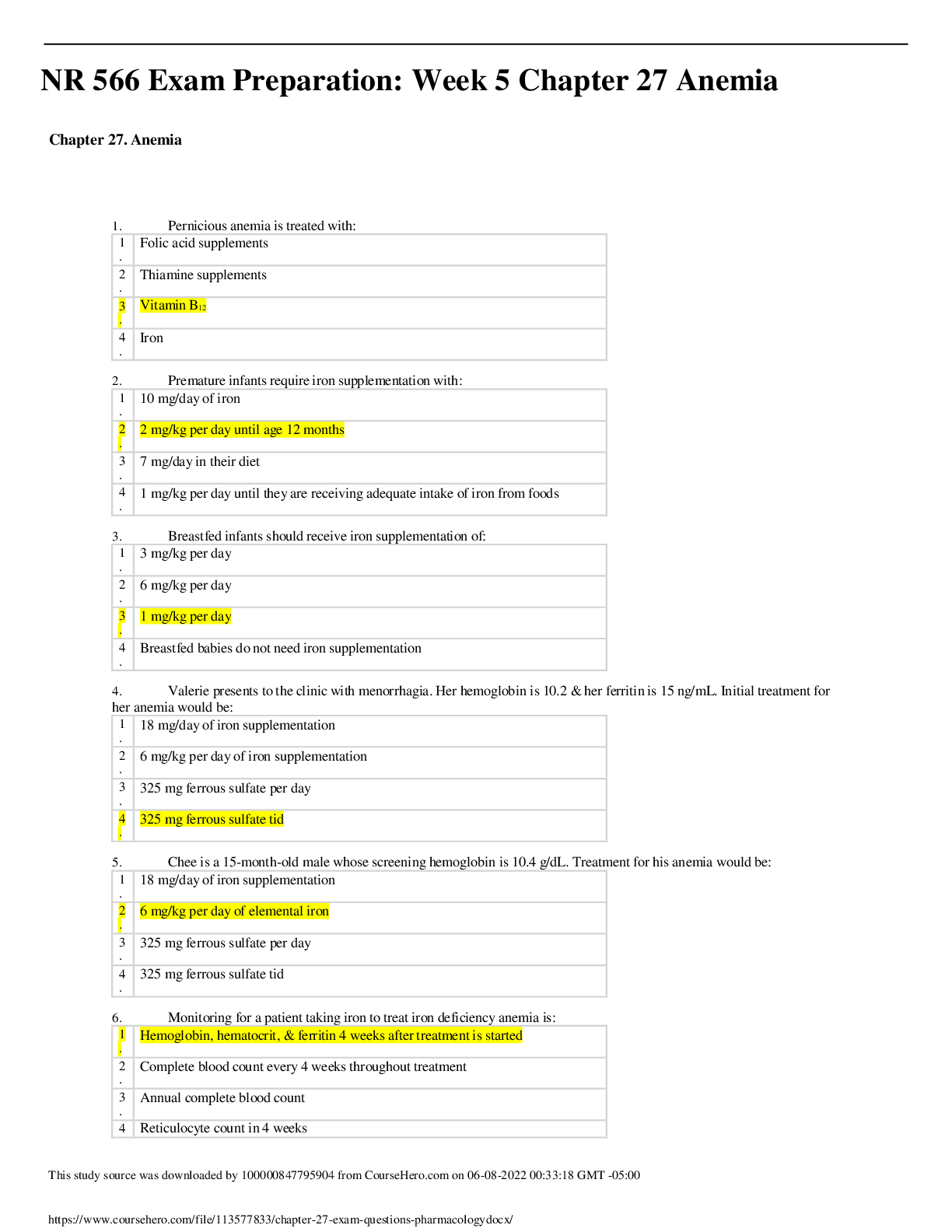
MULTIPLE CHOICE
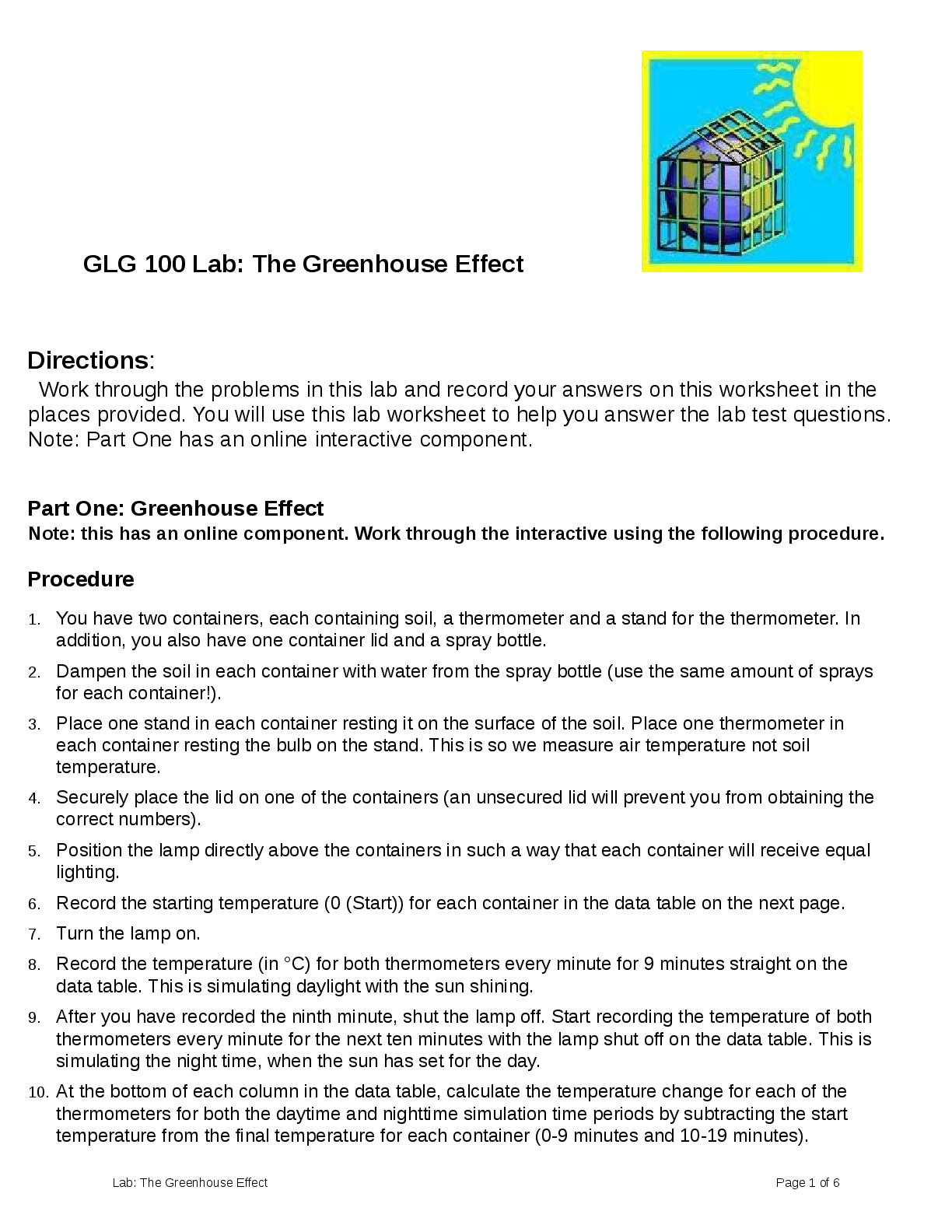
1. This organism was isolated from a sinus aspirate of a patient with chronic sinusitis and respiratory allergies. Identify the organism shown below.
(Courtesy CDC Public Health Image Library (PHI
...
MULTIPLE CHOICE
1. This organism was isolated from a sinus aspirate of a patient with chronic sinusitis and respiratory allergies. Identify the organism shown below.
(Courtesy CDC Public Health Image Library (PHIL); http://phil.cdc.gov/phil/home.asp.)
a. Candida albicans
b. Alternaria spp.
c. Mucor spp.
d. Pseudallescheria boydii
B
Although these isolates may be recovered from virtually any source, they are primarily implicated in chronic fungal sinusitis. Patients are often misdiagnosed and treated for an extended time for bacterial sinusitis. Microscopic evaluation reveals short conidiophores bearing conidia in chains that lengthen in an acropetal fashion. Multicelled conidia have angular cross walls and taper toward the distal end. Alternaria spp. are dematiaceous, rapidly growing fungi with colonies ranging from shades of gray, to brown, to black. CDC PHIL #3962
REF: 610 OBJ: Level 3: Synthesis
2. A bone marrow transplant patient develops an abrupt fever with respiratory distress. Despite treatment with antifungal medications, the patient dies on the fifth day. The following organism was isolated from both sputum and blood cultures. Identify the organism shown below.
(Courtesy CDC Public Health Image Library (PHIL); http://phil.cdc.gov/phil/home.asp.)
a. Rhizopus spp.
b. Mucor spp.
c. Histoplasma capsulatum
d. Aspergillus spp.
D
Aspergillus spp. are the second most isolated fungus after Candida spp. Neutropenia is the single most predictive factor for developing aspergillosis. Aspergillus spp. are the most frequent cause of disease in the bone marrow transplant recipient in addition to other cancer and transplant patients. Infection is initiated following inhalation of fungal conidia. In the lung air spaces, conidia germinate and invade the tissue. Within a few days, the patient develops a severe fever that fails to respond to antifungal therapy.
CDC PHIL #300
REF: 608 OBJ: Level 3: Synthesis
3. A graduate student returning from a month-long fieldwork experience in South America develops a dark nodular lesion on his left hand. Microscopic examination of the nodule reveals dark, coinlike inclusions. The following organism grows in culture. This organism is most likely:
(Courtesy CDC Public Health Image Library (PHIL); http://phil.cdc.gov/phil/home.asp.)
a. Cladosporium spp.
b. Candida spp.
c. Coccidioides immitis.
d. Cryptococcus neoformans.
A
An infrequent cause of disease, Cladosporium is primarily recovered as a laboratory contaminant. Infections are typically confined to the sinuses or following traumatic inoculation. Cladosporium forms brown, to olive, to black hyphae and conidia. Conidiophores are erect and may branch into several conidiogenous cells. This disease is diagnosed by the presence of characteristic lesions accompanied by microscopic sclerotic bodies, often referred to as copper pennies because of their shape and staining properties in tissue sections. CDC PHIL #3214
REF: 611 OBJ: Level 3: Synthesis
4. A Seattle woman with HIV complains of headaches and also displays some memory loss. Because of her history, the physician collects cerebrospinal fluid (CSF) and sends it to the laboratory for analysis. The following image is seen using India ink stain. What is the most likely identification of this organism?
(Courtesy CDC Public Health Image Library (PHIL); http://phil.cdc.gov/phil/home.asp.)
a. Coccidioides immitis
b. Blastomyces dermatitidis
c. Cryptococcus gattii
d. Candida albicans
C
Cryptococcus spp. are important causes of meningitis, pulmonary disease, and septicemia. C. neoformans, the most notable pathogen in this genus, has become a major cause of opportunistic infection in patients with acquired immunodeficiency syndrome (AIDS). Cryptococcus spp. express a capsule that produces the characteristic mucoid colony. The capsule can be detected surrounding the budding yeast in spinal fluid with the aid of India ink or Nigrosin. Cryptococcus gattii is an emerging pathogen, particularly in the Pacific Northwest of the United States. CDC PHIL #3771
REF: 612 OBJ: Level 3: Synthesis
5. The following organism was isolated from an immunocompetent patient with chronic sinusitis. This organism is most likely:
(Courtesy CDC Public Health Image Library (PHIL); http://phil.cdc.gov/phil/home.asp.)
a. Mucor spp.
b. Rhizopus spp.
c. Alternaria spp.
d. Curvularia spp.
D
Curvularia isolates are most often implicated in chronic sinusitis in immunocompetent patients. Multicelled conidia are produced on sympodial conidiophores. This genus is easier to identify because of the frequently crescent-shaped conidia with three to five cells of unequal size, with an enlarged central cell. CDC PHIL 4270
REF: 611 OBJ: Level 3: Synthesis
6. A patient returning from a trip to tropical Africa comes to his physician noting scaly patches of skin along the waistband, which are lighter in color than his normal skin. A skin scraping reveals budding yeast and short septate hyphal elements, with some branching present. This organism, shown below, is most likely:
(Courtesy CDC Public Health Image Library (PHIL); http://phil.cdc.gov/phil/home.asp.)
a. Malassezia furfur.
b. Trichophyton schoenleinii.
c. Microsporum audouinii.
d. Trichophyton rubrum.
A
The M. furfur complex causes tinea versicolor, a disease characterized by patchy lesions or scaling of varying pigmentation. M. furfur may be identified by visualizing skin scrapings from characteristic lesions in a potassium hydroxide preparation or by observing yellow fluorescence with a Wood’s lamp upon examination of the infected body site. Microscopic examination of the direct smear in potassium hydroxide (KOH) preparations reveals budding yeasts, approximately 4 to 8 m, along with septate, sometimes branched, hyphal elements. The microscopic appearance has gained M. furfur the term “the spaghetti and meatballs” fungus. CDC PHIL #2916
REF: 594 OBJ: Level 3: Synthesis
7. A patient who wears long-term contact lenses comes to her physician after noting a small spot on her cornea. Collection of the lesion for culture produced a fast-growing mold with a slight pink color on the surface. Microscopic review showed the following organism. This organism is most likely:
(Courtesy CDC Public Health Image Library (PHIL); http://phil.cdc.gov/phil/home.asp.)
a. Sporothrix schenckii.
b. Fusarium spp.
c. Mucor spp.
d. Geotrichum spp.
B
Fusarium is frequently seen in mycotic dermatitis. In bone marrow transplant patients, mortality from infections caused by the fusaria approaches 100%. Normally, abundant macroconidia with fewer microconidia are produced on vegetative hyphae. Macroconidia are banana- or canoe-shaped and are formed singly, in small clusters, or they may cluster together in mats termed sporodochia. CDC PHIL #4011
REF: 609 OBJ: Level 3: Synthesis
8. The following organism was isolated from the sputum culture of a cancer chemotherapy patient. This organism is most likely:
(Courtesy CDC Public Health Image Library (PHIL); http://phil.cdc.gov/phil/home.asp.)
a. Mucor spp.
b. Penicillium spp.
c. Geotrichum spp.
d. Alternaria spp.
C
Geotrichum has been implicated in pulmonary disease in immunocompromised patients. Microscopic evaluation reveals abundant arthroconidia formed from the vegetative hyphae that occur either single or branched. CDC PHIL #3056
REF: 609 OBJ: Level 3: Synthesis
9. A recreational cave explorer develops a productive cough and fever. A sputum culture is sent to the laboratory for bacterial, fungal, and mycobacterial cultures. A white mold colony develops on Sabouraud’s agar after 5 days at 22° C. Identify the organism shown below.
(Courtesy CDC Public Health Image Library (PHIL); http://phil.cdc.gov/phil/home.asp.)
a. Histoplasma capsulatum
b. Toxoplasma gondii
c. Microsporum tonsurus
d. Paracoccidioides brasiliensis
A
Histoplasmosis is acquired by the inhalation of the microconidia of Histoplasma capsulatum var. capsulatum. Histoplasmosis is also known as reticuloendothelial cytomycosis, cave disease, spelunker’s disease, and Darling’s disease. H. capsulatum grows as a white to brownish mold. Early growth of the mycelial culture produces round to pyriform microconidia measuring 2 to 5 m. As the colony matures, large echinulate or tuberculate macroconidia, a characteristic of the species, are formed.
CDC PHIL #10958
REF: 604 OBJ: Level 3: Synthesis
10. A young boy develops itchy, dry scaly lesions on his leg with a raised, red, outer edge. A culture of the lesion reveals the following organism in a lactophenol cotton blue stain. This organism is most likely:
(Courtesy CDC Public Health Image Library (PHIL); http://phil.cdc.gov/phil/home.asp.)
a. Trichophyton spp.
b. Mucor spp.
c. Microsporum canis.
d. Blastomyces dermatitidis.
C
Each ringworm lesion is the result of a local inoculation of the skin with the etiologic agent. Lesions enlarge with time, usually with most inflammation occurring at the advancing edge of the lesion. Macroconidia are spindle shaped with echinulate, thick walls; measuring 12 to 25 m by 35 to 100 m and have 3 to 15 cells. The tapering, sometimes elongated, spiny distal ends of macroconidia are key features that distinguish this species. CDC PHIL #15472
REF: 597 OBJ: Level 3: Synthesis
11. The following organism is isolated from a case of tinea capitis. The organism grew rapidly producing tan mold colonies. This organism is most likely:
(Courtesy CDC Public Health Image Library (PHIL); http://phil.cdc.gov/phil/home.asp.)
a. Trichophyton rubrum.
b. Epidermophyton audouinii.
c. Fonsecaea compacta.
d. Microsporum gypseum.
D
The fusiform, moderately thick-walled conidia typical of M. gypseum can have as many as six cells. Colonies that form tan to buff conidial masses are typical of fresh isolates, but this species tends to develop pleomorphic tufts of white, sterile hyphae in aging cultures and after serial transfers. CDC PHIL # 15079
REF: 597 OBJ: Level 3: Synthesis
12. This organism was isolated from a diabetic patient with a sinus infection. The mold was extremely fast growing with a dirty white surface. Microscopic observation revealed wide aseptate hyphae without rhizoids. This organism is most likely:
(Courtesy CDC Public Health Image Library (PHIL); http://phil.cdc.gov/phil/home.asp.)
a. Mucor spp.
b. Microsporum spp.
c. Sporothrix schenckii.
d. Malassezia furfur.
A
Mucor spp. have been implicated in rhinocerebral zygomycosis in addition to disseminated disease. Mucor spp. are commonly isolated from the environment worldwide. Rhizoids, typical of some Mucorales, are not found in Mucor spp. Mucor spp. grow rapidly and form cottony, dirty white colonies that become mousy-brown to gray with age. CDC PHIL # 3961
REF: 607 OBJ: Level 3: Synthesis
13. A student working for the summer at a plant nursery has a nodular lesion on his forearm that develops a draining tract. The following organism is isolated from the culture at 22C. Identify the organism shown below.
(Courtesy CDC Public Health Image Library (PHIL); http://phil.cdc.gov/phil/home.asp.)
a. Trichosporon boydii
b. Sporothrix schenckii
c. Trichophyton rubrum
d. Microsporum canis
B
Sporothrix schenckii commonly presents as a progressive lymphocutaneous infection beginning with a single draining lesion and progressing along the limbs via the lymphatic system forming multiple draining lesions. In temperate countries such as France, Canada, and the United States, most cases of sporotrichosis are associated with gardening, particularly with exposure to rose thorns (rose-handler’s disease) and sphagnum moss. Microscopic examination from culture reveals thin, delicate hyphae bearing conidia developing in a rosette pattern at the end of delicate conidiophores. CDC PHIL #10603
REF: 594 OBJ: Level 3: Synthesis
14. A patient with thickened and discolored nails was diagnosed with onychomycosis. A scraping of the nail was sent for culture. Microscopic analysis of the mold colony growth revealed the following morphology. The most likely identity of this organism is:
(Courtesy CDC Public Health Image Library (PHIL); http://phil.cdc.gov/phil/home.asp.)
a. Microsporum canis.
b. Sporothrix schenckii.
c. Aspergillus fumigatus.
d. Trichophyton mentagrophytes.
D
Onychomycosis, infection of the nails, is most often caused by dermatophytes but also may be the result of infection by other fungi. Nails become thick, discolored, and flakey. Some common agents that infect the nails are Trichophyton rubrum, T. mentagrophytes, and T. tonsurans, as well as Epidermophyton floccosum. Trichophyton mentagrophytes makes both microconidia and macroconidia. Microconidia are primarily globose but may appear tear-shaped. Microconidia are found primarily in clusters described as grapelike or “engrape.” Macroconidia are thin walled, smooth, and cigar shaped, with four to five cells separated by parallel cross walls. CDC PHIL # 15105
REF: 597 OBJ: Level 3: Synthesis
15. After returning from a vacation trip to the Amazon River delta in Brazil, a man notices a white dirtlike substance on his axilla hair that does not wash off in the shower. His health care provider samples the hairs and sends them to the laboratory for culture. Straw-colored yeastlike colonies show the following microscopic morphology. This is most consistent with an identification of:
(Courtesy CDC Public Health Image Library (PHIL); http://phil.cdc.gov/phil/home.asp.)
a. Malassezia furfur.
b. Trichosporon spp.
c. Microsporum audouinii.
d. Trichophyton rubrum.
B
Trichosporon spp. produce both arthroconidia and blastoconidia. White piedra occurs on the hair shaft and is characterized by a soft mycelial mat surrounding hair of the scalp, face, and pubic region. White piedra is endemic in tropical areas of South America, the Far East, and the Pacific. CDC PHIL #3936
REF: 595 OBJ: Level 2: Interpretation
16. A young man notices a darkened area of skin on the palm of his hand. It seems to be increasing in size over the past 2 weeks. His health care provider collects a skin scraping for microscopic examination, which shows septate hyphal elements and budding cells. This organism is most likely:
(Courtesy CDC Public Health Image Library (PHIL); http://phil.cdc.gov/phil/home.asp.)
a. Malassezia furfur.
b. Mucor spp.
c. Hortaea werneckii.
d. Microsporum canis.
C
Tinea nigra, characterized by brown to black nonscaly macules that occur most often on the palms of the hands and soles of the feet, is caused by H. werneckii. The disease is endemic in the tropical areas of Central and South America, Africa, and Asia. Diagnosis of tinea nigra can be made by direct examination of skin scrapings placed in 10% to 20% KOH. Microscopic examination shows septate hyphal elements and budding cells. CDC PHIL #4206
REF: 596 OBJ: Level 3: Synthesis
17. All of the following are examples of dimorphic fungi except:
a. Candida albicans.
b. Coccidioides immitis.
c. Histoplasma capsulatum.
d. Sporothrix schenckii.
A
These dimorphic fungi include a mould phase and either a yeast or spherule phase. The yeast or tissue state is seen in vivo or when the organism is grown at 37 C with increased CO2. The mould phase is seen when the organism is grown at room temperature. Thermally dimorphic fungal species associated with human disease include Blastomyces dermatitidis, C. immitis, H. capsulatum var. capsulatum, Paracoccidioides brasiliensis, S. schenckii, and Penicillium marneffei.
REF: 592 OBJ: Level 1: Recall
18. A patient with very pale patches on his arms and legs is examined at his physician’s office. His physician orders a fungal culture. The fungus shows a spaghetti-and-meatball appearance on the direct smear. What organism is it?
a. Microsporum canis
b. Malassezia furfur
c. Trichophyton rubrum
d. Epidermophyton floccosum
B
Microscopic examination of the direct smear in potassium hydroxide (KOH) preparations reveals budding yeasts, approximately 4 to 8 m, along with septate, sometimes branched, hyphal elements. This microscopic appearance has gained M. furfur the term spaghetti and meatballs fungus.
REF: 595 OBJ: Level 3: Synthesis
19. What is the causative agent of black piedra?
a. Microsporum canis
b. Trichosporon beigelii
c. Piedraia hortae
d. Sporothrix schenckii
C
P. hortae is the causative agent of black piedra, an infection that occurs on the hairs of the scalp.
REF: 595 OBJ: Level 1: Recall
20. What is the causative agent of tinea nigra?
a. Microsporum canis
b. Trichosporon beigelii
c. Piedraia hortae
d. Hortaea werneckii
D
Tinea nigra, characterized by brown to black nonscaly macules that occur most often on the palms of the hands and soles of the feet, is caused by P. werneckii.
REF: 596 OBJ: Level 1: Recall
21. All of the following organisms cause cutaneous mycoses except:
a. Sporothrix schenckii.
b. Trichophyton beigelii.
c. Microsporum canis.
d. Epidermophyton floccosum.
A
Three genera of fungi, Trichophyton, Microsporum, and Epidermophyton, are etiologic agents of dermatophytosis. Species within these genera are keratinophilic—that is, they are adapted to grow on hair, nails, and cutaneous layers of skin that contain the scleroprotein keratin.
REF: 594 OBJ: Level 1: Recall
22. Chromoblastomycosis is caused by all the following organisms except:
a. Fonsecaea compacta.
b. Penicillium marneffei.
c. Fonsecaea pedrosoi.
d. Phialophora verrucosa.
B
Chromoblastomycosis is caused by several infectious agents, namely Fonsecaea compacta, F. pedrosoi, Phialophora verrucosa, Cladophialophora carrionii, and Rhinocladiella aquaspera.
REF: 598 OBJ: Level 1: Recall
23. What organism is found in the San Joaquin Valley region of California?
a. Sporothrix schenckii
b. Penicillium marneffei
c. Coccidioides immitis
d. Paracoccidioides brasiliensis
C
Although morphologically identical, genetic evaluation has differentiated between C. immitis and C. posadasii. It appears that the species can be traced to specific geographical locations. C. immitis is encountered in the San Joaquin Valley region of California, whereas C. posadasii is found in the desert southwest of the United States, Mexico, and South America.
REF: 604 OBJ: Level 2: Interpretation
24. What organism frequently presents in fungus balls?
a. Fonsecaea compacta
b. Penicillium marneffei
c. F. pedrosoi
d. Aspergillus fumigatus
D
Neutropenia is the single most predictive factor for developing aspergillosis. Another frequent presentation is “fungus balls” in the lungs of agricultural workers who routinely are in contact with fungal conidia from environmental sources.
REF: 608 OBJ: Level 1: Recall
25. What organism is one of the primary opportunistic infections in AIDS patients?
a. Pneumocystis jirovecii
b. Hortaea werneckii
c. Coccidioides immitis
d. Paracoccidioides brasiliensis
A
Since the early 1980s, P. jirovecii (nee P. carinii) has remained one of the primary opportunistic infections found in patients with AIDS.
REF: 623 OBJ: Level 1: Recall
26. For potassium hydroxide (KOH) to work properly, what is done to speed up the dissolution of the keratin and skin layers?
a. Mix gently.
b. Let the slide sit for 30 minutes before reading.
c. Heat gently, then cool.
d. Let sit under an ultraviolet light for 20 minutes.
C
In this procedure, a drop of the KOH preparation is added to a slide. Nail scrapings, hair, skin scales, or thin scales of tissue are added to the drop, and a coverslip is added. The slide is then gently heated and allowed to cool for approximately 15 minutes. The KOH breaks down the keratin and skin layers so that observers can more easily visualize any fungi that may be present in the specimen.
REF: 617 OBJ: Level 2: Interpretation
[Show More]


 Med Surg test Latest Verified Questions and all Correct Answers with Explanations Problems Chapter 27 Management of Patients with Coronary.png)